 |
| About Bioline | All Journals | Testimonials | Membership | News |
|
||||||
|
||||||
East and Central African Journal of Surgery, Vol. 13, No. 2, September, 2008, pp. 87-90 Brachial Plexus Lesions: Anatomical Knowledge as an Essential Diagnostic Tool in a Situation of Limited Imaging Facilities J. Gashegu1, M. Nyundo2, F. Ntirenganya2, M. Perez2, I. Kakande2 1Clinical Anatomy Laboratory, 2Department Of Surgery, National University of Rwanda Code Number: js08038 Diagnosing brachial plexus lesion is a challenge in countries where imaging facilities are not well developed. Here we report 3 cases of different lesions of the brachial plexus sustained after a road traffic crush. The first case presented with a lesion of the 3 primary trunks of the right brachial plexus. The clinical examination showed paralysis of all terminal nerves of the brachial plexus and the collateral branches of both anterior and posterior brachial plexus. The second patient showed paralysis of all muscles of the shoulder and muscles of the anterior compartment of the arm. This clinical feature is in accordance of the upper brachial trunk lesion. The third patient had paralysis of muscles of the hand being innerved by median nerve or ulna nerve. He showed also paralysis of muscles of the anterior compartment of the forearm and anaesthesia of the medial region of hand and forearm. This clinical feature was in conformity with a lesion of inferior primary trunk. All diagnoses were made based on the clinical findings. These cases demonstrate the significance of a through anatomical knowledge in the clinical examination if one has to avoid confusing the signs of terminal nerves lesion with the trunk’s lesion. These cases underscore the importance of applied anatomy in clinical situations. Introduction Several Terms are used to describe Brachial Plexus Injuries depending on which trunk is injured and include Erb's Palsy (an upper trunk injury), Klumpke's Palsy (a lower trunk injury), Brachial Plexus Palsy, Erb-Duchenne Palsy, Horner's Syndrome (when facial nerves are also affected), and "Burners" or "Stingers" (usually associated with sports-related brachial plexus injuries). The "stinger" or "burner" syndrome is classically characterized by unilateral weakness and a burning sensation that radiates down an upper extremity. The condition may last less than a minute or as long as 2 weeks, with the latter duration described as a chronic burner syndrome. Torticollis is another term sometimes used in conjunction with brachial plexus injuries. Injuries to the Brachial Plexus can involve:
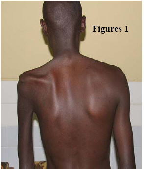
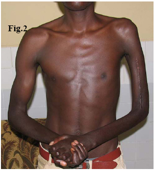
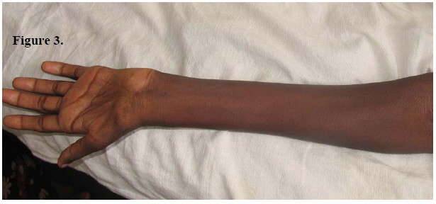
Injury of the brachial plexus is becoming more common because of the increasing number of motorcycle accidents1,2 and are usually associated with multisystem trauma that can delay the recognition of brachial plexus lesion when the patient is in a coma state3. The severity of the injuries can range from palsies that recover spontaneously to avulsion of the nerve roots with permanent loss of function3. The diagnosis of BPI has improved as a result of the modern medical imaging mostly the MRI. Electromyography (EMG), neurophysiology with compound muscle action potential (CMAP) and somatosensory evoked potential (SEP) are important to determine the level of the lesion2,3,4. Anatomical knowledge is very essential for the investigation being clinical or imaging. In conditions where imaging and physiological investigations are not available, clinical examination is the only mean and requirement of anatomical knowledge is crucial. In this paper, we present 3 cases of brachial plexus injury (BPI) seen at Butare University Hospital following trauma. Case reports Case 1. A 24-year-old male soldier presented to the consultation surgical clinic with a history of a persistent left upper limb weakness, sensory loss and paresthesia. He reported that one year prior to consultation he was a victim of an ambush of the military convoy and he found himself lying behind the car with his left upper limb on this back. He had a humerus fracture which was operated. Physical examination showed a paralysed left upper limb with atrophy of all muscles of the upper limb (Figures 1 and 2). The pectoralis major muscle and the muscles of rotator cuff were paralysed and atrophic. Rhomboid muscle, serratus muscle, the low part of the trapezus as well as dorsalis major were also atrophic. There was loss of sensation of the entire left upper limb. The patient had also a pain beginning on the neck and irradiated to the upper limb which was found to be the Tinel sign. This pain was triggered by palpation at the lateral side of the left neck where a small mass was visible and was estimated to be of grade 3 that is intolerable pain sufficient to awaken the patient from sleep5. The motor deficit was related to palsy all terminal nerves of the brachial plexus namely the musculo-cutaneous, median, ulnar, radial and axillary nerves and the most prominent collateral braches (pectoral, suprascapular, long thoracic, sub scapular nerves). Based on the anatomical background we concluded that it is the lesion of the three primary trunks. Case 2 .A 19-old male patient was received at outpatient consultation with weakness of the right upper limb. Four months earlier, he had been involved in a motorcycle accident. The details of the accident were not very clear. After two days of accident he felt weakness of the upper limb and sought for help at a health centre. He had received same medication without improvement so he came to consultation. The physical examination showed paralysis of rotator cuff muscles, the deltoid and muscles of the anterior compartment of the arm. The patient was not able to abduct and externally rotate of the shoulder. Neither was he able to he able to do the flexion of the elbow but cpuld extend elbow joint. There was marked atrophy of rotator cuff muscles, deltoid muscle and the anterior compartment of the right arm. The pectoralis major, trapezus, rhomboid, dorsalis major muscles were normal. The muscles and movements of forearm and hand were also normal. There was a sensory deficit at the deltoid region and part of the lateral area of the forearm. He had also a Tinel sign trigger by palpation on the scaleneus region. A diagnosis of injury of the upper primary trunk of the brachial plexus (C5-C6) was made. Case 3. It is a 46-year-old male patient was referred to our department of surgery with weakness of the left upper limb following involvement in an accident a one month before. He had also received some medicines without a improvement. On physical examination, the patient was unable to flex his fingers and left wrist. However he could extend his left fingers and wrist joint. The movements of the left elbow as well as the left shoulder were normal. He had atrophy of muscles of the thenar and hypothenar eminences of the left hand (Figure 3). There was also atrophy of muscles of the anterior compartment of forearm. Sensory deficit was found on the medial aspects of the hand and forearm. A positive Tinel sign was also found on palpation at the scalenous region of the neck, the pain was evaluated as grade 2: severe pain. A diagnosis of a lesion of the inferior primary trunk of the left brachial plexus was reached. Discussion The three cases of brachial plexus lesions reported demonstrate the importance of a vast knowledge of anatomy and clinical skills. Injuries to the Brachial Plexus can result in full to partial paralysis of one or both arms with a temporary or, when the nerve cannot completely heal, a life time injury. The Brachial Plexus can be damaged in a number of different ways including accidents involving high impact conditions (automobiles, motorcycles, snowmobiles, sports) but most brachial plexus injuries occur during birth with a condition called Shoulder Dystocia (SD). Brachial plexus injuries are the most common peripheral nerve injuries seen in athletes. True rate of brachial plexus injuries is difficult to determine due to significant underreporting. Many stingers last briefly and players do not seek medical attention. Clancy et al6 reported that 33 of 67 college football players (49%) sustained at least 1 burner during collegiate play. Sallis et al7 surveyed Division III college football players and reported that 65% experienced brachial plexus injuries. Complete traumatic brachial plexus palsy is a severe neurological condition that affects mostly the young adults and leaves them with important sequels. It is usually presented as upper limb motor and sensitive deficits. The avulsion of the brachial plexus roots are believed to be of bad prognostic sign5,8. The case of complete brachial plexus injury we present carried a poor prognosis with almost no possibility of recovery. The injury of upper trunk alone of the brachial plexus is believed to be the most frequent lesion of the brachial plexus9. It counts for one third of all brachial plexus injury4. The mechanism of injury is the closed traction on the upper limb. More specifically, the injury of the upper trunk is caused by downward traction with abduction and internal rotation of the shoulder3. Clinical feature of the case we present showed the impairment of the shoulder abduction, external rotation and the flexion. There are the main symptoms of the injury of the upper trunk of the brachial plexus4,9. Injury of the low trunk is a rare condition. The mechanism of lesion is the upward traction with abduction and internal rotation which results mostly in lesions of lower cervical roots. It is generally recognized that limited avulsion of lower cervical roots has a better prognosis. The case we present had impairment of intrinsic muscles of the hand and muscles of the anterior compartment of the forearm. These symptoms can also suggest the injury of both ulnar and median nerves. We ruled out this condition for two reasons. Firstly, it is unusually that two different nerves are simultaneously injured3. Secondly, the sensory examination showed that the patient lost the sensation of light touch and pinprick was impaired on the medial region of hand and forearm. This distribution is the cutaneous territory of the low trunk of the brachial plexus and not of the ulnar and median nerves9. Conclusion: Knowledge of applied anatomy is vital for a good neurological examination in brachial plexus injury particularly in situations where the imaging facilities are poor. There is need to emphasize applied anatomy clinical skills during undergraduate and posrtgraduate traing of medical students and surgery residents. The three cases described represent the three main types of avulsions of brachial plexus roots and have been diagnosis clinically based on the deep knowledge of anatomy. References
© 2008 East and Central African Journal of Surgery The following images related to this document are available:Photo images[js08038f3.jpg] [js08038f1.jpg] [js08038f2.jpg] |
| |||||||||
