 |
| About Bioline | All Journals | Testimonials | Membership | News |
|
||||||
|
||||||
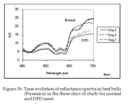
Meat colour of fighting bulls Color de la carne de toros de lidia B. Hernández, G. Lizaso, A. Horcada, M.J. Beriain1 and A. Purroy Escuela Técnica Superior de Ingenieros Agrónomos, Universidad Pública de Navarra, Campus Arrosadía, 31006 Pamplona, Spain Recibido Marzo 06, 2006. Aceptado Junio 07, 2006. Code Number: la06017 ABSTRACT. Meat colour (CIE L*a*b*) from 14 fighting bulls was analysed. Meat from these animals (Longissimus dorsi muscle) presented high pH values (6.37±0.28, 24hours post mortem) and can be considered, in view of its special treatment prior to slaughter, as stressed meat. In spite of this fact, only two animals exhibited the typical behaviour of DFD meat. Upon comparing these results with commercial beef from bulls of the Pirenaicabreed sacrificed in a slaughterhouse, it was seen that the colour of meat from fighting bulls is darker, redder, more purple and more saturated than that of beef from the commercial Pirenaica breed. Keywords: fighting bull/ beef/ breed/ meat colour/ DFD meat. RESUMEN. En este artículo se analiza el color (CIE L*a*b*) de la carne de 14 toros de lidia. La carne de estos animales (Longissimus dorsi muscle) ha presentado valores de pH elevados (6.37±0.28, 24horas post-mortem) y puede considerarse debido al especial tratamiento recibido antes de su muerte como carne estresada. A pesar de ello solo dos de los animales han mostrado el comportamiento típico de la carne DFD. Al comparar esta carne con la de ternera comercial, procedente de 15 machos de raza Pirenaica sacrificados en un matadero, se ha visto que la carne de toro de lidia tiene un color rojo más púrpura y más saturado que la carne de ternera comercial de raza Pirenaica. Palabras clave: toro de lidia/ carne bovina/ raza/ color de carne/ carne DFD Introduction Fighting bulls are very special animals, which have been selected for their beauty and behaviour in the bullring independent of their later use as meat. Fighting bulls are subjected to both physical and emotional stress before their death, conditions known to affect quality of meat. Meat from stressed or excessively fatigued animals has a high pH (>6.0 as opposed to 5.4-5.8 in normal meat) and a dark colour (Renerre, 1990; Seideman et al., 1984). The three main factors affecting meat colour are the myoglobin content, the relative proportions of its derivatives, and the ultimate pH (MacDougall, 1982; Seideman et al., 1984). The effect of pH on meat colour stability is important from the standpoint of both ultimate pH in postrigor muscle, and the rate of pH decline in prerigor postmortem condition. In general low pH values favour the oxidation of myoglobin (Faustman and Cassens, 1990). High pH meat, known as DFD (Dark, Firm, Dry) or dark cutting meat in beef, does not bloom when exposed to oxygen (Egbert and Cornforth, 1986). Beef has the highest concentration of myoglobin and is therefore darker than meat of other species. In the absence of oxygen, myoglobin is in the form of deoxy or reduced myoglobin (Mb) that has a purple-red colour. On exposure to air, it is oxygenated to form oxymyoglobin (MbO2), which imparts to the muscle a bright red colour that consumers find attractive. Finally there is an oxidised form, metmyoglobin (MMb) which has a dull, brown colour that consumers associate with loss of quality (Jeremiah et al., 1972; Kropf et al., 1986; Lynch et al., 1986; MacDougall, 1982). Many authors have found a positive correlation between the red coordinate a* and the assessment of acceptability by consumers (Chan et al., 1995, 1996; Johansson, 1989; Renerre and Mazuel, 1985; Strange et al., 1974). The decrease in readness (lower a*) is due to an increased oxidation of myoglobin (Hernandez et al., 1999) and indicates a decrease in colour acceptability (Moore and Young, 1991). In addition, chroma (C*) has been described as a good parameter to characterize colour changes, because this value decreases as the brown colour appears (Lizaso, 1998; MacDougall, 1977; Renerre and Mazuel, 1985; Smulders and Van Laack, 1989). Colour together with myoglobin content has been suggested as a useful means to characterise beef from different breeds of Spanish cattle (Insausti et al., 1999; Lizaso, 1998). The effect of breed on meat colour can be explained by the maturing rate of animals (Renerre, 1982). In this sense unstable colour as compared to beef cattle (Faustman and Cassens, 1990; Lanari and Cassens, 1991). The aim of this work is to characterise the meat of fighting bull, study how ageing changes meat colour and investigate the stability of colour or shelf life of this kind of meat in comparison with other typical commercial beef breed. Material and Methods Animals Fourteen 4 yr old running bulls selected from the Fiesta of San Fermin (Pamplona, Spain) were studied. These animals were from an extensive breeding system and for the last year they had eaten commercial concentrate. They had run in the traditional Bull Run in the morning and were slaughtered by a sword wound in the bullring that afternoon. After slaughter, carcasses were kept for 4 hours at ambient temperature (18ºC) and subsequently chilled for 20 hours at 2ºC and 98% relative humidity. These animals were compared with another beef group consisting of 15 bulls of Pirenaica breed slaughtered at 12-13 months of age. Pirenaica cattle are the most important source of beef in the western Spanish Pyrenees. This breed is included in a quality denomination following the EEC policy of promoting and guaranteeing the quality and authenticity of products due to some differential characteristics. The conventional Pirenaica animals were weaned at approximately 5-6 months of age; before weaning they were fed with maternal milk and commercial starter concentrate. From weaning to slaughter the animals received a commercial concentratesimilar to that fed to the fighting bulls and ammonium-treated barley straw, both ad libitum. MethodsLongissimus dorsi muscle (pars lumborum) was removed from the left half of the carcass 24 hours post-mortem. This muscle was cut into steaks (2.0 cm thick) that were individually placed in plastic foam trays overwraped with oxygen permeable film and kept in the refrigerator (2ºC) during the time of study. The pH of Longissimus dorsi muscle was measured at 24 hours post-mortem with a pH meter (Crison 507) with a penetration probe. Reflectance spectra (R (%), wavelength interval 400-700 nm, wavelength resolution = 10 nm) and CIE L* a* b* coordinates, hue (h*) and chroma (C*) were determined using a MINOLTA CM-2002 with a D65 illuminant and 10º observer at three different periods of time: (1) after 24 hours of oxygenation, (5) 5 days post mortem and (7) 7 days post mortem. Colour was also determined through chemical parameters, i.e. content of myoglobin derivatives (deoxymyoglobin, oxymyoglobin and metmyoglobin). Relative proportions of myoglobin derivatives were estimated using the method described by Stewart et al. (1965) following the guidelines for meat colour evaluation (Hunt et al., 1991). Total content of myoglobin was determined (Hornsey, 1956) using a SHIMAZU UV-2001 PC spectrophotometer. Statistical analysis Analysis of variance was performed using the SPSS 6.1.2 (SPSS, 1995) statistical package. The following model was used: Yijk = m + Ti + Bj+ Ti x Bj +e ijk where Yijk= pH, myoglobin content, CIE L*a*b* coordinates, hue (h*) and chroma (C*); Ti = fixed effect due to the periods of time; Bj = fixed effect due to the breed; Ti x Bj = effect due to the interaction between periods of time and Breed; e ijk = residual effect. Results and Discussion Remarkable differences where found in the meta colour between fighting bulls and the group of commercial beef cattle (P<0.001). Fighting bulls showed a content of heme-pigments more than double that of the other breed (10.96±0.20 mg of myoglobin /g of fresh tissue versus 4.17 ±0.19) (P<0.001; Table 1). In general colour of meat from fighting bulls was darker, redder and more purple than that of beef from the commercial Pirenaica breed (P<0.001). pH values in fighting bulls correspond, in theory, to DFD meat (6.37±0.28) as described by Lawrie (1988) versus a normal meat (5,47±0.04) observed in the beef bulls. This last fact is in agreement with what could be expected in meat that has suffered such a stressing treatment. Averaged carcass weights of fighting bulls and Pirenaica bulls were similar. Colour evolution of meat of fighting bulls is shown in Table 2. Colour coordinates CIE L*, a*, b*, hue h* and chroma C* were measured after a period of oxygenation of 24 hours (day 1), five days post mortem (day 5) and seven days post mortem (day 7). In bull fighting and beef Pirenaica. Evolution of colour was similar in fighting bulls and beef Pirenaica bulls. Lightness L* remained constant during the days of the study. The red coordinate a* and the chroma C* decreased between day 1 and day 7 (P<0.05). The higher values of blue-yellow coordinate (b*) were observed at day 1. Thereafter, no differences were observed. Highest Hue h* was observed on day 1 in fighting bulls and day 7 in Pirenaica commercial breed bulls. This evolution is in agreement with previous studies performed in beef (Hernandez, 1994; Lizaso, 1998) that showed similar evolution of colour coordinates with time. However, MacDougall (1982) report that lightness increases with meat ageing. Meat of fighting bulls has L* values lower than those of beef from the Pirenaica group at different periods of time (P<0.001) due to its greater content of pigments and high pH values. Therefore, fighting-bulls meat is darker than meat of Pirenaica bulls at all ageing times. This high content of myoglobin is due to the production system, the breed itself and their older age at slaughter (four years in fighting bulls versus about one year in the Pirenaica breed). More active animals, such as those on pasture, will produce meat with higher concentrations of myoglobin and darker in colour than less active animals. Furthermore myoglobin concentration increases with increasing chronological age (Renerre, 1982). With the same quantity of myoglobin, meat is darker when it has high pH values. Violent excitement immediately before slaughter influences ultimate pH in ruminant animals and the muscle fibers of dark cutters are swollen and tightly packed together forming a barrier to the diffusion of oxygen and the absorption of light (Seideman et al., 1984). Table 1. Values of live weight, myoglobin content and pH in Longissimus dorsi muscle of Fighting bulls and Pirenaica breed bulls.
***, P<0.01; NS, P> 0,05 Table 2. Evolution of colour coordinates CIE L*, a*, b*, hue h*, and chroma C*(± standard deviation), in fighting bulls and Pirenaica breed bulls (n=15) after 24 hours of oxygenation (Day 1) five days post mortem (Day 5) and seven days post mortem (Day 7).
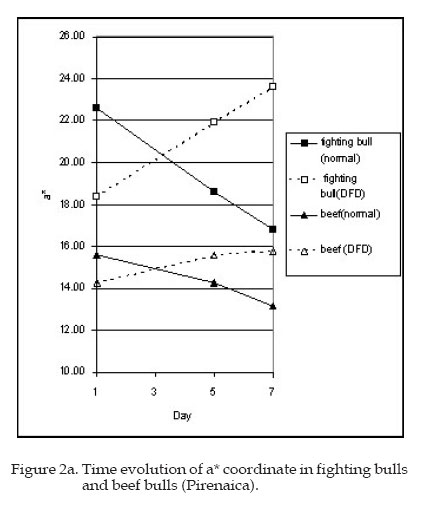
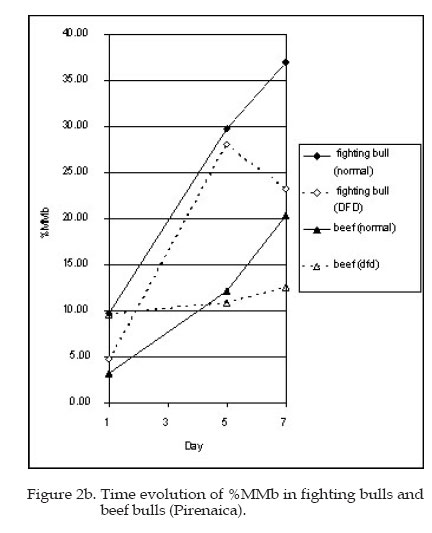
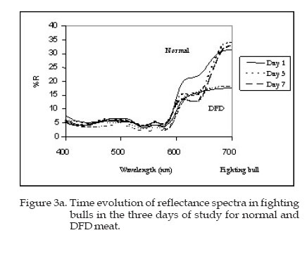
Values in rows with different letter: are significantly different P<0.05 between periods of time In Figure 1, combinations of a* and b* coordinates of fighting bulls and Pirenaica bulls are represented. These two coordinates show that fighting-bulls meat is more purple red or saturated and less red than Pirenaica beef. Meat with pH values between 5.5 and 5.8 is considered normal whereas that with pH ³5.8 is considered DFD meat. Different authors suggest that DFD meat is characterized by a dry, firm and dark aspect which causes problems of health security during packaing and is linked to a decrease in the price (Renerre and Valin, 1979; Warner et al., 1986; Wheeler and Koohmaraie, 1994). Normal meat blooms when it is exposed to air, developing a bright red colour due to the oxygenation of myoglobin to form oxymyoglobin as the predominant surface pigment, whereas DFD does not bloom when exposed to air (Egbert and Cornforth, 1986; Seideman et al., 1984). However in fighting bulls, meat with high pH values has a normal behaviour. This fact can be seen in a decrease of the a* coordinate (Figure 2a) as well as an increase of the metmyoglobin content with time (Figure 2b). In normal meat the a* coordinate decreases and the proportion of MMb increases with time. In DFD meat a* increases with time due to the slow oxygenation of pigments. If the evolution of reflectance spectra is examined (Figures 3a and Figures 3b), a similar conclusion can be extracted. Colour evolution in DFD meat is quite different from the evolution in normal meat. In normal meat the characteristic peaks of oxymyoglobin in the central part of the spectrum clearly appear from the very first hours of exposure to air. The characteristic depression between 610nm and 630nm wave length of metmyoglobin becomes deeper with time due to the increasing proportion of oxidised pigment on the surface. On the contrary, in DFD meat the oxymyoglobin peaks and the metmyoglobin spectrum becomes depression are poorly marked and the shape of the reflectance spectrum does not appreciably change with meat ageing (Hernandez, 1994). The reflectance spectra of fighting bulls have shown changes in DFD meat practically imperceptible, whereas in normal meat spectra the characteristic depression of metmyoglobin is marked in a progressive manner. Surprisingly, meat from only two fighting bulls with individual pH values of 6.1 and 6.6 were considerated as having a DFD behaviour. All the animals catalogued as DFD in the beef breed group have meat with pH>6 while all those catalogued as normal have pH<6, whereas in the fighting bull group the normal also animals have meat pH values comprised between 5.8 and 6.3. Then, there are animals with high pH values and normal behaviour despite the special pre-slaughter treatment. In Figures 2b it can be also seen that the proportion of metmyoglobin in fighting bulls is higher than in beef bulls. If the value of 20% of metmyoglobin pointed out by Renerre and Mazuel (1985) as a criterion of rejection by consumers, or in other words, shelf-life is considered, meat from fighting bulls reached this level in three days versus seven days for normal meat of the Pirenaica breed. In conclusion, despite the Bull Run earlier in the day and the special conditions of slaughter resulting in high pH, meat from fighting bulls can not be regarded as DFD meat. The muscular tissue of these animals seems to resist well this kind of stress treatment, maybe due to genetic causes. This breed deserves further careful study as a meat-producing animals. Literature Cited
© 2006 ALPA. Arch. Latinoam. Prod. Anim. The following images related to this document are available:Photo images[la06017f3b.jpg] [la06017t1.jpg] [la06017f3a.jpg] [la06017f2b.jpg] [la06017f1.jpg] [la06017t2.jpg] [la06017f2.jpg] | |||||||||||||||||||||||||||||||||||||||||||||||||||||||||||||||
| |||||||||