 |
| About Bioline | All Journals | Testimonials | Membership | News |
|
||||||
|
||||||
East African Journal of Public Heath, Vol. 5, No. 2, August, 2008, pp. 122-125 Molecular Epidemiology Of Chikungunya Virus In Vellore District, Tamilnadu, India in 2006 Deepika.T. Lakshmipathy & Dharumadurai Dhanasekaran 1 Division of Environmental Biotechnology, School of Biotechnology, Chemical and Biomedical Engineering, VIT University, Vellore-632014,
India. Received 14th February 2008, Revised 24th July 2008, Accepted 30th July 2008 Code Number: lp08023 Abstract Objective: The present
study was carried out with the aim of evaluating the epidemiology of
Chikungunya virus (CHIKV) in Vellore district and also to identify the most
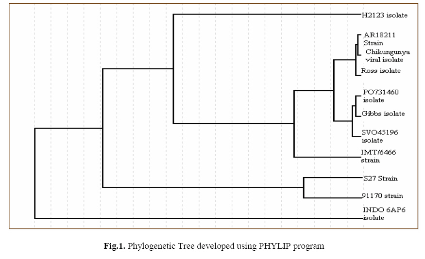
susceptible sex and age group to CHIKV infection. Keywords: Chikungunya virus E1 gene, Phylogenetic analysis, African genotype, RNA secondary structure, Restriction sites. Introduction Chikungunya fever is a mosquito-borne illness of humans caused by the Chikungunya virus. The virus is currently causing one of the largest reported outbreaks of CHIKV fever in the last 40years (1). CHIKV is enzootic in many countries in Asia and throughout tropical Africa (2). Chikungunya virus, a member of Alpha virus genus in the family Togaviridae is an enveloped, single, positive strand RNA virus about 70nm in diameter and spherical in shape (3). Surface projections are distinctive glycoprotein spikes covering evenly the surface. The complete genome is 11,824 nucleotides long. The symptoms of Chikungunya include fever which can reach 39°C, a petechial or maculopapular rashes, and arthralgia or arthritis affecting multiple joints, headache, conjunctival infection, and slight photophobia. Dermatological manifestations such as lichenoid eruption, hyperpigmentation and multiple ecchymotic spots were observed in a recent outbreak of CHIKV fever (4). There is neither specific treatment nor vaccine currently available. The US Centers for Disease Control and Prevention (CDC) fact sheet on Chikungunya advises against using Aspirin. Ibuprofen, Naproxen and other non-steroidal anti-inflammatory drugs are recommended for arthritic pain and fever (5). Chikungunya is believed to have originated in Africa (6). It was first reported in 1952 from Makonde plateaus. It was first isolated in 1953 by R.W.Ross during an epidemic in Newala district,Tanzania; East Africa and described in 1955 by Marion Robinson and WHR Lumbsdenin (3). The virus has been reported from Congo, Uganda, East Africa, Senegal, Central African Republic, Cameroon, Mayotte, Portugal, Guinea, Philippines, Malaysia and Reunion Islands (7-9). Since Tanzania outbreak in 1952, Chikungunya virus has caused outbreaks in East Africa (Tanzania and Uganda) (10), in Austral Africa (Zimbabwe and East Africa) (11), in West Africa (Senegal) and in Central Africa (Central African Republic of the Congo) (12). Since the documented Asian outbreak in 1958 in Bangkok, Thailand, outbreaks have been reported in Cambodia, Vietnam, Laos, Myanmar, Malaysia, Philippines and Indonesia (13-15). Chikungunya outbreak was reported in Calcutta and Chennai in 1963-1965 and in 1973, an epidemic was documented in Barsi, Maharashtra (5). Since the beginning of 2005, CHIKV has emerged in the islands of the South-western Indian Ocean and the outbreak was first reported in Comoros. Later in the same year, the virus had circulated to the other islands and countries – Mayotte, Seychelles, Reunion, Mauritius, Madagascar and India (16). The current outbreak in India started in the end of 2005 (17). A total of 129 districts in 8 states had been reported of being affected by the virus (5). Tamil Nadu recorded the highest number of districts affected with CHIKV infection with nearly 300 cases being reported in the Vellore district. The aim of this study was to determine the epidemiology of CHIKV in Vellore district. The prevalence of CHIKV infection in Vellore district is significantly maximum, hence the present study is planned to identify the most predominat strain of CHKV and the factors most likely responsible for the incidence and spread of CHKV infection in Vellore district ,Tamilnadu,India. Materials and Methods Area of sampling and collection of blood samples Totally five locations were selected in and around Vellore district, Tamil Nadu, India (12.93°N, 79.13°E) namely Vellore, Katpadi, Thiruvallam, Sathuvachari and Gudiyatham. 5ml blood was collected from CHIKV infected patients in 15 ml centrifuging tubes containing EDTA (anticoagulant) and these samples were brought to the laboratory and refrigerated until use for further investigation. The CHIKV blood samples were processed in biological safety cabinet in Department of Biotechnology, VIT University,Vellore,Tamilnadu,In dia. RNA isolation and Gel electrophoresis The total RNA was isolated from the plasma using the TRIZOL reagent (Invitrogen). The viral RNA pellet was either stored at -70°C until use or dissolved in 50μl of DEPC treated distilled water immediately. The isolated RNA was later analyzed in 1% denatured as well as native agarose gels. Reverse Transcription-polymerase Chain Reaction The isolated RNA was transcribed into complementary DNA (18) using the primer 5’TTACGAATTCACGCGT25V 3’. PCR amplification was performed on the first strand cDNA using the polyT primer (5’TTACGAATTCACGCGT25V 3’) and a forward primer (5’TACCCNTTYATGTGGGG 3’).The following parameters were used for PCR amplification: 30 cycles of denaturation at 95°C for 30 seconds, primer annealing at 50°C for 30 seconds and extension at 72°C for 3 minutes. A final extension of 10 minutes was used to ensure complete product synthesis. Sequencing and phylogenetic analysis The PCR products were analyzed by running a 1% agarose gel stained with ethidium bromide and the confirmed amplicon was sent for sequencing (First-Base, Singapore). Specific CHIKV primers (5’GCRACAAACCCSGTAAG 3’; 5’ACTGGCTRAAAGAACGAGG 3’) were provided along with the samples for sequencing. The nucleotide sequence obtained was compared with the CHIKV sequences available in the NCBI database using the bioinformatics tool BLAST. A phylogenetic tree was constructed using the program PHYLIP. The sequence was later deposited in the GenBank of NCBI and accession number procured. Prediction of RNA secondary structure, Restriction sites of CHIKV The secondary structure and the restriction sites in the E1 gene of CHIKV were predicted using the bioinformatics tools Genebee and NEBCutter (version 2.0) respectively. Survey on Chikungunya fever A survey on the outbreak of CHIKV fever was carried out in and around Vellore district with the objective of finding out the most commonly affected age group among the infected individuals. The parameters of the survey include age, sex, educational status of the patient, sanitary conditions of the locality and preventive measures undertaken by the patients. Also the major symptom of the persistent infection was found out in the survey analysis. The results were predicted by a percentage analysis of the samples. Results Sequencing and phylogenetic analysis The nucleotide sequence of the E1 gene of the CHIKV obtained on sequencing (1447 bp), on performing a BLAST showed 87% similarity to that of the AR18211 strain in the NCBI (Table 1). Phylogenetic analysis also showed that the isolate under investigation belonged to AR18211 strain (Fig.1). The Accession number of our sequence is EF559252. Table 1. BLAST results showing similarity of E1 CHIKV nucleotide sequence with that of other Strains
Table 2. Results of survey analysis on the outbreak of CHIKV infection in Vellore district
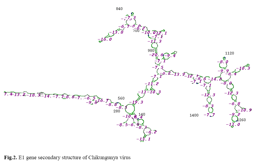
RNA secondary structure and Restriction sites in E1 gene of CHIKV The RNA secondary structure of E1 gene of CHIKV predicted showed the free energy of the predicted structure to be -329.6 kkal/mol, threshold energy to be -4.0, Cluster factor to be 2, Conserved factor also to be 2 and compensated factor to be 4 (Fig.2). Similarly the restriction sites in the E1 gene of CHIKV predicted showed the sites for various commercial and NEB restriction enzymes such as AciI, BsrBI, Sth132I, SetI etc. Also the restriction site analysis showed the GC and AT content of the E1 gene to be 71% and 29% (Fig.3). Survey on Chikungunya fever The survey results showed that amongst the infected people, 52% were in the age group between 20 and 40 years and a striking 53% were males with the remaining 47% constituting the females. The results of the other parameters considered are summarized in Table 2. Discussion The slower mobility rate of the RNA on agarose gel is suggestive of the higher molecular size of Chikungunya virus genomic RNA which is ~11,827 nucleotides (19). The obtained sequence of the E1 gene of the Chikungunya virus (1447 bp) was compared with the available sequence in the NCBI database using the software BLAST. Since the obtained nucleotide sequence showed 87% similarity to AR18211 and only 86% similarity to Ross strain, 77% to PM2951isolate, 53% to RSU1 strain and S27 strain, 20% to IND06AP6, IND06MS1, IND06MS2, IND05KA1 isolates and 18% similarity to MALho289 isolate, the most prevalent strain in Vellore district was identified to be AR18211 strain of Chikungunya virus. Phylogenetic tree constructed in order to know the root of origin, also revealed the similarity of our isolate with the existing strains in the NCBI. All the current outbreaks in India including the Yawat outbreak (2000) were caused by African genotypes (20) in contrast to the Asian genotypes during the 1963-73 outbreaks. The present study results matched with that of the findings of earlier studies made by Prasanna et al in the year 2006. The results were also similar to that of the results of Dhinakar raj et al, 2006. Studies on the secondary structure of CHIKV RNA carried out by earlier investigators revealed that the nucleotide sequences at the 5’ termini of alpha viruses were more conserved in potential secondary structure than in sequence and that the secondary structure was important for viral RNA replication (21). Also various restriction sites in the E1 gene of CHIKV were found out using NEBCutter tool. Similar study had been carried out earlier the results of which showed sites for various restriction enzymes in the CHIKV RNA (22). The survey analysis carried out in the present study was in contrast to the results of the study carried out by earlier workers (20) where the analysis was based on Chi-squared test whereas in the present study, the survey was based only on percentage analysis. The study by Prasanna et al, (20) focused only on age group whereas in the present study other parameters such as sex, sanitary condition of the locality and preventive measures were also undertaken and considered. The serosurvey carried out in Kolkata in 1995 reported that sero-positivity was highest in above 50 years age group (23). The present study concludes that the strain responsible for CHIKV infection in Vellore district was AR18211, the most susceptible age group to be between 20 and 40 years, the most susceptible individuals are male population compared to female population and the major symptom of CHIKV infection is swelling of the joints. Acknowledgement The authors thank the management of VIT University for providing the facilities to carry out this study. The financial support to Dr.D.Dhanasekaran under L-RAMP student project scheme by L-RAMP foundation was gratefully acknowledged. The financial support by TNSCST (Students project scheme) to the authors was acknowledged. References
The following images related to this document are available:Photo images[lp08023f2.jpg] [lp08023f1.jpg] [lp08023f3.jpg] |
| |||||||||
