 |
| About Bioline | All Journals | Testimonials | Membership | News |
|
||||||
|
||||||
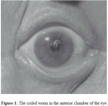
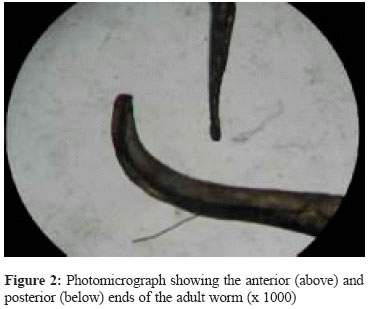
Indian Journal of Medical Microbiology, Vol. 23, No. 1, January-March, 2005, pp. 59-60 Case Report Loa loa in the anterior chamber of the eye: A case report Barua P, Barua N, Hazarika NK, Das S Department of Microbiology, Gauhati Medical College, Narakasur Hill, Guwahati - 781 032, Assam Code Number: mb05014 ABSTRACT An unusual case of loiasis from Assam is reported here. Loa loa is a subcutaneous filarial parasite of man and is transmitted to humans by chrysops flies. The patient presented with foreign body sensation and visual disturbances of the right eye. Examination revealed a white coiled structure in the cornea.. Routine blood and other investigations were within normal limits. A live adult worm was extracted and identity was confirmed by microscopy to be Loa loa. Patient was treated with diethylcarbamazine and steroid. We found this case interesting as the worm was present in the anterior chamber - an unusual site and there were no other positive findings besides the lone worm.Key words: Loa loa, anterior chamber, amicrofilaraemia Loa loa is a subcutaneous filarial parasite of man. This nematode is transmitted to humans by chrysops (mangrove flies) and produces a disease called loiasis. It has been associated with Calabar or fugitive swellings that are areas of angioedema due to migration of the adult worms through the subcutaneous tissues[1] and are often found at the extremities especially around joints. Eye infections may occur when the adult worm meanders into the subconjunctival tissues prompting the synonym "eye worm". The infection mostly remains asymptomatic for a long period. The microfilaria of Loa loa may not be often demonstrated in these cases, probably because the worms are not mature or only males are present in infection.[2] CASE REPORT A 48-year-old man presented to the Regional Institute of Ophthalmology, Gauhati, Medical College, Guwahati with complaints of foreign body sensation, itching, redness, pain, lacrimation and blurring of vision of the right eye for the last three months. The symptoms were insidious in onset and progressive in nature. Patient did not give history of fever or swelling in any part of the body. No history of travel to known endemic area could be found. The patient belonged to a nearby rural area and was a police constable by profession. He had visited a couple of physicians over the last three months and was prescribed topical antibiotics and steroid but to no avail. On general examination, the patient appeared to be healthy and no subcutaneous nodules or swelling on any part of the body was seen. There was no hepato-splenomegaly or lymphadenopathy. On ophthalmic examination, conjunctival hyperaemia and a white-coiled structure in the anterior chamber was observed. Vision and intraocular pressure were within normal limits. Lens and fundus were also normal. Slit lamp examination revealed a worm with cuticle partially embedded in the cornea [Figure - 1]. Routine blood examination was within normal limits. Differential eosinophil count was 360/µL. both thick and thin peripheral blood smears were negative for microfilaria on two separate occasions at daytime and at night. Aqueous humour showed no cellularity or microfilaria. C-reactive protein was raised to 2.4 mg/dL. Skin snip biopsy showed a few non-specific chronic inflammatory cells but no microfilaria. Other investigations like ESR, liver function tests, chest x-ray, sputum, stool and urine examinations were normal. A live adult actively motile worm was extracted through superior-temporal limbal stab incision and sent to the microbiology department for identification. Macroscopically, the worm was a thin, whitish, thread like semitransparent, cylindrical structure measuring 25mm in length. On microscopy the body of the worm was found to be covered with a cuticle with knob like structures. The anterior extremity had a simple mouth and the posterior end was rounded [Figure - 2]. The above findings suggested the identity of the worm to be an adult male Loa loa. The absence of microfilaria in the peripheral blood further corroborates the identity of the worm to be a male. After surgical removal of the worm, the patient was treated with diethylcarbamazine 500mg daily for three weeks and oral prednisolone 60mg for one week, tapered over next three weeks. DISCUSSION Loa loa was first extracted from the eye of a West Indian by Mongin in 1770,[1] and Guyot reported the first case from Angola in Africa in 1777.[3] It received its present name in 1913.[1] Though endemic in the Central and West Africa, sporadic cases have been reported from other parts of the world including India. Most of these cases describe the presence of Loa loa in the subconjunctival tissue,[4] though a few cases have been reported from India with worm in the anterior chamber of the eye.[2] However, to the best of our knowledge, this is the first case report of loiasis of anterior chamber of the eye from Assam in the northeastern part of India. The worm in this case, identified as adult male Loa loa, might have migrated to the anterior chamber in its larval stage either from the blood through ciliary vessels and grown there or may have burrowed through the coats of the eyeball. Amicrofilaraemia may be due to the presence of a lone adult male incapable of reproduction. Absence of inflammatory cells in the aqueous humour is explained by the use of topical steroids. In summary, this is a case of loiasis encountered in a non-endemic area, characterized by the presence of an adult worm in the anterior chamber of the eye without the usual features of microfilaraemia, eosinophilia and Calabar swelling. REFERENCES
Copyright 2005 - Indian Journal of Medical Microbiology The following images related to this document are available:Photo images[mb05014f1.jpg] [mb05014f2.jpg] |
| |||||||||