 |
| About Bioline | All Journals | Testimonials | Membership | News |
|
||||||
|
||||||
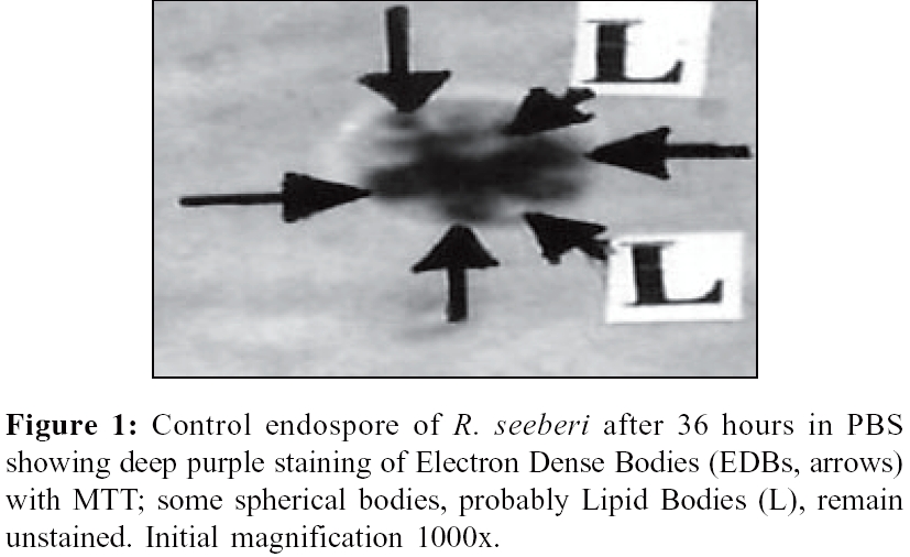
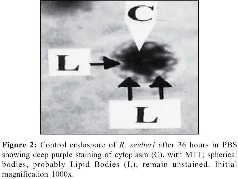
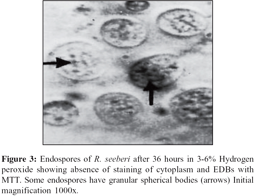
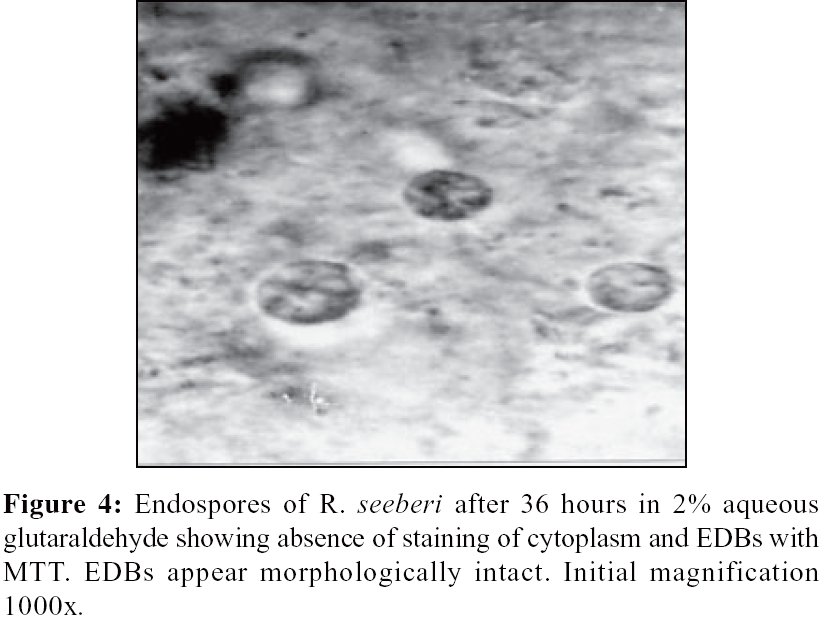
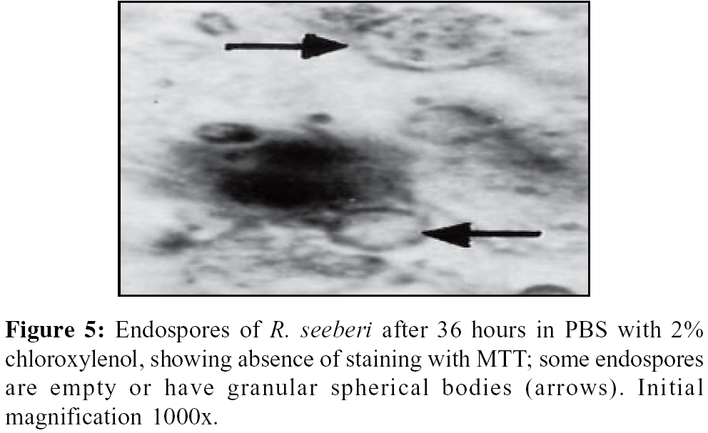
Indian Journal of Medical Microbiology, Vol. 24, No. 2, April-June, 2006, pp. 85-91 Special Article The effects of biocides (antiseptics and disinfectants) on the endospores of Rhinosporidium seeberi *SN Arseculeratne, DN Atapattu, P Balasooriya, R Fernando Department of Microbiology (SNA, DNA), Faculty of Medicine, University of Peradeniya and ENT Division (PB, RF), General Hospital, Kandy, Sir Lanka Code Number: mb06027 Abstract No data exists on the activity of biocides (antiseptics and disinfectants) on Rhinosporidium seeberi that causes rhinosporidiosis in humans and animals. On account of the inability to culture R. seeberi, in vitro , dyes were used to assess the morphological integrity and viability of biocide-treated endospores that are considered to be the infective stage of this pathogen. Evan's Blue (EvB) identifies the morphological integrity of the endospores while MTT (3-[4, 5-dimethylthiazol-2yl]-2, 5-diphenyl tetrazolium bromide) identifies metabolic activity through its reduction by cellular dehydrogenases to microscopically visible deposits of insoluble formazan. MTT-negativity has earlier been shown to correlate with absence of growth of yeast and mycelial fungi in culture and could thus indicate the loss of viability of MTT-negative rhinosporidial endospores. Hydrogen peroxide, glutaraldehyde, chloroxylenol, chlorhexidine, cetrimide, thimerosal, 70% ethanol, iodine in 70% ethanol, 10% formalin, povidone-iodine, sodium azide and silver nitrate were tested on freshly-harvested endospores and all biocides caused metabolic inactivation with or without altered structural integrity as shown by absence of MTT-staining after 3, 24 or 36 hour after exposure, while EvB stained only the endospores treated with sodium azide, ethanol, thimerosal, chloroxylenol, glutaraldehyde and hydrogen peroxide. With clinically useful biocides - chlorhexidine, cetrimide-chlorhexidine, 70% ethanol, povidone-iodine and silver nitrate, a total period of exposure of endospores to the biocide, for seven minutes, produced metabolic inactivation of the endospores. Anti-rhinosporidial antiseptics that could be used in surgery on rhinosporidial patients include povidone-iodine in nasal packs for nasal and naso-pharyngeal surgery, chlorhexidine and cetrimide-chlorhexidine on the skin, while povidone-iodine and silver nitrate could have application in ocular rhinosporidiosis.Keywords: Rhinosporidium seeberi, sensitivity, biocides Rhinosporidiosis caused by Rhinosporidium seeberi in humans and animals, has been known for over a hundred years. Outbreaks of ocular and nasal disease in humans[1] and in swans[2] were recently described and reports of sporadic cases, in temperate and tropical countries, continue to appear. This pathogen was for long regarded as a fungal-like organism[3] but with molecular biological tools and phylogenetic analysis, it was recently assigned to a new Protoctistan clade, the Mesomycetozoea, that includes pathogens of fish and amphibian.[4],[5] Although no evidence of human-to-human transmission of rhinosporidiosis has been documented, while the putative source of the pathogen is thought to be natural ground waters, disinfection of laboratories, wards, theatre and instruments, yet remains mandatory; ".emotive connotations for hospital staff." that occur with rabies,[6] probably exist in rhinosporidiosis as well, with its chronic, recurrent, disfiguring lesions that occasionally disseminate to the viscera. A need also exists clinically for the use of topical antiseptics to prevent recurrence of the disease due to the occurrence of ′auto-inoculation′of endospores[7] that will inevitably contaminate adjacent mucosal surfaces during surgery, with the consequent development of satellitic rhinosporidial polyps. No reports have been published on the assessment of sensitivity to anti-microbials, including biocides, of R. seeberi or of members of its new Clade Mesomycetozoea . Undoubtedly, the reason for this lack has been the inability to culture R. seeberi, in vitro, or to establish a model of rhinosporidiosis in experimental animals. The ontogenic development of endospores to sporangia in aqueous suspensions, has been described.[8],[9] (Arseculeratne, 1996, unpublished observations), but this phenomenon as an index of viability could not be used as the basis of a test for inactivation on account of the difficulty of standardising the optimum conditions needed for the development of endospores to sporangia as evidence of viability and development. The salt MTT (3- [4, 5-dimethylthiazol-2yl]-2, 5-diphenyl tetrazolium bromide) is reduced by mitochondrial dehydrogenases in viable mammalian and fungal cells, to a purple-coloured formazan that is quantifiable spectrophotometrically after extraction from the cells or is detected microscopically in the intact microbial cell. The tetrazolium ring is cleaved in active mitochondria and so the reaction occurs only in living cells, dead cells are almost completely negative.[10] This assay has also been used for the determination of the viability of fungi after treatment with anti-fungal drugs.[11],[12] The endospore of R. seeberi has prominent intra-endosporial spherical bodies, some of which have been termed lipid bodies (LB), while others with distinct differences in structure and ultrastructural appearances are the electron dense bodies (EDB).[2],[3],[13] The endospore′s cytoplasm and some spherical bodies that are probably the bodies that were regarded as EDBs, have been shown to be sites of MTT-reduction,[14] a reaction that was also used to assess the viability of rhinosporidial endospores that had been exposed to inactivating agents such as heat and formaldehyde.[14],[15] Further confirmation of the probable mitochondrial nature of the spherical bodies, the putative EDBs, of rhinosporidial endospores was their intense staining with tetramethyl rhodamine ethyl ester (TMRE).[14],[15] This is considered to be a specific stain for mitochondria.[16] Evan′s Blue (EvB) was also shown previously to stain the spherical bodies within the endospores although this stain does not, unlike MTT, identify metabolically inactive endospores but is capable only of showing morphological and tinctorial integrity of the endospores and their spherical bodies.[14] This report describes the use of the MTT-reduction test in the assessment of the sensitivity of rhinosporidial endospores, that are tentatively regarded as the infective units of R. seeberi , to a variety of biocides that are in clinical or laboratory use. Materials and Methods Buffer Endospores Endospores from freshly homogenised rhinosporidial tissues (obtained immediately after surgery) or such endospores that had been stored at 4°C for not more than a week, were found to react with MTT and were used in all the tests. Endospores from fresh, tissue homogenates after storage at - 20o C had lost the ability to reduce the MTT[15] and were not included. Rhinosporidial tissues were minced aseptically in distilled water and homogenised in a motor-driven Teflon -glass homogeniser. The distilled water served to lyse osmotically labile host cells such as red blood cells, to make microscopic visualisation of the endospores easier. The coarse tissue particles were deposited on brief standing and the supernatant was centrifuged at 400 × g at room temperature (RT, 28o C ambient) to deposit the endospores that were then washed thrice in PBS and finally resuspended to a density of approximately 2-3 × 10 6 endospores/mL, as counted in an Improved Neubauer blood cell counting chamber. The homogenates contained mainly endospores with a small proportion of juvenile sporangia. Replicate tests were done with endospores from homogenates of rhinosporidial tissues from different patients; each set of tests included a biocide free control of endospores from the preparation that was used for the biocide tests. Although freshly prepared endospores after Percoll purification[17] and storage at 4°C are equally well stained with MTT as are fresh endospores in tissue homogenates,[15] fresh tissue homogenates, sieved but not Percoll purified, were used to simulate clinical conditions with the presence of tissue debris that would have provided a more realistic estimate of antimicrobial efficacy, as in the Chick-Martin test rather than in the classical Rideal-Walker test for biocides. Some biocides, such as chloroxylenol[18] and chlorhexidine[19] are inactivated in the presence of organic matter. In the concentration response studies where counts of the stained or unstained endospores were made, the tissue homogenate was sieved through nylon mesh with a pore size of 25 µm through which endospores (10-15 µm) were separable from sporangia and tissue fragments; the latter would otherwise have prevented accurate counting of endospores. Biocides The biocide tests Approximately 100 µL of the endospore suspension was placed in 1.5 mL microfuge tubes and centrifuged at 400 × g for 10 minutes to deposit the endospores. Approximately 0.5 mL of the biocide solution was added, the tubes were side-tapped for dispersion of the deposit and the capped tubes were incubated at room temperature (RT, 28°C ambient) for, 3, 24 and 36 hours. The commonly used, clinically useful biocides, chlorhexidine, cetrimide-chlorhexidine, 70% ethanol, povidone-iodine and 1% silver nitrate, were tested with exposure times of approximately 7 minutes (5 minute incubation at RT and 2 minute during the first centrifugation for removal of the biocide). A concentration-response test was done with chlorhexidine-cetrimide in concentrations of ranging from 0.0002 to 2% with an exposure duration of 18 hours at room temperature. After incubation the tubes were centrifuged and the endospore deposit was washed thrice with PBS, or with distilled water for silver nitrate treated samples. Three washes of the treated deposit of endospores were necessary to avoid possible interference with the MTT reaction, caused by residual biocide. Dye tests Approximately 50 µL of a 0.2% of EvB in PBS was added to the thrice washed pellet of endospores, the mixture was agitated by side tapping of the tube and then incubated at RT for 45 minutes. A drop of the mixture was placed under a cover slip on a slide for microscopy and photography at a magnification of x1000, under oil. The MTT dye reduction test The endospore suspensions in the microfuge tubes were centrifuged at 400 × g for 10 minutes, washed thrice with PBS and to the deposit was added approximately 50 µL of MTT (Chemicon International, Inc, Temecula, CA, USA). The endospores were dispersed by side tapping of the tubes and the mixtures were incubated at 37o C in air for 3 hours. A drop of the dye endospore suspension was placed under a cover slip on a slide for microscopy and photography at a magnification of x1000 under oil. Scoring of the effect of biocides With EvB, the intensity of staining of the endospore′s cytoplasm and the EDBs was used as an indicator of morphological integrity and with MTT, the intensity of the purple staining of the cytoplasm and the EDBs was used as an indicator of metabolic activity of the endospores, since MTT reduction is dependent on enzymic (dehydrogenase) activity. Abnormal morphology of the endospore′s wall and disintegration or absence of the EDBs were also taken as indicators of the anti microbial effects of the biocides. The effects on EDBs (disintegration, absence, loss of staining) were considered more significant end points than the degree of staining of the cytoplasm, on account of the evidencethat these bodies are the ultimate generative unit of R. seeberi.[3],[13],[14],[23] Except with sieve-purified endospores, counts of endospores in crude homogenates were not made as tissue fragments in the latter could make accurate counting of endospores difficult; with these samples only qualitative assessments of the morphology and staining of the endospores were made. Microscopy of the MTT treated endospores rather than extraction of the formazan and spectrophotometry was used to determine the site of MTT reduction.[24] The tests were replicated six times; an initial test was done over a test period of three hour, replicated once more over 24 hours and replicated thrice at 36 hours. The response to both EvB and MTT were determined. The tests on sieve-purified endospores with selected, clinically useful biocides, chloroxylenol, chlorhexidine + cetrimide, 70% ethanol, povidone-iodine and silver nitrate were done with an exposure period of 7 minutes and the response to only MTT was determined. Results Control endospores Biocide-treated endospores After 36 hour exposure to 3-6% hydrogen peroxide, the EDBs were abnormal and granular; the EDBs and the cytoplasm stained with EvB but with MTT, both EDBs and cytoplasm remained unstained [Figure - 2]. After 36 hour exposure to glutaraldehyde , EDBs with normal morphology were identifiable and both cytoplasm and the EDBs were stained with EvB. Both EDBs and cytoplasm remained unstained with MTT [Figure - 2]. After 7 minutes exposure to chloroxylenol, the endospores were morphologically abnormal and unstained with MTT [Figure - 2]. After 36 hours many endospores were empty of spherical bodies but in others, they were recognisable though granular and unstained [Figure - 2]. Evan′s Blue stained the endospores. After 7 minutes exposure to chlorhexidine with cetrimide, the endospores were uniformly unstained with MTT and the spherical bodies appeared granular or were absent [Figure - 3]. After 36 hours, the EDBs were either absent or abnormal in both Evan′s Blue and MTT-stained preparations [Figure - 3]. The concentration response test (20 hours, RT) gave 0% of MTT stained endospores exposed to 2%, 0.2% and to 0.02% of this biocide while 6.9% were positively stained after exposure to 0.02%. This value differed significantly ( P < 0.0001) from that of the control endospores (95.3%). Endospores that were exposed to 0.0002% biocide showed staining of 94.8% which was not significantly different ( P > 0.8) from that of control (biocide-free) endospores 95.3% of which showed staining. The end-point of the titration was sharply defined and the minimum sporicidal concentration of chlorhexidine-cetrimide lay between 0.02% and 0.002%. After 36 hour exposure to chlorhexidine, the EDBs were either absent or when present were, as with the cytoplasm, unstained with MTT; these appearances were similar to those with chlorhexidine containing cetrimide. The walls of some endospores showed swelling or thickening [Figure - 3]; EvB staining was also absent. Cetrimide alone, at 36 hours, produced gross abnormalities of the endospores in which the EDBs were absent or unstained with MTT, as was the cytoplasm [Figure - 3]. Staining with EvB was also absent. Thimerosal produced less marked effects on the morphology of the endospore after 36 hours exposure; EDBs were identifiable but some were abnormal with a reduction in size and granularity. As in the cytoplasm, MTT staining of the EDBs was reduced or absent [Figure - 4], though staining with EvB was retained. After 7 minute exposure to 70% ethanol, the endospores including those within immature sporangia, were unstained with MTT [Figure - 4]. After 36 hours exposure, EDBs were identifiable but abnormal with absence of staining with MTT [Figure - 4]; the cytoplasm was also unstained. Evan′s Blue staining of the EDBs was of normal intensity though the morphological abnormalities seen in MTT treated endospores were also identified on the EvB stained endospores. Effects of 1% iodine in 70% ethanol were similar to those observed with 70% ethanol in endospores treated with iodine-ethanol for 36 hours [Figure - 4]; the cytoplasm and EDB were unstained with MTT. In contrast to the effects of 70% ethanol alone, the iodine ethanol combination produced an absence of staining with EvB indicating a greater degree of damage than with the ethanol alone. Formalin (1% and 10%) produced similar defects in the endospores, though with 10% formalin, more endospores showed an absence of EDBs [Figure - 5]. At both concentrations, the EDBs that remained were granular and unstained with MTT, as was the cytoplasm [Figure - 5]. The structural damage to the EDBs was apparently severe enough to cause absence of staining with EvB. After 7 minute exposure to povidone iodine, the EDBs were abnormal and unstained with MTT; the endospore′s walls were thickened [Figure - 5]. After 36 hours exposure, the EDBs were swollen and abnormal, with loss of staining with MTT; the cytoplasm too showed no staining with MTT [Figure - 5]; EvB also failed to stain the cytoplasm and EDBs. After 7 minute exposure to silver nitrate, the endospores were totally unstained with MTT in both cytoplasm and EDBs. Staining with EVB was however retained. After 36 hours exposure , the EDBs and the cytoplasm showed a loss of staining with MTT [Figure - 5]. In summary, loss of staining with both MTT and with EvB was seen in endospores treated with chlorhexidine, chlorhexidine-cetrimide, cetrimide, 10% formalin, iodine-ethanol and povidone-iodine. Only MTT staining was absent in endospores treated with sodium azide, 70% ethanol, thimerosal, chloroxylenol, glutaraldehyde and hydrogen peroxide and silver nitrate, though these agents did not abolish staining with EvB; the EvB staining of endospores treated with the latter biocides illustrates the inability of this dye to identify non-viable (MTT-negative) endospores, although it was capable of identifying their morphological and tinctorial integrity. Discussion Evan′s blue was previously found[15] to stain only a proportion, probably the EDBs, of the endospore′s spherical bodies, leaving spherical bodies of a similar size, regarded as the LBs unstained. EvB was capable of identifying the structural integrity of the endospores but not their viability, whereas MTT, on account of the need for mitochondrial dehydrogenase activity for its reduction to the purple-coloured formazan, was regarded as capable of indicating the metabolic activity and hence the viability of the endospores.[15] The absence of an in vitro culture system for R. seeberi and the lack of information on its metabolic pathways preclude the use of some of the conventional methods for assaying of microbicidal activity. The MTT dye-test described in this report provides a simple technique for the assessment of sensitivity of R. seeberi to biocides. The applicability of this test to rhinosporidial endospores depends on the presence of mitochondria in the endospore′s cytoplasm;[23] morphological elements of the nature of mitochondria have however not yet been described in the EDBs although their MTT-staining, indicative of enzymic reduction to produce the formazan and hence their deep-purple staining, indicates the presence of such enzymic activity in the EDBs that form a proportion of the spherical bodies in rhinosporidial endospores.[15] Endospores are a stage in the life-cycle of R. seeberi and are metabolically active elements. Although they possess a thick wall that contains cellulose and chitin, the endospores are not comparable with bacterial spores or the encysted stages of parasites, that are resistant to biocides; examples of the latter are the oocysts of Cryptosporidia that resist 70% ethanol, iodophors, quarternary ammonium compounds and aldehydes.[6] Cysts rather than pre-encystment trophozites of Acanthamoeba castellani are the most resistant form. Cyst forms are the most resistant to chemical disinfectants probably because, as with bacterial spores, they take up fewer disinfectant molecules from solution than vegetative forms[25] and the thick-walled, encysted amoeba is resistant to anti microbial agents.[26] The data on the action of biocides on bacterial endospores and encysted parasites cannot therefore be extrapolated to the rhinosporidial endospores on account of differences in structure and function of these forms. Biocides in general, with multiple targets, have a broader spectrum of activity than antibiotics; some (e.g, chlorhexidine, phenolic compounds, mercury salts at high concentrations) are able to coagulate the cytoplasm while antibiotics have specific intracellular targets.[25] This difference probably explains why the end points with biocides were clearer - complete loss of integrity and loss of MTT staining of the EDBs with these effects detectable in almost all the endospores, than with therapeutic antimicrobial agents such as amphotericin B and dapsone.[27] The remarkable sensitivity of rhinosporidial endospores to all the biocides tested was thus understandable. On the basis of the correlation of MTT negativity with non-viability in culture of mycelial fungi that were treated with a variety of anti fungal agents[11],[12] the MTT negativity of the biocide treated rhinosporidial endospores suggests that these agents were rhinosporicidal rather than rhinosporostatic. Our results confirm that differences exist between EvB and MTT[15] in that only MTT was able to identify the metabolically damaged endospores, the tinctorial integrity of which were however retained to be identified by EvB. Glutaraldehyde provides excellent preservation of cellular ultrastructure[28],[29] and it is noteworthy that although this agent abolished metabolic activity as shown by MTT negativity, the EDBs were identifiable with normal morphology. The contrasting appearances with EvB and MTT again indicated that only the MTT could detect metabolically damaged endospores, while EvB could identify only morphological integrity.[15] The proportionality between the MTT-staining and concentration of chlorhexidine+cetrimide indicated that it was this agent and not any other factor that caused the negativity of MTT-staining. Iodophores are advantageously used as a sporicidal antiseptic on the skin,[6] in which role its demonstrated efficacy on rhinosporidial endospores in the 7 minutes test could prevent their pathogenicity through "auto-inoculation" on the skin and mucosa of the nasal passages (with iodophor-soaked nasal packs), that would result in the development of satellitic polyps after trauma or surgery in these two sites. It could also be used, after dilution in aqueous benzalkonium chloride, in post operative ocular rhinosporidiosis. The total negativity of staining of formalin-treated endospores with both MTT and Evan′s Blue indicated their inactivation and structural damage caused by this agent. It is thus a useful disinfectant in a laboratory where rhinosporidial tissues are processed. Maintenance of EvB-staining of glutaraldehyde-treated EDB contrasts with the loss of staining with EvB and loss of morphological integrity after treatment with formalin. Glutaraldehyde is used as fixatives for electron microscopy while formalin is used as a fixative for paraffin-embedding of tissue for light microscopic histopathology. The loss of staining with the vital dye Evan′s Blue (EvB) of formalin treated EDBs is also in contrast to the tinctorial integrity of the endospores and their EDBs to haematoxylin in the conventional haematoxylin and eosin staining procedure and to periodic acid-Schiff stains of paraffin-embedded histopathological sections of formalin-fixed rhinosporidial tissue. Silver nitrate acts on enzymes with thiol groups. Topically-applied silver nitrate was used to prevent Ophthalmia neonatorum in infants delivered by mothers with gonococcal infection of the reproductive tract; ciprofloxacin is now recommended for this purpose but on account for the inactivity of ciprofloxacin on rhinosporidial endospores (Arseculeratne, 2003, unpublished data) silver nitrate, especially with its activity in the 7-minute test, could be used topically on eyes to prevent development of satellitic ocular rhinosporidial polyps through "autoinoculation" of endospores from the traumatised polyp. The use of 2% silver nitrate to cauterize the base of the polyp has been recommended.[30] On account of the irritant effect of this agent, povidone-iodine that is also effective against endospores, may be preferred when diluted with aqueous benzalkonium chloride. Acknowledgements We thank Dr. N. R. de Silva for a gift of MTT, Prof. Malik Peiris for suggestions, Dr. P. V. R. Kumarasiri for help with statistics and Mr. N.B. Eriyagama for technical assistance.References
Copyright 2006 - Indian Journal of Medical Microbiology The following images related to this document are available:Photo images[mb06027f5.jpg] [mb06027f4.jpg] [mb06027f3.jpg] [mb06027f2.jpg] [mb06027f1.jpg] |
| |||||||||