 |
| About Bioline | All Journals | Testimonials | Membership | News |
|
||||||
|
||||||
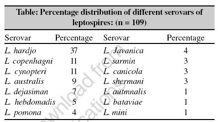
Indian Journal of Medical Microbiology, Vol. 24, No. 4, October-December, 2006, pp. 302 Correspondence A preliminary study on the prevalence of leptospira serovars among suspected cases of leptospirosis at Tirupati, Andhra Pradesh Sharma KK, Gururajkumar A, Mohan A, Sivakumar V, Kalawat U Sri Venkateswara Institute of Medical Sciences, Tirupati - 517 507, Andhra Pradesh Date of Submission: 18-Apr-2006 Code Number: mb06090 Dear Editor, Leptospirosis, transmitted by animals to human, is an emerging infectious disease. Although rats are the common reservoir of the leptospires, cattle are the most common reservoir of Leptospira hardjo , worldwide. Humans acquire the disease through accidental, occupational or recreational contact with contaminated water or through contact with urine, fluids or tissue of infected animals. It has protean manifestations, but fever, headache, prostration, myalgia and conjunctival suffusion are seen in most cases. The classical signs of haemorrhage, jaundice, meningitis and renal failure are rare.[1],[2],[3] While some studies from southern states of India have shown that autumnalis, australis, grippotyphosa and icterohaemorrhagiae are common circulating serovars in these zones; to the best of our knowledge, no true incidence of human leptospirosis in Andhra Pradesh has been reported. This could be due to lack of awareness on the part of treating physicians or lack of diagnostic facilities. Tirupati, Andhra Pradesh, is non-endemic area for leptospirosis and there is no authentic reporting from this area from the literature available to us. We report here the results of a hospital based prospective study from February 2001 to September 2003. Four hundred and seventy-nine blood samples were analyzed from suspected cases of leptospirosis by dark field microscopy (DFM), Leptospira IgM enzyme-linked immunosorbent assay (ELISA) and microscopic agglutination test (MAT). Four hundred and thirty-one (90%) serum samples were found to be positive by both DFM and Leptospira IgM ELISA. Twenty serum samples were found to be negative with MAT. Four hundred and fifty-nine samples showed agglutination reaction and MAT titer varied from less than 1:50 to 1: 6400. MAT titer 1:50 or more than 50 was found in 109 samples. Predominant serovar was L. hardjo (39%) followed by L. copenhagni (11%) and L. cynopteri (11%). The percentage distribution of different serovars of leptospires is shown in the table. In this study we considered the sera having MAT titer 1: 50 or more than 50 with the signs and symptoms of leptospirosis as positive. A MAT titer of 1:50 or > 50 to any of serovars was considered as evidence of leptospiral infection and individuals were defined as seropositive cases.[4] Most common isolate was L. hardjo in the area where there were numerous dairy farms. Seasonal variation was observed and the highest incidence of leptospirosis was during the rainy seasons. We noticed a wide variation between Leptospira IgM ELISA and MAT. MAT positivity was observed in about 22 % cases out of 479 ELISA positive cases. These discrepancies among IgM ELISA and MAT may be due to prolonged transportation time, which may have leads to contamination of serum samples as well as deterioration of immunoglobulins. References
Copyright 2006 - Indian Journal of Medical Microbiology The following images related to this document are available:Photo images[mb06090t1.jpg] |
| |||||||||