 |
| About Bioline | All Journals | Testimonials | Membership | News |
|
||||||
|
||||||
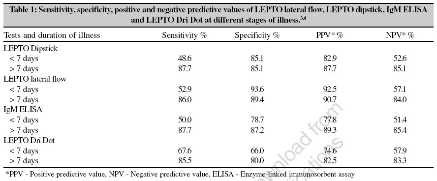
Indian Journal of Medical Microbiology, Vol. 24, No. 4, October-December, 2006, pp. 320-322 Supplement Recent advances in the laboratory diagnosis of leptospirosis and characterisation of leptospires Vijayachari P, Sehgal SC Regional Research Centre, WHO Collaborating Centre for Diagnosis, Research, Reference and Training in Lepospirosis, Regional Medical Research Centre (ICMR), Port Blair, Andaman and Nicobar Islands Code Number: mb06101 Leptospirosis is an emerging zoonotic disease of worldwide distribution . The disease can occur in the form of several syndromes[1] such as an acute febrile illness associated with headache and severe muscular pain, jaundice with nephritis (Weil′s disease), pulmonary hemorrhage[2] with respiratory distress, myocarditis with arrhythmias, meningitis and meningoencephalitis. In view of varied clinical manifestations, laboratory support is needed for confirmation of the diagnosis of this disease. Laboratory diagnosis of leptospirosis is an area ill-understood by many of the workers involved in leptospirosis diagnosis and surveillance. Selection of the right specimens and tests and the correct interpretation of test results are important in order to provide better patient care. Laboratory diagnostic tests are broadly divided into two categories viz, direct evidences (isolation of organism or demonstration of leptospires or amplification of specific fragment of leptospiral DNA) and indirect evidences (detection of antibodies to leptospires). Alternatively, the different methods used in the laboratory can be categorized into bacteriological, microscopic, immunological/serological and molecular techniques. Conventional bacteriological techniques such as isolation of the organism from clinical specimens or serology using microscopic agglutination test are laborious, time- consuming; need well-established laboratory facilities and skilled manpower. Since most of the institutions or hospitals may not have such facilities, more often than not, simple and rapid diagnostics are depended upon. IgM EIA, Micro-capsule agglutination test (MCAT), LEPTO Dipstick, Macroscopic slide agglutination test (Macroscopic SAT), LEPTO Lateral flow, Indirect haemagglutination assay (IHA) and LEPTO Dri Dot are some of the rapid tests used for the diagnosis. LEPTO Dipstick and LEPTO Lateral flow are IgM immunoassays whereas LEPTO Dri Dot is a latex agglutination test. The principle of MCAT is similar to that of latex agglutination assay. All these tests detect antibodies and the sensitivity of these tests is usually low during first week of illness. However these tests have acceptable sensitivities during the second week of the disease. The indices of validity and utility of some of these techniques at different time intervals of illness are summarized in the table below [Table - 1]. One of the molecular techniques used for the early diagnosis of leptospirosis in recent years is the amplification of specific fragment of leptospiral genomic DNA in clinical samples using polymerase chain reaction (PCR). Since the test detects specific fragment of leptospiral DNA, the positive test result confirms the diagnosis. PCR on serum samples using two sets of primers (G1/G2 and B64I /B64II) showed 95.2% sensitivity and 91.4% specificity when compared with isolation and/or paired MAT. Although the test is difficult to perform at peripheral level, health care centers or small institutions, it has a significant role during the investigation of suspected outbreaks. Characterization of leptospires is essential for understanding the epidemiology of the disease. Serovar is the basic taxon of leptospires and it is defined based on surface antigenic make-up (antigenic classification). More than 300 serovars have been described in L. interrogans sensu lato whereas L. biflexa sensu lato contains 45 serovars.[5] The classification system based on genetic similarities is being used in conjunction with classical antigenic classification during the recent years. Based on genetic homology in DNA hybridization experiments, 15 genomic species ( L. interrogans, L. kirschneri, L. borgpetersenii, L. santarosai, L. noguchii, L. weilii, L. inadai, L. biflexa, L. meyeri, L. wolbachii, Genomo species 1, Genomo species 3, Genomo species 4 and Genomo species 5) have been described in the genus Leptospira whereas Leptonema and Turneria have one species each ( L. illini and T. parva respectively).[6] Genomic species is a group of Leptospiraceae serovars whose DNA show 70% or more homology at the optimal re-association temperature of 55°C or 60% or more homology at a stringent re-association temperature of 70°C and in which the related DNA contain 5% or less unpaired bases. Though DNA - DNA hybridization is considered to be the gold standard technique for species-level identification of leptospires, it is seldom used because of its complexity. Several PCR-based DNA fingerprinting methods have become popular and are being used routinely for characterization of leptospires. Random amplified polymorphic DNA (RAPD) fingerprinting, arbitrarily primed PCR (APPCR), single nucleotide polymorphism of specific PCR products are some of the examples. REP-PCR (repetitive extragenic PCR) and FAFLP (fluorescent amplified fragment length polymorphism) are recent methods in the characterization of leptospires. One of the well-characterized repetitive elements among a variety of prokaryotes is repetitive palindromic extragenic units. Because of the widely distributed nature of these repetitive DNA elements in genomes of various microorganisms, these sites are being used as binding sites for alleles multiplication to generate fingerprints. AFLP is a three-step procedure in which genomic DNA is restricted, ligated with oligonucleotides and the ligated DNA fragments are amplified for generation of genetic markers. FAFLP is a technique that combines the power of restriction fragment length polymorphism (RFLP) with the flexibility of PCR based technology by ligating primer recognition sequences (adapters) to the restricted DNA. References
Copyright 2006 - Indian Journal of Medical Microbiology The following images related to this document are available:Photo images[mb06101t1.jpg] |
| |||||||||