 |
| About Bioline | All Journals | Testimonials | Membership | News |
|
||||||
|
||||||
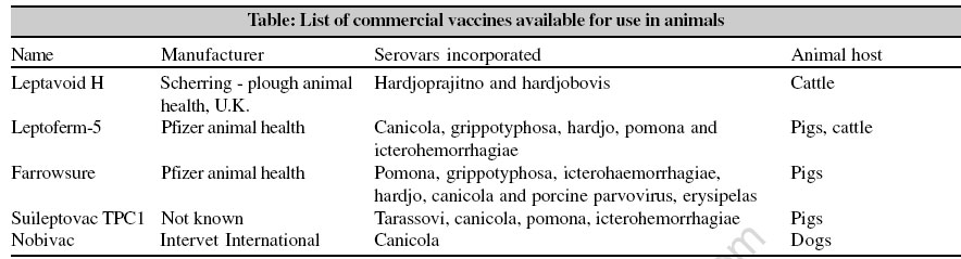
Indian Journal of Medical Microbiology, Vol. 24, No. 4, October-December, 2006, pp. 331-336 Supplement Prospects of developing leptospiral vaccines for animals Srivastava SK Division of Bacteriology and Mycology, IVRI, Izatnagar, Bareilly - 243122 Code Number: mb06105 Leptospirosis is a common disease of livestock, pet animals and wildlife throughout the world. Sporadic cases and outbreaks of the disease have been reported from USA, U.K, Australia, New Zealand, USSR and countries of Europe and Asia. The disease is common in cattle, buffalo, sheep, goat, dogs and equines and causes fever, jaundice, nephritis, reproductive disorders and death. In dairy animals, loss of milk and mastitis may be observed. In India, Taylor and Goyale for the first time in 1931 reported the isolation of Leptospira organisms from patients showing jaundice in Andaman Islands. Soon after this report, Ayyar[1] reported isolation of Leptospira. interrogans serovar Icterohaemorrhagiae from dogs in Madras city. Since then several reports have emerged confirming the prevalence of leptospirosis in various animal species and man in India.[2],[3],[4],[5] Isolation of Leptospira serovars from diseased and carrier animals including cases of leptospirosis are commonly reported from coastal regions of Kerala, Tamil Nadu, Gujarat, Maharashtra and Andaman Islands. Adinarayanan et al reported natural outbreaks of bovine leptospirosis in U.P. due to serogroup Hebdomadis.[2] Antibodies against serovars Pomona, Autumnalis, Grippotyphosa, Javanica and Poi have been reported from Tamil Nadu.[6] Srivastava et al[7] detected agglutinins in 7% of 1839 cattle and 14.3% of 393 buffaloes belonging to various states. Agglutinins were most common against Pomona, Patoc, Icterohaemorrhagiae and Grippotyphosa. Ratnam et al[8] reported antibodies to Sejroe, Autumnalis and Pomona in cattle in Tamil Nadu. Srivastava and Kumar[9] reported seropositivity in 15.8% of 2601 cattle sera, mostly belonging to Andhra Pradesh. Long-term control strategies of the disease include adoption of hygienic measures, rodent control and vaccinations. Commercial Leptospira vaccines are available in many countries for cattle, dogs and swine but vaccination may prove only partially effective due to generation of serovar specific immunity, prevalence of several Leptospira serovars and the tendency of the vaccinated animals to become carriers of the organisms. A successful vaccination programme requires that epidemiological studies should be continued to assess the extent of the problem and the knowledge of involvement of different Leptospira serovars in causing the disease in a given population. If in a particular region, leptospirosis is considered economically a priority disease, which has to be tackled by vaccination, the target animal population must be specified, as it may not be possible to vaccinate all the animals. In this review various attempts made in the past and the efforts being made presently on the development of Leptospira vaccines are given. Inactivated whole culture vaccine Over the past 50 years, a number of Leptospira vaccines have been developed and evaluated in laboratory animals, cattle, swine, dogs, sheep and horses. Most of these have been bacterins containing inactivated organisms by various chemical means and heat. Broom[10] demonstrated some protection of hamsters against Canicola given phenol-inactivated cultures in high doses. Brunner and Meyer[11] reported the successful immunization of hamsters and dogs with lyophilized Canicola or Icterohaemorrhagiae bacterin and showed the serovar specificity of their vaccines as no cross-protection was provided against other serovars. Thus, multivalent vaccines have been prepared incorporating several serovars (Copenhageni, Pomona, Hardjo, Grippotyphosa, Autumnalis and Icterohaemorrhagiae) and satisfactory results have been obtained in guinea pigs and hamsters.[12],[13] Brown et al[14] reported the development of formalin-inactivated, adjuvant containing bacterin. Calves vaccinated with this preparation developed significant agglutination titres, whereas previous bacterins without adjuvant had generated lower titers. In addition, the vaccinated calves successfully resisted challenge with Leptospira infected guinea pig blood. Hoag and Bell[15] developed an acetic acid heat extracted Pomona bacterin, which rendered vaccinated calves resistant to leptospiremia, leptospiruria and kidney lesions. Bolin et al[16] demonstrated that steers given two of pentavalent vaccines containing serovar Hardjo bovis or Hardjoprajitno, failed to protect against the challenge infection as demonstrated by the shedding of organisms in urine though their MAT titres were higher. The study suggested that the efficacy of a vaccine should not be based on serology alone. Palit et al[17] reported that a trivalent bovine leptospirosis vaccine against Pomona, Copenhageni and Hardjo was immunogenic and capable of inducing high titres. However, the efficacy of the vaccine remained doubtful as no challenge studies were conducted. Dhaliwal et al[18] on the other hand reported that the efficacy of a vaccine could best be measured by observing fertility performance and milk yield. Leptospiral bacterins, when introduced into domesticated species, often failed to induce significant levels of agglutinating antibodies, especially in swine in spite of the use of adjuvant.[19] Hodges et al[20] described the use of a commercial bivalent (Pomona, Hardjo) vaccine in New Zealand, which protected pigs against the infection and prevented leptospiruria as well. Francois et al[21] demonstrated that a single vaccination of swine with a killed leptospirosis vaccine did not induce MAT titres in 96% of animals after 15 days. After 28 days the titres were 1:100 when a booster was given on day 15-post vaccination. These failures led scientists toward evaluating the type of immunoglobulins produced in response to antigenic stimulation, as animals were often refractory to infection in the absence of an agglutinating titer.[22] Currently a polyvalent anti-leptospiral vaccine called Suileptovac TPCI is available for vaccinating swine. This vaccine contains serovars Tarassovi, Canicola, Pomona and Icterohaemorrhagiae. Multivalent vaccines are useful in covering infections caused by a variety of serovars, however, the efficacy of such vaccines against diseases caused by all the serovars incorporated in the vaccine is doubtful. There are a few reports, which have demonstrated that the vaccinated animals may not develop antibody titres against all the serovars incorporated in the vaccine.[23] Bramel and Scheidy[24] evaluated the effect of re-vaccination on horses and calves with a Pomona bacterin. Results in horses indicated a local swelling at the injection site that persisted for three to four days. One vaccination had no adverse reaction on the horse. Bivalent bacterins for dogs that contain Canicola and Icterohaemorrhagiae have been in the market since the 1950s.[25] They are prepared from chemically inactivated whole cells, which make them relatively allergenic. Because immunity after vaccination is highly serovar specific, immunized dogs are not protected against other serovars that are common in many areas and that may infect dogs. They may also suppress the immune response in young puppies and thus vaccination of pups less than 9 or 10 weeks of age is not recommended. In addition, the vaccine-induced immunity in dogs is often less than six months; repeated vaccination in endemic areas would thus be essential for protection.[26] In recent years, vaccines against canine leptospirosis in USA have been prepared using immunogenic subunits from most commonly disease causing serovars, Grippotyphosa and Pomona. This is used in addition to the conventional vaccine using Canicola and Icterohaemorrhagiae. The vaccine induces MAT titres not only against Pomona and Grippotyphosa but also against Autumnalis, leading to misdiagnosis of the disease caused by serovar Autumnalis.[27] Live attenuated vaccine One of the first attenuated live vaccines used in cattle was a Pomona vaccine.[28] The leptospires were attenuated by passage through eggs and the vaccine was used in an aborting cattle herd. Results from this field study indicated that both, live and attenuated vaccines protected cattle and reduced the abortion rate. Hubert and Miller[29] developed a live attenuated vaccine containing gamma irradiated Icterohaemorrhagiae cells (less than 70,000 rad). Guinea pigs vaccinated with the irradiated leptospires were capable of producing agglutinating antibodies and showed protection from death and renal infection. Control animals and those receiving formalin-killed, sonically disrupted and acid-heat-extracted water-soluble bacterins, failed to provide adequate protection from clinical disease. Contradicting results were reported by Babudieri et al[30] that irradiated vaccines were no more effective in preventing either death or persistent renal infection than chemically inactivated vaccine. In both studies, the vaccines were serovar-specific. Stalheim[31] either irradiated or exposed serovar Pomona to 60 μg/ ml dihydrostreptomycin and carried out vaccination-challenge studies in hamsters and swine. Protection was seen in both groups of animals. However, when vaccinated hamsters were challenged with a non-lethal Pomona serovar, which was capable of establishing renal infection, only 60% (6 of 10) were protected from renal infection. Leptospires could not be detected in the kidneys of challenged pigs given the experimental vaccines 14 days previously. In the early 1970s, Fish and Kingscote[32] immunized swine with a live avirulent Pomona vaccine. Upon challenge, only control pigs were infected. Sentinel pigs did not have demonstrable agglutinins whereas the vaccinated pigs had low transient titers. Sera from vaccinated pigs with no titer were capable of passively protecting guinea pigs. Even though the attenuated live vaccines were capable of stimulating higher titers than chemically inactivated leptospiral bacterins, they have not gained wide acceptance or use among veterinary biologic manufacturers. This is probably due to the difficulty of maintaining the viability upon storage as well as the possible reversion to the virulent state in the host animal. Acellular Vaccines From killed bacterins, the focus of Leptospira vaccine research shifted towards defining the primary structural component responsible for protection upon challenge. In a previous work Anderson and Johnson[33] had noted that the primary target for antibody complement action on the leptospires was in the outer envelope (OE) as evidenced by electron microscopy. Also, the virulent leptospires were more resistant to the action of antibody complement than avirulent, leaving the impression that virulent leptospires possessed some component in the OE that may be associated with virulence. Subsequently, Auran et al[34] removed the OE of serovar Canicola by solubilizing it in sodium dodecyl sulfate (SDS) as evidenced by electron microscopy. Once the OE was removed the protoplasmic cylinder showed loss of intracellular organization. Immunization of hamsters followed by challenging with live organisms after 14 days was protective; 10 μg wet weight per animal of either formalinized whole cells (WC) or OE, protected hamsters from death and kidney infection. Bey et al[35] quantitatively compared the immunogenicity of lyophilized leptospiral WC and OE vaccines from virulent and avirulent strains in hamsters, using the criteria of 50% protection from death (PD50D) and kidney protection (PD50K). Leptospira serovars evaluated were Canicola, Icterohaemorrhagiae, Pomona and Grippotyphosa. Results from these studies revealed that the OE and WC from virulent strains were more immunogenic and protective than the OE or WC from avirulent strains. Combining the OE from virulent strains of these four serovars and Hardjo formed a pentavalent OE vaccine, which had satisfactory immunogenic potency in hamsters and cattle.[36] In 1975, Takashima and Yanagawa[37] evaluated the immunogenicity of leptospiral OE and the cell wall extracted with SDS. Results of these studies conducted in guinea pigs supported that the OE was a better immunogen than lyophilized WC, which was better than the cell wall extracted with SDS. The protein component of the OE was least protective. In addition, it took more immunogen to protect guinea pigs from renal infection than death. Siddique and Shah[38] evaluated outer envelope hexavalent (Pomona, Canicola, Hardjo, Icterohaemorrhagiae, Grippotyphosa, Swajizak) vaccine in hamsters. Animals given 10 μg dose developed higher MAT titres and more protection against challenge with Pomona as compared to those given 1 μg dose. The leptospiral structural component that lies under the OE is the protoplasmic cylinder (PC) . Hamsters immunized with various doses of PC from virulent leptospires and challenged 14 days later had similar PD50D values as the OE and WC.[39] Further, dogs immunized with one mg dry weight PC had the greatest number of sensitized lymphocytes, as monitored by blast transformation. Outer membrane proteins OMPs as a whole or fractionated, conserved proteins among pathogenic leptospires that can generate cross protection against various serovars has become a major focus of current leptospirosis vaccine research. Zhang et al[40] prepared an OMP vaccine of 39 kDa molecular weight hydrophobic protein and tested in guinea pigs. The vaccine was found to confer a solid immunity against challenge similar to that observed with a whole cell vaccine. Haake et al[41] examined the immunoprotective capacity of leptospiral transmembrane porin, Omp L1 and Lip41 lipoprotein from Grippotyphosa in hamsters. Hamsters immunized with E. coli membrane expressing both the proteins protected against challenge than vaccinated with either protein alone, showing synergistic activity of the proteins. A steric hindrance of OmpL1 by LipL41 has been proposed for this activity.[42] Srivastava and Tiwari[43] demonstrated that OMP from Leptospira spp. was cross-reactive. Zhang et al[44] identified BMD - 3A and BMD - 10 genes in L. interrogans serovar Lai responsible for the expression of antigens important in the generation of immune response against leptospiral infection. Later, Jiang et al[45] reported the cloning and expression of two proteins of approximate molecular mass of 68 and 23 kDa from L. interrogans serovar Lai; p68 elicited a strong immune response (MAT titres 1:524, 288) in guinea pigs and generated a titre of 1:262, 144 in rabbits. The same workers reported that the vaccination of guinea pigs with DNA recombinant plasmid for p68 protected against virulent L. interrogans serovar Lai. The level of protection in p68-vaccinated group was 100% as against 75% in DNA vaccinated group.[46] Haake et al[47] demonstrated LipL32 protein as the most abundant protein expressed by a conserved gene of pathogenic leptospires. This is also called hemolysis-associated protein (Hap-1). OMP L1 in combination with Lip32 had no protective activity in contrast to Lip41 reported earlier.[48] However, studies using gene for vaccination have revealed that LipL32 could be a protective immunogen.[48] Adenovirus expressed Lip32 protein has been reported to induce significant level of protection in gerbils.[49] Other virulent-associated surface exposed outer membrane proteins include the Lig proteins; Lig A consists only of Ig-like domains, whereas Lig B has an additional unique domain at the C-terminal.[50] Serum samples collected from Leptospira infected dogs or rats showed reactivity with LigA and LigB proteins while serum samples from vaccinated animal showed no reactivity, indicating that these proteins are up-regulated during infection.[51],[52] Further, in a mouse model the Lig proteins elicited protective immunity against challenge with the homologous serovar Manilae as well as the heterologous serovar Icterohaemorrhagiae.[51] [Table] A novel approach to enhance the immunogenicity of the proteins shown to be exhibiting synergism is to attempt eukaryotic expression of fusion gene encoding these protein antigens. Yan et al[53] constructed L. interrogans LipL32/ 1-ompL1/ 1 fusion gene and achieved the expression in Pichia pastoris The recombinant protein was able to react with antibodies against this fusion protein raised in rabbit. The same group[54] later demonstrated the immunogenicity of this fusion protein using rabbit antisera and human patients′ sera. Lipopolysaccharide The variation in the carbohydrate composition of LPS reflect the antigenic diversity among pathogenic leptospires. Reports have shown that it generates serovar specific immunity. Faine et al[55] isolated a lipopolysaccharide from leptospires, designated F4. This material was capable of protecting mice from death only when given in doses of 50 mg/ animal in Freund′s incomplete adjuvant. Subsequently, Adler and Faine[56] found that rabbit antisera against leptospiral LPS protected hamsters from infection only if agglutinins were concomitantly present, indicating that the lipopolysaccharide may not be an important protective antigen. This was also supported by the findings that cattle vaccinated with pentavalent vaccine were vulnerable to the infection by serovar Hardjo despite the presence of high anti-LPS antibody titres.[57] Contrary to this, a recent report indicates that LPS from L. biflexa serovar Patoc was cross protective to hamsters when challenged with L. interrogans serovar Manilae.[58] Immunization protected hamsters from infection and prevented renal carrier stage. Srivastava[59] reported that purified LPS from serovar Pyrogenes was able to generate antibodies in rabbits responsible for lysis of live leptospires. Though this is indicative of the protective efficacy of LPS, more studies are required to arrive at a firm conclusion. DNA Vaccines The use of a DNA construct encoding leptospiral proteins is a promising new approach for vaccination against leptospirosis. Several studies using flaB2 gene have been done.[60],[61],[62] The cpG motif, found within the gene and the vector was immunostimulatory and was thought to act as an adjuvant for DNA vaccines. Another study with DNA vaccine expressing Hap 1/LipL32 of L. interrogans serovar Autumnalis or Grippotyphosa, used in gerbils showed cross-protection against challenge with Canicola.[49] Constraints in leptospirosis vaccine development and use Since the vaccines largely confer serovar-specific immunity, continuous epidemiological monitoring of the prevalence of Leptospira serovars in a zone or region is desired to select the correct serovars for incorporating into the vaccine. MAT may be useful in investigating serovar-distribution provided antibodies are monitored at regular intervals, especially when new animals have been introduced in the herd. While investigating the seroprevalence, care should be taken in interpreting the results since no test can differentiate antibodies generated in animals after the vaccination from those developed after an infection. When more than one serovar is chosen to prepare a vaccine, scientific data should be available to exclude any possibility of suppression of immune response to any of the serovar(s) added in the vaccine. Repeated booster immunization may not provide sufficient immunity against such serovars. Development of leptospirosis vaccines is an arduous and time-consuming process. Unlike many other bacteria, spirochetes require fastidious growth conditions. Bulk promotion of organisms may not be achieved satisfactorily. Use of bio-fermentor may or may not enhance the growth of several serovars, e.g., Canicola. The potency testing of leptospirosis is a cumbersome process. According to the current United States Standard requirements for Veterinary Biologicals, it requires large number of hamsters. This is time-intensive and has large associated test variances. Due to increased awareness and social concern, use of animals is being restricted prompting scientists to device in vitro tests. The Center for Veterinary Biologics in USA (USDA-APHIS) has developed an antigen capture ELISA for testing of leptospirosis vaccines containing Canicola, Grippotyphosa and Icterohaemorrhagiae and Pomona. The monoclonal antibody, used in ELISA detects lipo-oligosaccharide moiety of leptospiral antigens. This is another constraint since in recent years membrane proteins have been increasingly found to be conferring protection in animals. References
Copyright 2006 - Indian Journal of Medical Microbiology The following images related to this document are available:Photo images[mb06105t1.jpg] |
| |||||||||