 |
| About Bioline | All Journals | Testimonials | Membership | News |
|
||||||
|
||||||
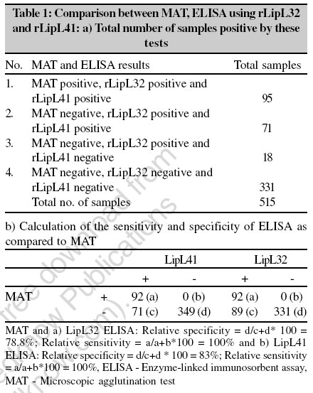
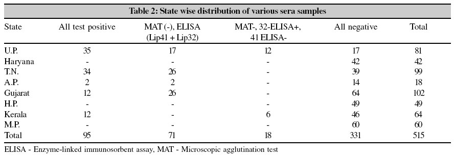
Indian Journal of Medical Microbiology, Vol. 24, No. 4, October-December, 2006, pp. 346-348 Supplement Evaluation of recombinant Leptospira interrogans serovar canicola outer membrane proteins as diagnostic antigen Srivastava SK, Chaudhuri P, Thangapandian E, Mariya R, Amutha R Division of Bacteriology and Mycology, Indian Veterinary Research Institute, Izatnagar, Uttar Pradesh - 22 Code Number: mb06110 Leptospirosis of animals and man is an important re-emerging infectious disease worldwide. In domestic animals it is an important cause of abortion, stillbirth, infertility, decreased milk production and death.[1],[2] In dairy cattle, it is responsible for great economic losses as a consequence of agalactia, abortion, stillbirth, birth of weak calves and reduced fertility.[3],[4] The standard serological test, such as microscopic agglutination test (MAT) is time-consuming and requires technical skill. The demonstration of sero-conversion by using this test requires paired acute and convalescent serum samples limiting it to a few reference laboratories.[5] The other available serological methods uses whole cell antigen preparations[6],[7] for diagnosis as well as for screening of the cases of leptospirosis. The whole-cell antigen preparations possess broadly reactive immunodominant epitopes[8] present in both, pathogenic and non-pathogenic leptospires as well as a diverse group of non-leptospiral species.[9] Recently, recombinant antigens based sero-diagnostic strategies have been developed to conduct seroprevalence studies in human leptospirosis.[10] The important protein antigens residing in the outer membrane of leptospires reported in recent years are LipL32, LipL41 and OmpL1; they are expressed in-vivo during infection.[11],[12] LipL32 and LipL41 are important and surface-exposed leptospiral outer membrane proteins expressed only in pathogenic Leptospira spp.[13] In this study, recombinant LipL32 and LipL41 proteins were tested an antigens in ELISA against serum bovine samples. Materials and Methods Growth and maintenance of bacterial strains The leptospiral strains were maintained in semi-solid EMJH medium. LB broth and LB agar medium were used for E. coli cultures. Preparation of antisera The hyper-immune serum against serovar Canicola cells was raised in New Zealand white rabbits of about one kilogram body weight according to the method described by Faine.[14] For preparation of antiserum against the recombinant proteins, 0.25 mg of the protein in 0.5 ml PBS was mixed with Freund′s incomplete adjuvant (FIA, DIFCO Laboratories, USA) and inoculated subcutaneously at two to four sites on the back into each of the two rabbits. The first booster 0.25 mg protein in FIA was given after 14 days of first inoculation followed by two more inoculations of the protein (0.25 mg each) at seven days intervals. Blood was collected three weeks after the first inoculation and titrated using MAT. Production of rLipL32 and rLipL41 antigens The primers were designed with restriction sites from the previously reported gene sequences of L. kirschneri serovar Grippotyphosa.[13] The genes encoding LipL32 and LipL41 were amplified from the genomic DNA of Leptospira interrogans serovar Canicola and cloned into pDrive (Qiagen) cloning vector and sequenced. The nucleotide sequence was submitted to GenBank under the accession number AY642287. The genes was sub-cloned into the pPROEXHTb (LipL41) and pPROEXHTc (LipL32) expression vector (Life Technologies) and transformed into Escherichia coli DH5a cells. The recombinant clones were screened using 1mM IPTG (Isopropyl-a-D-thiogalactopyranoside). The polyhistidine (6X-His) tagged fusion proteins were purified under denaturing conditions by Nickel chelating affinity chromatography. The recombinant proteins were dialyzed and the concentration was determined by Lowry method (Bangalore Genei, India) Bovine serum samples A total of 515 bovine serum samples obtained from various parts of the country were screened for the presence of anti-leptospiral antibodies. These animals had the history of reproductive problems like abortion, stillbirth, repeat breeding, weak calves and mastitis. Microscopic agglutination test The microscopic agglutination test (MAT) was carried out according to Faine[14] using reference strains of eight different leptospiral serovars viz, Leptospira interrogans serovars Hardjo (Hardjoprajitno), Pomona, Pyrogenes, Icterohaemorrhagiae, L. borgpetersenii serovars Tarassovi, Javanica, Sejroe, Ballum. Reciprocal agglutination titres of greater than or equal to 100 were considered positive. Enzyme-linked immunosorbent assay (ELISA) ELISA was done as per standard protocols,[15] with initial standardization done with 10 test serum samples and an equal number of known negative sera. Serum was used at 1:100 dilution. Individual serum samples were tested in duplicate on three different plates. The cut-off O.D. value was calculated as double of the mean value of negative control plus twice the S.D. The relative sensitivity and specificity of the ELISA were evaluated in comparison to MAT as described below. Sensitivity = a / (a+b) x 100, where ′a′ is the number of sera positive by ELISA and MAT, ′b′ the number of sera positive by MAT but negative by ELISA. Specificity = d / (c+d) x 100, where′d′ is the number of sera negative by ELISA and MAT, ′c′ the number of sera negative by MAT but positive by ELISA. Results and Discussion The optimum concentration of each of the purified recombinant antigens, which showed maximum difference between the positive and negative sera, was determined to be 100 ng/well. Of the total 515 samples, 331 were negative as tested by MAT and ELISA to these recombinant antigens. 95 serum samples were positive by MAT, recombinant(r) LipL32 ELISA and rLipL41 ELISA; 89 sera were negative by MAT of which 71 were positive by both rLipL32 as well as rLipL41 ELISA and 18 were positive by rLipL32 ELISA. The sensitivity and specificity of rLipL32 ELISA as against MAT was calculated to be 100% and 78.8% respectively, whereas using rLipL41 ELISA it was 100% and 83% [Table - 1]. The relative sensitivity using either of the antigens was 100% suggesting they were as effective as M at0 in detecting the true positive cases. However, the specificity was less than MAT. The observation that ELISA was positive in MAT negative cases could be due to the fact that MAT was done with a limited number of leptospiral serovars. Thus our findings support the prevailing view that the ELISA is more sensitive than MAT.[16],[17] The seroprevalence of leptospirosis based on MAT in animals of various states is given in [Table - 2]. Maximum positive samples were from Uttar Pradesh followed by Tamil Nadu, Gujarat and Kerala. A high prevalence of the disease was observed in animals suggesting that control measures are to be adapted in future to contain the infection. One of the methods of control is the use of a suitable vaccine in the affected population. Before any such attempt is envisaged, information on the prevalence of various serovars in a given population is essential. The study indicated that commonly occurring serovars in animals were Canicola, Pomona and Pyrogenes. Predominance of these serovars in animals has been reported earlier in the country. Development of a vaccine incorporating these serovars will be of advantage for future vaccination studies. Acknowledgements The authors are thankful to the Director, IVRI for providing necessary facilities for conducting the research. Thanks are also due to the Department of Biotechnology, Government of India for the financial support. References
Copyright 2006 - Indian Journal of Medical Microbiology The following images related to this document are available:Photo images[mb06110t2.jpg] [mb06110t1.jpg] |
| |||||||||