search
for
 About Bioline  All Journals  Testimonials  Membership  News


Indian Journal of Medical Microbiology
Medknow Publications on behalf of Indian Association of Medical Microbiology
ISSN: 0255-0857 EISSN: 1998-3646
Vol. 25, Num. 1, 2007, pp. 10-17

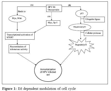
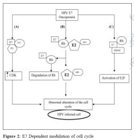
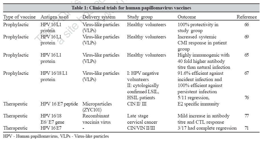
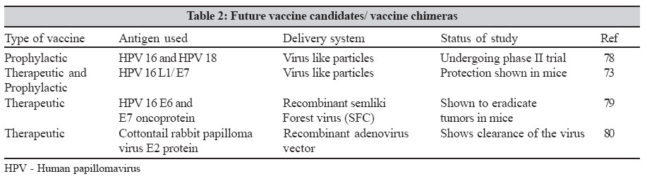
Indian Journal of Medical Microbiology, Vol. 25, No. 1, January-March, 2007, pp. 10-17

Review Article

An overview of human papillomaviruses and current vaccine strategies

Department of Clinical Virology, Christian Medical College, Vellore - 632 004, Tamil Nadu
Correspondence Address:Department of Clinical Virology, Christian Medical College, Vellore - 632 004, Tamil Nadu
priyaabraham@cmcvellore.ac.in

Date of Submission: 07-Feb-2006
Date of Acceptance: 04-Apr-2006

Code Number: mb07005

Abstract

Cervical cancer is one of the most common cancers in women worldwide, particularly in developing countries. The viral origin of cervical cancer has been proven beyond any reasonable doubt. Persistent infection with certain subsets of human papillomaviruses is recognized as a necessary cause for the development of cervical cancer. Persistence of oncogenic HPVs, immunodeficiency, high HPV viral load and cofactors like smoking, multiple sex partners and poor nutrition predispose to cervical cancer. Prophylactic vaccines using HPV virus-like particles containing capsid protein L1 have shown protection against disease in animals and are currently undergoing clinical trials. Therapeutic vaccines using HPV E6 and E7 proteins are also being investigated for their ability to remove residual infection.

Keywords: Cervical cancer, human papilloma virus, vaccines

Epidemiological evidence has proved beyond doubt that human papillomavirus (HPV) infection is the most important risk factor for cervical intraepithelial neoplasia and invasive cervical cancer.[1],[2] HPVs infect the basal epithelium and are grouped as cutaneous and mucosal types. Based on the association with cervical cancer, genital HPVs are further grouped into high risk types (16, 18, 31, 33, 35, 39, 45, 51, 52, 56, 58, 59, 68, 73 and 82), probable high-risk types (26, 53 and 66) and low risk types (6, 11, 40, 42, 43, 44, 54, 61, 70, 72, 81 and CP6108).[3] Of the high risk types, HPV 16 is the most prevalent type and is seen in at least 50% of invasive cervical cancer patients globally followed by types 18, 45, 31, 33, 52 and 58.[4] Various studies have looked at the prevalence of HPV genotypes in Indian women[5],[6],[7] and the most common genotypes reported are HPV 16 and 18.

Basic Virology

HPV are members of the family Papillomaviridae.[8] They are small, non-enveloped DNA viruses, 55 nm in diameter.[9] They have an outer icosahedral capsid made of two structural proteins, L1 and L2. The genome of HPV is circular and approximately 8 Kb long.[8] The genome has eight open reading frames coding for six early proteins (E1, E2, E4, E5, E6 and E7) and two late proteins (L1 and L2). E1 and E2 genes are involved in viral replication.[10] E2 protein also plays an important role as a repressor of E6 and E7 gene expression. E4 gene codes for protein E1-E4 that disrupt the cytoplasmic keratin network.[11] The E4 protein is also found to contribute to regulation of host cell cycle control.[12] E5 protein is known to aid in cellular transformation and plays a significant role in viral replication.[13] The late proteins L1 and L2 make up the viral capsid.

E6 and E7 proteins play a major role in the malignant transformation of cervical cells as can be seen in studies using various cell lines.[14],[15] The E6 protein forms a complex with the cell cycle regulator protein p53[16] using cellular ubiquitin ligase E6AP.[17],[18] This ubiquitination was found to lead to accelerated degradation of p53.[19] The E6 protein also down regulates p53 activity by targeting the co-activator of p53, CBP/p300.[20] Inactivation of p53 leads to loss of cell cycle control such as cell cycle arrest and apoptosis. E6 protein associates with Myc/Mak proteins and Sp-1 and this complex binds to the promoter region of the catalytic subunit of telomerase enzyme, hTERT.[21],[22],[23] This leads to an increase in telomerase activity, thereby facilitating immortalization [Figure - 1].

The E7 protein associates with retinoblastoma family of proteins. The E7 protein binds to the phosphorylated retinoblastoma protein (Rb) and separates it from E2F/DP1 complex.[24] The E7 protein then mediates degradation of the Rb protein through the ubiquitin-proteosome pathway. E7 protein also binds to various other cellular proteins like cyclin dependent kinase (cdk) inhibitor p21, possibly leading to loss of cell cycle control.[25] The E7 protein also associates with another group of proteins called histone deacetylases (HDACs) independent of Rb protein and this results in the expression of E2F/DP1 inducible genes. E2F proteins activate expression of various genes necessary for cell cycle progression and DNA replication[26] [Figure - 2]. Thus the combined effect of these two viral oncoproteins and above mentioned cellular factors contribute to the malignant transformation of the cervical cells.

Epidemiology

Human papillomavirus is transmitted by sexual contact. However a non-sexual mode of transmission through fomites has also been proposed.[27] Epidemiological evidence also points to an association between cervical cancer and various risk factors. Impaired cell mediated immunity is a major risk factor for cervical cancer. Evidence for this is derived from studies showing a high incidence of HPV infection in human immunodeficiency virus seropositive women.[28] High HPV viral load is a risk factor associated with high stages of cervical disease.[29] Persistence of high-risk types of HPV is another major risk factor in the development of cervical cancer. A higher risk of acquiring HPV was attributed to decreasing age of first sexual intercourse, multiple sexual partners and clinical history of other venereal diseases.[30] High parity was found to increase the risk of infection.[31] Long-term use of oral contraceptives[32] is also a significant cofactor in the development of cervical cancer.

Most Indian studies have reported HPV 16 and 18 as the major genotypes. There is evidence that there are other oncogenic HPV types circulating in our population. In our center, the predominant HPV types detected were HPV 16 and 18 followed by 52, 33, 58, 35, 31, 45, 51 and 56 (unpublished data).

More recently, naturally occurring HPV variants are gaining importance as viral markers of epidemiology and pathogenesis. Based on sequence analysis of the E6, L1, L2 and long control region, HPV 16 is grouped into five major phylogenetic clusters. They are European (E), Asian (As), Asian-American (AA), African-1(Af1) and African-2 (Af2).[9] A study from our center has shown a higher prevelance (92%) of European (E) variant class of HPV 16 in Indian population.[33]

Pathogenesis

HPVs infect the basal layers of the cervical squamous epithelium through mild abrasions or trauma. HPV is thought to enter the cell after attaching to cell surface receptor heparan sulphate.[34] The virus life cycle depends on the differentiation of the host squamous epithelium. In the basal layers of the epithelium, the virus genome is episomal and establishes itself at low copy numbers. The early protein E1 in association with E2 is thought to trigger the replication process. At this stage, the expression of the viral oncoproteins E6 and E7 are kept in check by the E2 protein, which acts as a transcriptional repressor. In the terminal layers, the virus switches from theta replication to rolling circle mode of replication, resulting in a high copy number of viral DNA.[35] At this stage, the virus manages to integrate its DNA into the host chromosome. This disrupts the E2 open reading frame leading to excess synthesis of the E6 and E7 oncoproteins. This eventually is thought to lead to malignant transformation of the epithelial cells. Studies from our centre have shown that HPV 16 E6 and E7 transcripts are seen in all cases of invasive cancer but not in cases of cervical intraepithelial neoplasia (CIN) I and II. The presence of episomal E2 DNA in high propotion (52.4%) of patients with invasive carcinoma and the presence of significant variations in the E2 gene suggests that there are alternate mechanisms of E6 and E7 gene expression.[36],[37] Finally, capsid proteins L1 and L2 are synthesized and mature virions are produced. The release of HPV virions is thought to be facilitated by the E1-E4 protein.

Laboratory Diagnosis

The high incidence of cervical cancer in developing countries emphasizes the need for proper screening to reduce this global burden. The primary tools for diagnosis of HPV infection are cytology, histology and recently, detection of HPV DNA.

Cytology and histology

The Papanicolou (Pap) smear, introduced by Papanicolau and Traut in 1943, identifies changes in cells of the transformation zone of cervix caused by HPV infection.[38] Abnormal cells are vacuolated with a pyknotic nucleus surrounded by a halo and are termed as "koilocytes". The current interpretation of Pap smear is based on the Bethesda system.[39] However, cytology has its limitations. Inadequate sampling, poor sensitivity with false negative results, contaminants in the sample, have been reported.[9] Automated cytologic tests using PapNet (Neuromedical systems, Suffern, NY) and Autopap 300 QC (Neopath, Redmond, Wash) have been approved by the Food and Drug Administration, USA for screening smears to identify false negative smears. Fluid based technology also reduces false negative smear results. The specimen is collected in a preservative solution, debris is removed thereby aiding clear visualization of the cells.[40] Colposcopy and colposcopy directed biopsies are done in patients with abnormal pap smears. Visual inspection is done with the naked eye after application of 3% acetic acid solution (VIA).[9] Dysplastic cells appear as acetowhite lesions. Visual inspection with Lugol′s iodine (VILI) is another approach where the cervix is viewed with the naked eye after application of iodine solution. Normal squamous epithelial cells appear brown or almost black in colour whereas abnormal cells appear colourless, pale or mustard yellow in colour.[41] Cervical biopsy is then done to conform malignancy. Cervical biopsy is considered the gold standard for the detection of cervical neoplasia and HPV infection.

HPV DNA detection

HPV cannot be easily cultured in the laboratory. Therefore, molecular methods to detect HPV DNA are been used to confirm the presence to HPV in clinical specimens.

The currently used techniques for HPV DNA detection are the hybrid capture second generation (HC2) assay and polymerase chain reaction (PCR). The hybrid capture assay is the only commercial kit approved by the FDA for the detection of HPV. The hybrid capture assay detects 13 high-risk types (16, 18, 31, 33, 35, 39, 45, 51, 52, 56, 58, 59 and 68) and 5 low risk types (6, 11, 42, 43, 44). Hybrid Capture 2 assay was found to be a good screening tool for the detection of cervical intraepithelial neoplasia III and invasive cervical cancer.[42] The assay works on a hybridization/ signal amplification principle and uses chemiluminescence to detect the presence of HPV.[43] Recently a prototype version of Hybrid Capture 3 has been released. The hybrid capture 3 was found to be more sensitive and equally specific like its predecessor when compared to the PCR based assays.[44] But this method does not distinguish between the different HPV genotypes.

Most PCR protocols for the detection of genital HPVs use consensus primers GP5/6,[45] degenerate primers MY09/11 and its modified version PGMY09/11,[46] amplifying a wide spectrum of genital HPVs. HPV genotyping is done by nucleotide sequencing, restriction fragment length polymorphism,[47] reverse hybridization line probe assay[48] and line blot assay.[49] Quantification of HPV viral load has been done by real time PCR assays.[50]

Studies including those from this centre have shown an association between the presence of HPV DNA in plasma of patients with advanced stages of invasive cervical cancer and metastasis to distinct organs.[51],[52] HPV DNA in plasma can thus be used as a marker of poor disease progression.

Immunology

HPV infection is transient and 70% of women clear the infection in one year suggesting the role of an effective immune system in clearing the virus.[53] Immune response to HPV is however, weak and studies using HPV virus-like particles (VLPs) have shown that antibodies are detected approximately eight months after incident HPV infection.[54] The same study also shows that women who seroconverted were 5.7 times more likely to progress to HPV associated squamous intraepithelial neoplasia (SIL) than women who did not seroconvert. These results suggest that in natural infection, antibodies to HPV are not protective and serve as a marker of disease progression. Another study showed a seroprevalence of 46% in sexually active college women infected with HPV 16.[55]

The importance of cell mediated immunity in controlling HPV infection comes from studies showing increased prevalence of HPV in HIV seropositive women.[28] Cytotoxic T lymphocyte (CTL) response to HPV E6 and E7 proteins were detected more commonly in HPV 16 positive patients without CIN than in HPV 16 positive patients with (CIN), suggesting a protective role of CTL′s in HPV infection.[56] This CTL response was found to be mediated by both CD4-positive and CD8-positive T lymphocytes.[57] Lymphoproliferative responses to a HPV 16 E7 peptide 37-54 were found to be associated with regression of disease and loss of HPV infection suggesting a protective role of cell mediated immune response.[58]

Treatment

Treatment options for cervical neoplasia depend on the stage of the disease. Cervical non-invasive lesions are treated by ablative methods like cryotherapy, laser therapy, cold coagulation and diathermy or excisional methods like loop electrosurgical excision procedure (LEEP) and conization. In cryotherapy, abnormal tissue is frozen by an instrument and destroyed. Laser therapy uses a single laser beam to cut abnormal tissue. In loop LEEP, an electrically charged wire is used to excise the lesion. Early invasive cancers are treated with radical hysterectomy or internal/external radiation therapy. Advanced cancer patients are treated with internal/ external radiation therapy and chemotherapy.[9]

Additional therapies to treat HPV infection of the cervix have also been tried. Local application of antiviral agents like cidofovir has been shown to be partly effective against CIN III lesions.[59] Various immunomodulatory agents like recombinant interferon gamma,[60] imiquinod 5%[61] have also been shown to be efficacious in CIN treatment and management. In a preliminary study, antioxidant curcumin caused a down regulation of HPV 18 transcription in Hela cell line.[62] Further studies will establish the clinical efficacy of curcumin in treatment of cervical cancer.

Prevention

HPV screening

Screening programs have greatly reduced the incidence of cervical cancer in the developed world. Prevention programs should comprise of health education and periodic pelvic examinations, which may include direct visual inspection (DVI), VIA, VILI, pap smear and HPV DNA testing. However, large scale routine screening and treatment for cervical cancer is hard to achieve in a developing country like India. A cost-effective vaccine is therefore needed as an alternative to screening and treatment. There are two types of vaccines being developed for HPV infection, prophylactic vaccine and therapeutic vaccine. Some of the studies on vaccine trials and future candidate vaccines are summarized in [Table - 1][Table - 2].

Prophylactic vaccines prevent infection by inducing production of neutralizing antibodies. A good prophylactic vaccine should be safe and should induce long-lasting protection. Development of prophylactic vaccines for HPV is hindered by various factors. HPVs are difficult to grow in vitro . Moreover, HPVs are species specific, so vaccine evaluation in animal models is also not possible. Sub-unit vaccines using HPV VLPs, obtained by expressing capsid protein L1and L2 in both prokaryotic and eukaryotic cells are currently being used as candidate vaccines. In early animal models like rabbits[63] and canines,[64] these VLPs have shown to be highly immunogenic and also shown to elicit high titres of neutralizing antibodies. An early human trial using HPV 16 VLPs produced in insect cells showed high titres of neutralizing antibodies up to 40 times higher than in a natural infection.[65] Another randomized control trial was conducted using HPV 16 VLPs consisting of HPV 16 L1 protein expressed in yeast. This study showed complete protection from disease in the vaccinated group compared to the unvaccinated group.[66] This study has shown that HPV 16 VLPs used as prophylactic vaccines can reduce cervical cancer risk in women who are HPV 16 negative. Recently, a bivalent HPV 16/ 18 virus-like particle vaccine has been shown to be efficacious in preventing incident and persistent infections with HPV 16 and 18.[67] Quadrivalent vaccines using VLPs made of HPV 6, 11, 16, 18 have been tested for their efficacy in chimpanzees. The study shows that this vaccine elicits both neutralizing antibodies as well as a transient CTL responses.[68] Currently phase III trials are being undertaken to prove its efficacy. Another human trial demonstrated the ability of HPV VLP vaccines to induce both humoral as well as cell-mediated immune responses in healthy individuals as shown by increased neutralizing antibodies as well as cytokine response.[69]

Future vaccine candidates in our country should incorporate the important prevalent high-risk HPV types.

Therapeutic vaccines

Therapeutic vaccines are being developed to treat already established infections. As the early oncoproteins E6 and E7 are expressed through out the lifecycle of the virus and their presence necessary to maintain the transformed state in cell lines,[70] they are potential targets as therapeutic vaccines. A phase I trial using E7 peptide vaccine showed regression of disease in the study population.[71] Vaccination with long peptides of E7 protein of HPV 16 elicited a robust CD4-positive T helper cell response as well as a CD8-positive CTL response in mice.[72] DNA vaccines comprising the HPV 16 E6 gene were shown to elicit protective cell mediated immune response in mice.[73] Chimeric VLPs have also been developed by fusing non-structural proteins E7 or E2 into L2 protein. These chimeric VLPs elicit both humoral immune responses as well as cell mediated immune responses[74] and may be relevant for a population that has established HPV related cervical disease.

Conclusions

HPV infection has been reported to be a frequently occurring sexually transmitted disease.[9] It has also been implicated in causing cervical cancer, which is the most frequently occurring cancer in Indian rural women. India carries a fourth of world′s burden of cervical cancer.[75] It is important that health authorities place cervical cancer screening and HPV detection as a high priority in future cancer prevention health strategies[80].

References

1.Walboomers JM, Jacobs MV, Manos MM, Bosch FX, Kummer JA, Shah KV, et al . Human papillomavirus is a necessary cause of invasive cervical cancer worldwide. J Pathol 1999; 189: 12-9.  Back to cited text no. 1  [PUBMED]  
2.Munoz N. Human papillomavirus and cancer: The epidemiological evidence. J Clin Virol 2000; 19 :1-5.  Back to cited text no. 2  [PUBMED]  [FULLTEXT]
3.Munoz N, Bosch FX, de Sanjose S, Herrero R, Castellsague X, Shah KV, et al . Epidemiologic classification of human papillomavirus types associated with cervical cancer. N Engl J Med 2003; 348 :518-27.  Back to cited text no. 3  [PUBMED]  [FULLTEXT]
4.Clifford GM, Smith JS, Plummer M, Munoz N, Franceschi S. Human papillomavirus types in invasive cervical cancer worldwide: A meta-analysis. Br J Cancer 2003; 88: 63-73.  Back to cited text no. 4  [PUBMED]  [FULLTEXT]
5.Chatterjee R, Roy A, Basu S. Detection of type specific human papillomavirus (HPV) DNA in cervical cancers of Indian women. Indian J Pathol Microbiol 1995; 38 :33-42.  Back to cited text no. 5  [PUBMED]  
6.Menon MM, Sinha MR, Doctor VM. Detection of human papillomavirus (HPV) types in precancerous and cancerous lesions of cervix in Indian women: A preliminary report. Indian J Cancer 1995; 32 :154-9.  Back to cited text no. 6    
7.Das BC, Sharma JK, Gopalkrishna V, Das DK, Singh V, Gissmann L, et al . A high frequency of human papillomavirus DNA sequences in cervical carcinomas of Indian women as revealed by southern blot hybridization and polymerase chain reaction. J Med Virol 1992; 36 :239-45.  Back to cited text no. 7  [PUBMED]  
8.Howley PM, Lowy DR. Papillomaviruses and their replication, Chapter 65. In : Field's Virology, Volume 2, 4th ed. Knipe DM, Howley PM, editors. Lippincott Williamsand Wilkins: Philadelphia; 2001. p. 2197-229.   Back to cited text no. 8    
9.Burd EM. Human papillomavirus and cervical cancer. Clin Microbiol Rev 2003; 16 : 1-17.  Back to cited text no. 9    
10.Chiang CM, Ustav M, Stenlund A, Ho TF, Broker TR, Chow LT. Viral E1 and E2 proteins support replication of homologous and heterologous papillomaviral origins. Proc Natl Acad Sci USA 1992; 89: 5799-803.  Back to cited text no. 10    
11.Doorbar J, Ely S, Sterling J, McLean C, Crawford L. Specific interaction between HPV 16 E1-E4 and cytokeratins results in collapse of the epithelial cell intermediate filament network. Nature 1991; 352 : 824-7.  Back to cited text no. 11    
12.Nakahara T, Nishimura A, Tanaka M, Ueno T, Ishimoto A, Sakai H. Modulation of the cell division cycle by human papillomavirus type 18 E4. J Virol 2002; 76 : 10914-20.  Back to cited text no. 12    
13.Genther SM, Sterling S, Duensing S, Munger K, Sattler C, Lambert PF. Quantitative role of the human papillomavirus type 16 E5 gene during the productive stage of the viral life cycle. J Virol 2003; 77 :2832-42.  Back to cited text no. 13    
14.Halbert CL, Demers GW, Galloway DA. The E7 gene of human papillomavirus type 16 is sufficient for immortalization of human epithelial cells. J Virol 1991; 65 : 473-8.  Back to cited text no. 14    
15.Hudson JB, Bedell MA, McCance DJ, Laimins LA. Immortalization and altered differentiation of human keratinocytes in vitro by the E6 and E7 open reading frames of human papillomavirus type 18. J Virol 1990; 64: 519-26.  Back to cited text no. 15    
16.Werness BA, Levine AJ, Howley PM. Association of human papillomavirus types 16 and 18 E6 proteins with p53. Science 1990; 248: 76-9.  Back to cited text no. 16    
17.Cooper B, Schneider S, Bohl J, Jiang Y, Beaudet A, Vande Pol S. Requirement of E6AP and the features of human papillomavirus E6 necessary to support degradation of p53. Virology 2003; 306 :87-99.  Back to cited text no. 17    
18.Huibregtse JM, Scheffner M, Howley PM. Cloning and expression of the cDNA for E6-AP, a protein that mediates the interaction of the human papillomavirus E6 oncoprotein with p53. Mol Cell Biol 1993; 13 :775-84.  Back to cited text no. 18    
19.Hubbert NL, Sedman SA, Schiller JT. Human papillomavirus type 16 E6 increases the degradation rate of p53 in human keratinocytes. J Virol 1992; 66 :6237-41.  Back to cited text no. 19    
20.Zimmermann H, Degenkolbe R, Bernard HU, O'Connor MJ. The human papillomavirus type 16 E6 oncoprotein can down-regulate p53 activity by targeting the transcriptional coactivator CBP/p300. J Virol 1999; 73 :6209-19.  Back to cited text no. 20    
21.Veldman T, Horikawa I, Barrett JC, Schlegel R. Transcriptional activation of the telomerase hTERT gene by human papillomavirus type 16 E6 oncoprotein. J Virol 2001; 75 :4467-72.  Back to cited text no. 21    
22.Veldman T, Liu X, Yuan H, Schlegel R. Human papillomavirus E6 and Myc proteins associate in vivo and bind to and cooperatively activate the telomerase reverse transcriptase promoter. Proc Natl Acad Sci USA 2003; 100 :8211-6.  Back to cited text no. 22    
23.Oh ST, Kyo S, Laimins LA. Telomerase activation by human papillomavirus type 16 E6 protein: Induction of human telomerase reverse transcriptase expression through Myc and GC-rich Sp1 binding sites. J Virol 2001; 75 : 5559-66.  Back to cited text no. 23    
24.Munger K, Werness BA, Dyson N, Phelps WC, Harlow E, Howley PM. Complex formation of human papillomavirus E7 proteins with the retinoblastoma tumor suppressor gene product. Embo J 1989; 20 :4099-105.  Back to cited text no. 24    
25.Funk JO, Waga S, Harry JB, Espling E, Stillman B, Galloway DA. Inhibition of CDK activity and PCNA-dependent DNA replication by p21 is blocked by interaction with the HPV-16 E7 oncoprotein. Genes Dev 1997; 11 :2090-100.  Back to cited text no. 25    
26.DeGregori J, Leone G, Miron A, Jakoi L, Nevins JR. Distinct roles for E2F proteins in cell growth control and apoptosis. Proc Natl Acad Sci USA 1997; 94 :7245-50.  Back to cited text no. 26    
27.Roden RB, Lowy DR, Schiller JT. Papillomavirus is resistant to desiccation. J Infect Dis 1997; 176 :1076-9.  Back to cited text no. 27    
28.Sun XW, Kuhn L, Ellerbrock TV, Chiasson MA, Bush TJ, Wright TC Jr. Human papillomavirus infection in women infected with the human immunodeficiency virus. N Engl J Med 1997; 337 :1343-9.  Back to cited text no. 28    
29.Swan DC, Tucker RA, Tortolero-Luna G, Mitchell MF, Wideroff L, Unger ER, et al . Human papillomavirus (HPV) DNA copy number is dependent on grade of cervical disease and HPV type. J Clin Microbiol 1999; 37: 1030-4.  Back to cited text no. 29    
30.La Vecchia C, Franceschi S, Decarli A, Fasoli M, Gentile A, Parazzini F, et al . Sexual factors, venereal diseases and the risk of intraepithelial and invasive cervical neoplasia. Cancer 1986; 58 :935-41.  Back to cited text no. 30    
31.Munoz N, Franceschi S, Bosetti C, Moreno V, Herrero R, Smith JS, et al . Role of parity and human papillomavirus in cervical cancer: The IARC multicentric case-control study. Lancet 2002; 359: 1093-101.  Back to cited text no. 31    
32.Moreno V, Bosch FX, Munoz N, Meijer CJ, Shah KV, Walboomers JM, et al . Effect of oral contraceptives on risk of cervical cancer in women with human papillomavirus infection: The IARC multicentric case-control study. Lancet 2002; 359: 1085-92.  Back to cited text no. 32    
33.Sathish N, Abraham P, Peedicayil A, Sridharan G, Chandy C. HPV 16 E6 sequence variations in Indian patients with cervical neoplasia. Cancer lett 2005; 229: 93-9.  Back to cited text no. 33    
34.Giroglou T, Florin L, Schafer F, Streeck RE, Sapp M. Human papillomavirus infection requires cell surface heparan sulfate. J Virol 2001; 75 :1565-70.  Back to cited text no. 34    
35.Flores ER, Lambert PF. Evidence for a switch in the mode of human papillomavirus type 16 DNA replication during the viral life cycle. J Virol 1997; 71: 7167-79.  Back to cited text no. 35    
36.Sathish N, Abraham P, Peedicayil A, Sridharan G, John S, Chandy G. Human papillomavirus 16 E6/E7 transcript and E2 gene status in patients with cervical neoplasia. Mol Diagn 2004; 8: 57-64.  Back to cited text no. 36    
37.Sathish N, Abraham P, Peedicayil A, Sridharan G, Shaji RV, Chandy G. E2 sequence variations of HPV 16 among patients with cervical neoplasia seen in the Indian subcontinent. Gynecol Oncol 2004; 95: 363-9.  Back to cited text no. 37    
38.Shingleton HM, Orr JW Jr, editors. Carcinoma of the cervix: Historical aspects, epidemiology and screening, Chapter 1. In : Cancer of the cervix: Diagnosis and treatment . Churchill Livingston: New York; 1987.p. 1-15.  Back to cited text no. 38    
39.Solomon D, Davey D, Kurman R, Moriarty A, O'Connor D, Prey M, et al . The 2001 Bethesda system. Terminology for reporting results of cervical cytology. JAMA 2002; 287 :2114-9.   Back to cited text no. 39    
40.Spitzer M. Cervical screening adjuncts: Recent advances. Am J Obstet Gynecol 1998; 179 :544-56.  Back to cited text no. 40    
41.Sankaranarayanan R, Nene BM, Dinshaw K, Rajkumar R, Shastri S, Wesley R, et al . Early detection of cervical cancer with visual inspection methods: A summary of completed and on-going studies in India. Salud Publica Mex 2003; 45 :S399-407.  Back to cited text no. 41    
42.Sherman ME, Lorincz AT, Scott DR, Wacholder S, Castle PE, Glass AG, et al . Baseline cytology, human papillomavirus testing and risk for cervical neoplasia: A 10-year cohort analysis. J Natl Cancer Inst 2003; 95 :46-52.   Back to cited text no. 42    
43.Iftner T, Villa LL. Chapter 12: Human papillomavirus technologies. J Natl Cancer Inst Monogr 2003; 31 :80-8.  Back to cited text no. 43    
44.Castle PE, Lorincz AT, Scott DR, Sherman ME, Glass AG, Rush BB, et al . Comparison between prototype hybrid capture 3 and hybrid capture 2 human papillomavirus DNA assays for detection of high-grade cervical intraepithelial neoplasia and cancer. J Clin Microbiol 2003; 41 :4022-30.  Back to cited text no. 44    
45.Snijders PJ, van den Brule AJ, Schrijnemakers HF, Snow G, Meijer CJ, Walboomers JM. The use of general primers in the polymerase chain reaction permits the detection of a broad spectrum of human papillomavirus genotypes. J Gen Virol 1990; 71: 173-81.  Back to cited text no. 45    
46.Gravitt PE, Peyton CL, Alessi TQ, Wheeler CM, Coutlee F, Hildesheim A, et al . Improved amplification of genital human papillomaviruses. J Clin Microbiol 2000; 38: 357-61.  Back to cited text no. 46    
47.Bernard HU, Chan SY, Manos MM, Ong CK, Villa LL, Delius H, et al . Identification and assessment of known and novel human papillomaviruses by polymerase chain reaction amplification, restriction fragment length polymorphisms, nucleotide sequence and phylogenetic algorithms. J Infect Dis 1994; 170: 1077-85.  Back to cited text no. 47    
48.Kleter B, van Doorn LJ, Schrauwen L, Molijn A, Sastrowijoto S, ter Schegget J, et al . Development and clinical evaluation of a highly sensitive PCR-reverse hybridization line probe assay for detection and identification of anogenital human papillomavirus. J Clin Microbiol 1999; 37: 2508-17.   Back to cited text no. 48    
49.Gravitt PE, Peyton CL, Apple RJ, Wheeler CM. Genotyping of 27 human papillomavirus types by using L1 consensus PCR products by a single-hybridization, reverse line blot detection method. J Clin Microbiol 1998; 36: 3020-7.  Back to cited text no. 49    
50.Gravitt PE, Burk RD, Lorincz A, Herrero R, Hildesheim A, Sherman ME, et al . A comparison between real-time polymerase chain reaction and hybrid capture 2 for human papillomavirus DNA Quantitation. Cancer Epidemiol Biomarkers Prev 2003; 12: 277-84.  Back to cited text no. 50    
51.Pornthanakasem W, Shotelersuk K, Termrungruanglert W, Voravud N, Niruthisard S, Mutirangura A. Human papillomavirus DNA in plasma of patients with cervical cancer. BMC Cancer 2001; 1: 2-13.  Back to cited text no. 51    
52.Sathish N, Abraham P, Peedicayil A, Sridharan G, John S, Shaji RV, et al . HPV DNA in plasma of patients with cervical carcinoma. J Clin Virol 2004; 31: 204-9.  Back to cited text no. 52    
53.Ho GY, Bierman R, Beardsley L, Chang CJ, Burk RD. Natural history of cervicovaginal papillomavirus infection in young women. N Engl J Med 1998; 338: 423-8.  Back to cited text no. 53    
54.Carter JJ, Koutsky LA, Wipf GC, Christensen ND, Lee SK, Kuypers J, et al . The natural history of human papillomavirus type 16 capsid antibodies among a cohort of university women. J Infect Dis 1996; 174: 927-36.  Back to cited text no. 54    
55.Viscidi RP, Kotloff KL, Clayman B, Russ K, Shapiro S, Shah KV. Prevalence of antibodies to human papillomavirus (HPV) type 16 virus-like particles in relation to cervical HPV infection among college women. Clin Diagn Lab Immunol 1997; 4: 122-6.  Back to cited text no. 55    
56.Nakagawa M, Stites DP, Farhat S, Sisler JR, Moss B, Kong F, et al . Cytotoxic T lymphocyte responses to E6 and E7 proteins of human papillomavirus type 16: Relationship to cervical intraepithelial neoplasia. J Infect Dis 1997; 175 :927-31.  Back to cited text no. 56    
57.Nakagawa M, Stites DP, Palefsky JM, Kneass Z, Mosciki AB. CD4- positive and CD8- positive cytotoxic T lymphocytes contribute to human papillomavirus type 16 E6 and E7 responses. Clin Diagn Lab Immunol 1999; 6: 494-8.  Back to cited text no. 57    
58.Kadish AS, Timmins P, Wang Y, Ho GY, Burk RD, Ketz J, et al . Regression of cervical intraepithelial neoplasia and loss of human papillomavirus (HPV) infection is associated with cell-mediated immune responses to an HPV type 16 E7 peptide. Cancer Epidemiol Biomarkers Prev 2002; 11: 483-8.  Back to cited text no. 58    
59.Snoeck R, Noel JC, Muller C, De Clercq E, Bossens M. Cidofovir, a new approach for the treatment of cervix intraepithelial neoplasia grade III (CIN III). J Med Virol 2000 ; 60:205-9.  Back to cited text no. 59    
60.Sikorski M, Zrubek H. Recombinant human interferon gamma in the treatment of cervical intraepithelial neoplasia (CIN) associated with human papillomavirus (HPV) infection. Eur J Gynaecol Oncol 2003; 24: 147-50.  Back to cited text no. 60    
61.Diaz-Arrastia C, Arany I, Robazetti SC, Dinh TV, Gatalica Z, Tyring SK, et al . Clinical and molecular responses in high-grade intraepithelial neoplasia treated with topical imiquimod 5%. Clin Cancer Res 2001; 7: 3031-3.  Back to cited text no. 61    
62.Prusty BK, Das BC. Constitutive activation of transcription factor AP-1 in cervical cancer and suppression of human papillomavirus (HPV) transcription and AP-1 activity in HeLa cells by curcumin. Int J Cancer 2005; 113: 951-60.  Back to cited text no. 62    
63.Christensen ND, Reed CA, Cladel NM, Han R, Kreider JW. Immunization with virus- like particles induces long-term protection of rabbits against challenge with cottontail rabbit papillomavirus. J Virol 1996; 70: 960-5.  Back to cited text no. 63    
64.Suzich JA, Ghim SJ, Palmer-Hill FJ, White WI, Tamura JK, Bell JA, et al . Systemic immunization with papillomavirus L1 protein completely prevents the development of viral mucosal papillomas. Proc Natl Acad Sci USA 1995; 92: 11553-7.   Back to cited text no. 64    
65.Harro CD, Pang YY, Roden RB, Hildesheim A, Wang Z, Reynolds MJ, et al . Safety and immunogenicity trial in adult volunteers of a human papillomavirus 16 L1 virus-like particle vaccine. J Natl Cancer Inst 2001; 93: 284-92.  Back to cited text no. 65    
66.Koutsky LA, Ault KA, Wheeler CM, Brown DR, Barr E, Alvarez FB. A controlled trial of a human papillomavirus type 16 vaccine. N Engl J Med 2002; 347: 1645-51.  Back to cited text no. 66    
67.Harper DM, Franco EL, Wheeler C, Ferris DG, Jenkins D, Schuind A, et al . Efficacy of a bivalent L1 virus-like particle vaccine in prevention of infection with human papillomavirus types 16 and 18 in young women: A randomized controlled trial. Lancet 2004; 364: 1757-65.  Back to cited text no. 67    
68.Palker TJ, Monteiro JM, Martin MM, Kakareka C, Smith JF, Cook JC, et al . Antibody, cytokine and cytotoxic T lymphocyte responses in chimpanzees immunized with human papillomavirus virus-like particles. Vaccine 2001; 19: 3733-43.  Back to cited text no. 68    
69.Pinto LA, Edwards J, Castle PE, Harro CD, Lowy DR, Schiller JT, et al . Cellular immune responses to human papillomavirus (HPV)-16 L1 in healthy volunteers immunized with recombinant HPV 16 L1 virus-like particles. J Infect Dis 2003; 188: 327-38.  Back to cited text no. 69    
70.DeFilippis RA, Goodwin EC, Wu L, DiMaio D. Endogenous human papillomavirus E6 and E7 proteins differentially regulate proliferation, senescence and apoptosis in HeLa cervical carcinoma cells. J Virol 2003; 77: 1551-63.  Back to cited text no. 70    
71.Muderspach L, Wilczynski S, Roman L, Bade L, Felix J, Small LA, et al. A phase I trial of a Human papillomavirus (HPV) peptide vaccine for women with high-grade cervical and vulvar intraepithelial neoplasia who are HPV 16 positive. Clin Cancer Res 2000; 6: 3406-16.  Back to cited text no. 71    
72.Zwaveling S, Mota SC, Nouta J, Johnson M, Lipford GB, Offringa R, et al . Established human papillomavirus type 16-expressing tumors are effectively eradicated following vaccination with long peptides. J Immunol 2002; 169: 350-8.  Back to cited text no. 72    
73.Peng S, Ji H, Trimble C, He L, Tsai YC, Yeatermeyer J, et al . Development of a DNA vaccine targeting human papillomavirus type 16 oncoprotein E6. J Virol 2004; 78: 8468-76.  Back to cited text no. 73    
74.Greenstone HL, Nieland JD, DeVisser KE, De Bruijn ML, Kirnbauer R, Roden RB, et al . Chimeric papillomavirus virus-like particles elicit antitumor immunity against the E7 oncoprotein in an HPV 16 tumor model. Proc Natl Acad Sci USA 1998; 95 :1800-5.  Back to cited text no. 74    
75.Shanta V, Krishnamurthi S, Gajalakshmi CK, Swaminathan R, Ravichandran K. Epidemiology of cancer of the cervix: Global and national perspective. J Indian Med Assoc 2000; 98: 49-52.  Back to cited text no. 75    
76.Sheets EE, Urban RG, Crum CP, Hedley ML, Politch JA, Gold MA, et al . Immunotherapy of human cervical high-grade cervical intraepithelial neoplasia with microparticle-delivered human papillomavirus 16 E7 plasmid DNA. Am J Obstet Gynecol 2003; 188: 916-26.   Back to cited text no. 76    
77.Borysiewicz LK, Fiander A, Nimako M, Man S, Wilkinson GW, Westmoreland D, et al . A recombinant vaccinia virus encoding human papillomavirus types 16 and 18, E6 and E7 proteins as immunotherapy for cervical cancer. Lancet 1996; 347: 1523-7.  Back to cited text no. 77    
78.Billich A. HPV vaccine MedImmune/ GlaxoSmith Kline. Curr Opin Investig Drugs 2003; 4: 210-3.   Back to cited text no. 78    
79.Daemen T, Riezebos-Brilman A, Regts J, Dontje B, van der Zee A, Wilschut J. Superior therapeutic efficacy of alphavirus-mediated immunization against human papilloma virus type 16 antigens in a murine tumour model: Effects of the route of immunization. Antivir Ther 2004; 9: 733-42.  Back to cited text no. 79    
80.Brandsma JL, Shlyankevich M, Zhang L, Slade MD, Goodwin EC, Peh W, et al . Vaccination of rabbits with an adenovirus vector expressing the papillomavirus E2 protein leads to clearance of papillomas and infection. J Virol 2004; 78: 116-23.  Back to cited text no. 80    

Copyright 2007 - Indian Journal of Medical Microbiology


The following images related to this document are available:

Photo images

[mb07005f2.jpg] [mb07005t1.jpg] [mb07005t2.jpg] [mb07005f1.jpg]
Home Faq Resources Email Bioline
© Bioline International, 1989 - 2024, Site last up-dated on 01-Sep-2022.
Site created and maintained by the Reference Center on Environmental Information, CRIA, Brazil
System hosted by the Google Cloud Platform, GCP, Brazil