 |
| About Bioline | All Journals | Testimonials | Membership | News |
|
||||||
|
||||||
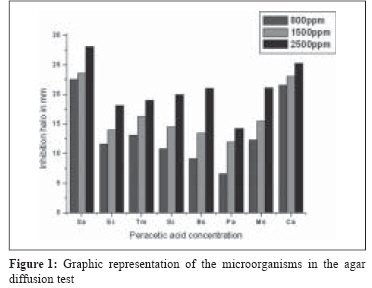
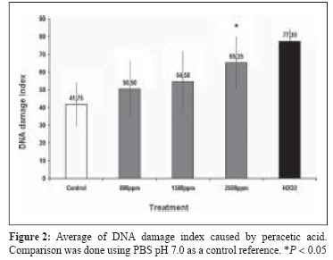
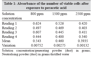
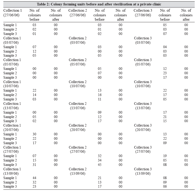
Indian Journal of Medical Microbiology, Vol. 26, No. 2, April-June, 2008, pp. 117-122 Original Article Evaluation of the effectiveness of peracetic acid in the sterilization of dental equipment Ceretta R, Paula MMS, Angioletto Ev, Meier MM, Mitellstadt FG, Pich CT, Junior SA, Angioletto E UNESC - Programa de Pós-Graduação em Ciências da Saúde Av. Universitária, 1105 ZIP 88806-000 SC Date of Submission: 04-Sep-2007 Code Number: mb08036 Abstract Purpose: To evaluate the effectiveness of peracetic acid in the microbiological sterilisation of dental materials.Methods: Peracetic acid solution was evaluated at concentrations of 800, 1500 and 2500 ppm. At these concentrations, it was determined whether peracetic acid caused corrosion to dental instruments and induced cellular mutagenicity and cytotoxicidity. In addition, the minimum inhibitory concentration (MIC), the minimum bactericidal concentration (MBC), agar diffusion and diffusion by well method, were also verified. Results: The corrosion rate, calculated from potentiodynamic assays was 10 -6 cm/year, indicating that the product does not damage equipment. The sterilisation capacity of peracetic acid at 2500 ppm was the best. The comet assay indicated genotoxic activity at 2500 ppm. Conclusions: This study demonstrated the effectiveness of peracetic acid for sterilizing dental equipment, providing another alternative for the prevention of infections in clinics. Keywords: Cytotoxicity, chemical agents, genotoxic activity, peracetic acid, sterilisation The dental work environment is a place of high contamination risk due to the type of work that is performed and the materials used. [1] In dental clinics, the greatest variety and concentration of microorganisms is found in the patient′s mouth. Given this reality, matters related to the control of infections and biosafety regulations have taken on a new focus. Procedures such as how to pick up materials from drawers, something previously considered harmless, should be reviewed and corrected. [2] Among the many causes of disease, are those of an infectious nature, [3] characterised by an inflammatory response against the presence of microorganisms or their tissue invasion. [4] Sterilisation process includes the elimination of all viable organisms, including spores. However, it should be highlighted that certain instruments cannot be sterilised by heat, one of the most commonly employed methods in dentistry. In these cases, sterilisation using chemical agents is required for materials like the gutta-percha cones used in root canal fillings, rubber sheets for absolute isolation and intra-oral positioners for X-rays, among others. Infections that can occur in dental clinics are similar to hospital infections, which are currently well-studied, since they represent serious risks to the patients undergoing treatment. Therefore, the surgeon dentist must control dental clinic infections rigorously, so that their own lives and the lives of their patients, their assistants and their relatives are not at risk. [5] It is of fundamental importance that the procedures established by the biosafety regulations are strictly followed and that cleaning and sanitisation of the location, as well as the equipment and work surfaces, comply with legal requirements. [6] A chemical agent which has been studied and used for the chemical sterilisation of materials and equipment is peracetic acid, due to its quick action at low concentrations. [7],[8],[9] Evaluation of the effectiveness of chemical sterilisation using peracetic acid on contaminated dental equipment constitutes the scope of the present work. Materials and Methods The formulation containing peracetic acid was supplied by FGM Produtos Odontológicos Ltda which is developing the product (Joinville/SC, Brazil). Solutions at concentrations of 800, 1500 and 2500 ppm were prepared by the dissolution of a powder formulation which generated peracetic acid under vigorous agitation in fixed volumes of distilled water. A neutralizing agent was added. For microbiological assays, the following microorganisms were used: Bacillus subtilis ATCC 6633, Escherichia coli ATCC 8739, Mycobacterium smegmatis ATCC 700044, Pseudomonas aeruginosa ATCC 9027, Salmonella choleraesuis ATCC 14028, Staphylococcus aureus ATCC 25923, Trichophyton mentagrophytes ATCC 9533 and Candida albicans. These were obtained from the Andrι Toselo Foundation (Brazil). Agar diffusion test Minimum inhibitory concentration assay Minimum bactericidal concentration assay Electrochemical measurements Genotoxicity study Comet preparation and analysis were carried out according to the procedures described by Malluf and Erdtmann, [13] with modifications. The slides were prepared by mixing 5 μl from each cell incubation with 95 μl of low melting point agarose (0.5%) and placing the mixture onto a fully-frosted slide. The coverslip was then gently removed and the slides were lowered into cold, freshly made lysing solution (2.5 M NaCl, 100 m M EDTA, 10 mM Tris, 1% Na Sarcosinate, to which 1% Triton X-100 and 10% DMSO was added). After at least 2 h at 4°C, the slides were gently removed from the lysing solution and immersed on electrophoresis buffer (300 mM NaOH, 1 mM EDTA, pH 13) until the liquid completely covered the slides, where they were left for 30 min. Electrophoresis was carried out for 15 min at 25 V (0.90 V/cm). After electrophoresis, the slides were treated three times with neutralising buffer (0.4 M) Tris, pH 7.5) and stained with silver nitrate (AgNO 3 ). One hundred cells per subject were analysed under a microscope (Nikon Ellipse E 200) with 100 times magnification for DNA damage evaluation. The cells were assessed visually and received scores from 0 (undamaged) to 4 (maximally damaged) according to tail intensity (size and shape). Thus, the total scores were from 0 (all undamaged) to 400 (all maximally damaged). Frequency of damage was also evaluated in order to determine the number of damaged cells within the 100 cell field. Cytotoxicity study Field test Solutions of peracetic acid at 2500 ppm were prepared and used soon after; prior to the first sterilisation of the day, with collections done after the first, third and sixth patient, regardless of the clinical procedure performed. Four sterile test tubes were used for collection, three containing 5 ml of saline solution and one containing 5 ml of BHI. After the dental procedure, the material used was washed in a conventional manner with soap and water and dried with towels. Later, the material was rubbed for thirty seconds with a sterile swab. Next, the swab was immersed in the medium (saline or BHI), sealed and, after the sixth patient, the test tubes with BHI were incubated at 35°C for 24 h. From the samples immersed in saline solution, 100 μl were removed and inoculated on a dish containing PCA with the aid of a Drigalski loop. The objective of this test was to prove contamination on the material used in dental procedures. Next, the material was completely submerged in a solution of peracetic acid at a concentration of 2500 ppm for 20 minutes; after this period, the material was removed from the solution with sterile tweezers and dried with sterile gauze. The procedure for the collection and laboratory preparation of the samples was similar to those previously described for sterilization. After 24 hours, colony counting was done on the plates. Results Agar diffusion test Observation revealed that for Pseudomonas aeruginosa , the mean inhibition zone was the smallest among all the microorganisms studied, even at a concentration of 2500 ppm. In contrast, Staphylococcus aureus presented the greatest inhibition zone. Peracetic acid presented bactericidal action for all the microorganisms studied and was most effective at a concentration of 2500 ppm [Figure - 1]. The data analysed presented statistical significance between concentrations at a 0.05% level of significance (anova). The results presented in the MBC tests indicate that, when Pseudomonas aeruginosa and Salmonella choleraesuis were inoculated at concentrations of 800 ppm and 1500 ppm, they still developed colonies. Corrosion assays Comet assay At the concentrations of 800, 1500 and 2500 ppm the index of DNA damage observed was 50.50 ± 16.13, 50 ± 17.41 and 65.25 ± 14.27, respectively. For the treatment with PBS (negative control) and for treatment with 150 μM with H2 O 2 (positive control) the observed index was 41.75 ± 12.42 and 77.33 ± 7.51. A statistical difference occurred at a concentration of 2500 ppm ( P < 0.05), considering a negative control containing PBS pH 7.0 as a reference for comparison. It should be noted that the index of DNA damage was determined by calculating the sum of damage level classification of 0, 1, 2, 3 or 4 multiplied by the number of cells with the damage, among 100 cells scored. The score varied from 0; where 0 was scored when all the cells observed presented no DNA damage, while 400 was scored when all the cells observed in the comet assay were damaged. Cytotoxicity test As observed in the T- test analysis, the values obtained were t = −1.63734 and P = 0.15267, with a 0.5% significance level. Comparison between the concentrations of 800 ppm and 1500 ppm presented no significant difference. T- test analysis between the concentrations of 1500 ppm and 2500 ppm presented t = −2.08364 and P = 0.08232. For a 0.5% level of significance, the results did not constitute a significant difference. One-way ANOVA analysis presented values of f = 5.80421 and P = 0.02404. For a 0.5% level of significance, the results showed a significant difference. The verification tests regarding the effectiveness of peracetic acid sterilisation were realised at a concentration of 2500 ppm, because at this concentration, the results in the laboratory were shown to be more efficient. Stage developed at dental clinics Discussion The differences observed between Staphylococcus aureus and Pseudomonas aeruginosa in the agar diffusion test could be due to the physiological differences between these microorganisms. Staphylococcus aureus is a gram positive bacterium, while Pseudomonas aeruginosa is a gram negative bacterium, which has an additional layer of protein in its cellular membrane, hindering the action of peracetic acid and making the microorganism less susceptible to acidic action. The results from MBC in this study are in agreement with the results observed in the MIC tests, which leads to the conclusion that lower concentrations (800 and 1500 ppm) enabled the growth of some of the microorganisms studied. Given these results, the concentration of 2500 ppm was chosen as the most suitable for field tests in the dental clinics. One of the important aspects that should be taken into account when using chemical sterilisation is the corrosive degradation of equipment. The corrosion rates in all ranges of concentration are very low in comparison with other active metals in acid media. Similar values are found for copper electrodes under identical conditions. Moreover, the results confirmed that the corrosion rate decreased for both electrodes with diminishing peracetic acid concentrations. These results suggest that peracetic acid solution at 2500 ppm is safe for use in aseptic procedures involving dental instruments, while causing minimal corrosive damage. Peracetic acid in direct contact with the cells can cause damage to DNA. At a lower concentration (800 ppm) its effect was smaller than at a concentration of 2500 ppm. This indicated that possible acid residues, which could come into contact with tissue after the material was rinsed, are probably diluted, thus reducing the DNA damage effect considerably; however, in the experiment, the solutions came into direct contact with exposed cellular DNA. Comparison between the concentrations of 800 ppm and 1500 ppm presented no significant difference. T- test analysis between the concentrations of 1500 ppm and 2500 ppm presented t = −2.08364 and P = 0.08232. For a 0.5% level of significance, the results did not constitute a significant difference. One-way ANOVA analysis presented values of f = 5.80421 and P = 0.02404. For a 0.5% level of significance, the results showed a significant difference. The verification tests regarding the effectiveness of peracetic acid sterilisation were realised at a concentration of 2500 ppm, because at this concentration, the results in the laboratory stage were shown to be more efficient. In the dental clinic, the data obtained in this study show the effectiveness of peracetic acid sterilisation at a concentration of 2500 ppm on the samples collected, with verification of the total absence of bacterial growth after sterilisation of the material. The results of the bactericidal analysis on samples from the private dental clinic were similar to those from the public clinic, proving the effectiveness of peracetic acid sterilisation and showing that complete sterilisation was achieved. Peracetic acid solutions at 2500 ppm in contact with surfaces for 20 minutes were shown to be effective at sterilising dental instruments for several types of microorganisms, without causing damage to the same. It should be noted that the sterilisation period was shorter than that in the autoclave process or than other sterilising solution such as glutaraldehyde. The product was shown to be cytotoxic at concentrations of 1500 ppm and 2500 ppm. However, the product is unlikely to have contact with patient tissue. We believe that the peracetic acid can be used in chemical sterilisation of dental instruments employing concentration of 2500 ppm, without corrosive damage. Acknowledgements The authors would like to thank FGM Produtos Odontológicos LTDA for donating peracetic acid and CNPq for the financial support.References
Copyright 2008 - Indian Journal of Medical Microbiology The following images related to this document are available:Photo images[mb08036f1.jpg] [mb08036f2.jpg] [mb08036t2.jpg] [mb08036t1.jpg] |
| |||||||||