 |
| About Bioline | All Journals | Testimonials | Membership | News |
|
||||||
|
||||||
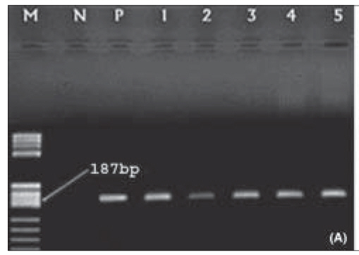
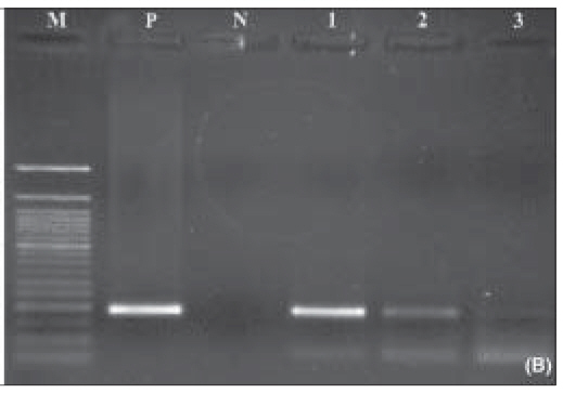
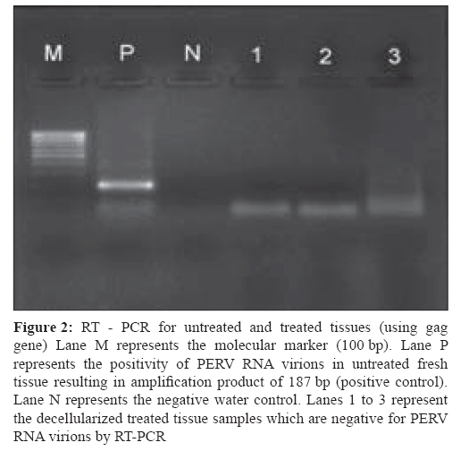
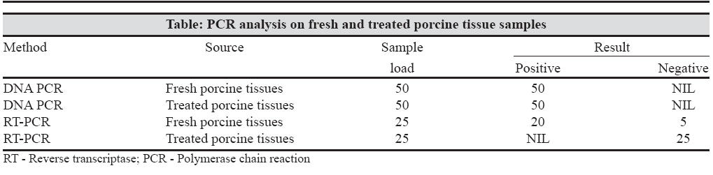
Indian Journal of Medical Microbiology, Vol. 26, No. 3, July-September, 2008, pp. 228-232 Original Article Existence of proviral porcine endogenous retrovirus in fresh and decellularised porcine tissues Prabha S, Verghese S Department of Microbiology, International Centre for Cardio Thoracic and Vascular Diseases, Frontier Lifeline Pvt Ltd, Dr. K.M. Cherian Heart Foundation, R-30-C, Ambattur Industrial Estate Road, Chennai - 600 101 Date of Submission: 05-Jan-2008 Code Number: mb08071 Abstract Purpose: Swine are expected to be utilized as xenograft donors for both whole organ and cellular transplantation. A major concern in using porcine organs for transplantation is the potential of transmission of porcine endogenous retrovirus (PERV). Tissue-engineered or decellularised heart valves have already been implanted in humans and have been marketed by certain companies after Food and Drug Administration (FDA) approval. The aim of this study was to examine the existence of porcine endogenous retrovirus (PERV) in fresh and decellularised porcine tissues.Methods: Porcine tissues (both fresh and decellularised) were analysed using validated assays specific for PERV: polymerase chain reaction (PCR), reverse transcriptase polymerase chain reaction (RT-PCR). Results: PERV specific GAG sequences were found in the porcine heart tissue samples using PCR for DNA and RT- PCR for RNA. All tissue samples (both fresh and treated tissues) like aortic valve, pulmonary valve and heart muscle showed the presence of PERV DNA. RT PCR for PERV was positive in all fresh tissues and was found to be negative in decellularised treated tissues. Conclusions: PCR is a rapid, specific test for the detection of PERV virus in xenografts. These findings have demonstrated that the presence of proviral DNA form of PERV in porcine tissues needs to be carefully considered when the infectious disease potential of xenotransplantation is being assessed. Keywords: Decellularised tissues, porcine endogenous retro virus (PERV), xenograft Among all species analysed, the domestic pig seems to be the most appropriate organ donor for xenotransplantation which offers chances to alleviate the shortage of human donor organs. Porcine endogenous retroviruses (PERVs) are present in genomes of all pigs and are capable of infecting human cells in vitro thus posing a serious threat for xenotransplantation procedures. The existence of porcine endogenous retrovirus (PERV), which are transmitted [1] and of porcine DNA viruses that can persist without symptoms in their natural host (e.g., herpes viruses) [2] strengthened objections to the clinical use of pig xenografts due to the possible development of xenozoonosis. PERV display approximately 50 proviral integration sites in the pig genome. [3] Virions observed in cell lines are morphologically related to C-type viruses. Genetically, three classes of PERV (class A, B, and C), which differ in their env genes, are known. Recent reports demonstrated that PERV which are released from different pig cell lines are able to infect human cells in vitro. [4] In a retrospective study, no cross - species transmission of PERV in 160 patients treated with pig tissue was observed. [5] The majority of porcine pathogens can be removed by keeping animals in specific pathogen free conditions. However, these methods do not eliminate those viruses transmitted within the genome, in particular, PERV which has been reproducibly shown to infect human cells in vitro . However, Specke et al demonstrated that, despite in vitro infection of cell lines derived from several species with PERV, none of the corresponding animal models tested showed evidence of PERV infection, regardless of the method of infection or the source of virus used. [6] Nevertheless, the threat of zoonotic potential in vivo has raised concerns about the presence of endogenous retrovirus sequences in the pig genome. Donor heart valves or animal-derived valves depleted of cellular antigens can be used as a scaffold material. Removing the cellular components results in a material composed of essentially extracellular matrix proteins that can serve as an intrinsic template for cell attachment. Examples of decellularisation techniques are freeze-drying, [7] treatment with trypsin/EDTA, [8] detergent treatment [9] and multi-step enzymatic procedures. To remove any residual DNA and RNA from the matrix, nuclease digestion steps are desirable. [9] The maintenance of mechanical properties depends on the decellularisation method used [10] and on the degree of cross-linking, which stabilises the collagen structure but decreases the ability of tissue in growth. Decellularised porcine scaffolds have been successfully used for cardiovascular tissue engineering. The use of xenograft and allograft tissue as part of bioprosthetic vascular devices such as heart valves and vascular grafts has long been the focus of research. The use of these natural biomaterials has typically required chemical or physical pretreatment aimed at preserving the tissue by enhancing the resistance of the material to enzymatic or chemical degradation, reducing the immunogenicity of the material, and sterilising the tissue. Multiple cross linking techniques have been explored in an attempt to find the ideal procedure to stabilise the collagen - based structure of the tissue while maintaining its mechanical integrity and natural compliance. In addition to the cross linking techniques, decellularisation approaches may reduce host immune response to bioprosthetics and generate natural biomaterials for use in cell seeding and tissue engineering applications. Natural derived materials offer many mechanical, chemical and biological advantages over synthetic materials and thus hold tremendous potential for use in tissue engineering therapies. In view of the significance of PERV for xenotransplantation, we investigated the presence or absence of porcine endogenous retrovirus in decellularised tissues. Materials and Methods Cell lineThe cell line used in this study PK-15 (Porcine Kidney Epithelial Cell Line) was obtained from the national centre for cell science (NCCS), Pune. This cell line contains PERV virus which is spontaneously released in the tissue culture supernatant. The cell line was maintained in minimum essential medium (Gibco) supplemented with 10% foetal bovine serum, 1 mM sodium pyruvate, 1% non essential aminoacids, 100U of penicillin per mL and 100 μg of streptomycin per mL. Porcine tissue Porcine tissues were placed in 1% sodium deoxycholate (DCA, Himedia) for 50 hours followed by enzymatic treatment (DNase and RNase, Gene I) for 24 hours. Another lot of tissues was placed in 1% triton x100 (Himedia) and 0.2% ethylenediaminetetraacetic acid (EDTA) in Dulbecco′s PBS (Himedia) for 50 hours followed by enzymatic digestion for 24 hours. In order to standardise the procedure, the detergents used as well as the enzymatic digestions were made to vary in the concentration along with variation in the time period of immersion of the concerned tissues in these chemicals. This procedure was executed under continuous shaking in a shaker. In both the procedures, decellularisation was followed by collagen cross-linking and sterilisation using 4% and later 10% formalin in stages for 15-24 hours. Heparin treatment was used overnight which prevents blood protein seepage in the decellularised matrix, as it conjugates with the collagen through various receptors. The processed tissues were finally preserved in 70% alcohol. Sample preparation for PCR analysis PERV proviral PCR assay Detection of PERV gag sequences in tissue by RT - PCR: PCR reaction mix of 50 µL containing 100ng of PRE TFI primer, 100ng of PRETRI primer, 1x PCR reaction buffer and 2.5U of Taq was added to each RT reaction tube. PCR amplification was performed as described above, and the amplified product was detected as for PERV proviral analysis. Control RT-PCR reactions that received no RT were included for each test run to confirm that a positive result was due to the presence of PERV RNA and was not the result of combination with residual PERV genomic DNA. Results Representative PCR test results of PERV DNA in untreated fresh porcine tissue and treated decellularised tissues are shown in [Figure 1A, 1B]. DNA extracted from tissue samples was screened by PCR for the detection of GAG gene sequences. Thus PERV proviral PCR assay detected the presence of PERV DNA in all the 50 treated and untreated porcine tissue samples tested.[Figure - 2] shows the representative RT PCR test results from untreated fresh and decellularised treated porcine tissue samples. The RT PCR assay detected presence of PERV RNA in fresh tissues (20/25) and the absence of PERV RNA virions in all the 25 decellularised treated tissue samples. Results of PCR and RT-PCR are summarised in the table. Discussion Endogenous retroviruses (ERV) are remnants of ancestral retroviral infections that have integrated into the germline DNA as a proviral genome, which is vertically transmitted from parent to offspring. Porcine endogenous retroviruses (PERV) may be present in all organs, as multiple copies of PERV can be integrated into germ-line DNA. New and more infectious groups of PERV are being identified, as well as their capacity to infect various types of human cells in vitro . [6] Although PERV are normally non-pathogenic to their natural host, they have been shown to propagate efficiently and can cause disease if they cross species barriers. Transmission of PERV by the porcine scaffold to the xenogenic host has been considered as a possible limitation of this concept.The described PCR based assays can be used to screen porcine tissues for PERV DNA and RNA sequences. The PERV assays were designed with conserved PERV oligomers to allow the detection of all known PERV variants. We have developed polymerase chain reaction (PCR) assays to detect proviral PERV GAG sequences by using specific primers. All these PCR assays gave positive results for proviral DNA on porcine tissues in all fresh and decellularised treated tissues. The provirus usually survives as part of the host genome rather than as an infectious agent. Over evolutionary time periods, most of these proviruses acquire mutations so that, with few exceptions, they become defective and incapable of producing protein. The safety of porcine tissue for guided tissue regeneration as well as for xenotransplantation has been questioned recently by Patience and Wilson who demonstrated that PERV is capable of infecting human cell lines in vitro . [1],[12] In this study, we also described transcriptionally active PERV by GAG RT-PCR, in treated and untreated porcine tissues. Since proviral DNA load does not necessarily correlate with viral RNA load we investigated whether the intact proviruses were dormant or biologically active. Our method included DNase pretreatment of RNA extracts, which was necessary to remove any residual PERV DNA that may originate from porcine cells. In addition, we included a control PCR reaction without RT to confirm that the positive RT PCR results in untreated fresh porcine tissues were due to PERV RNA alone. Negative test results seen in RT PCR with untreated fresh samples suggest the inability of the provirus to express functional viral RNA in porcine tissues. Previous studies have demonstrated PERV RNA transcripts in cellular RNA from various pig tissues. [1],[12],[13] Furthermore, we did not detect any RNA particles in treated porcine matrix. As the tissues are decellularised, presence of live viruses is unlikely. Retroviruses in porcine tissues were the major concerns and the formaldehyde treatment used in both the procedures removes the chance of any microbial infection. Walles et al, demonstrated that after chemical decellularisation of porcine tissue, up to 2% of native DNA is still detectable within the matrix. [14] Zeltinger et al, in his study observed residual cell remnants after chemical decellularisation of porcine heart valves. [15] In conclusion, although the presence of PERV DNA was detectable, PERV RNA sequences were absent in decellularised tissues. In contrast, complete cell removal was not achieved by the decellularisation procedure developed and so the PERV proviral DNA was still detectable in decellularised tissues. Whether chemical decellularisation of porcine tissue used for tissue engineering can prevent PERV-transfection has not been delineated so far. The risk of trans species retroviral infections should not be underestimated, since HIV as another harmless animal retrovirus may cause severe disease in man. The objective of this study was to demonstrate the presence of PERV in heart valve tissue and to study the effects of decellularisation procedures on PERV proviral DNA and RNA. However, further research is needed to clarify whether these decellularised porcine vascular scaffolds can cause cross-species transmission of PERV in transplant patients References
Copyright 2008 - Indian Journal of Medical Microbiology The following images related to this document are available:Photo images[mb08071f1a.jpg] [mb08071t1.jpg] [mb08071f1a&b.jpg] [mb08071f2.jpg] [mb08071f1b.jpg] |
| |||||||||