 |
| About Bioline | All Journals | Testimonials | Membership | News |
|
||||||
|
||||||
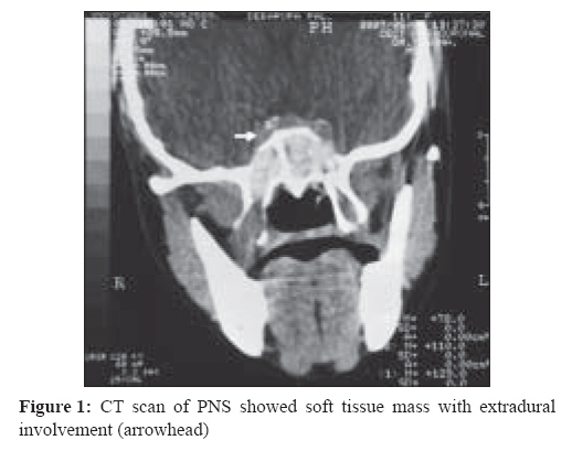
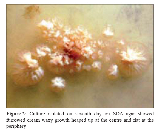
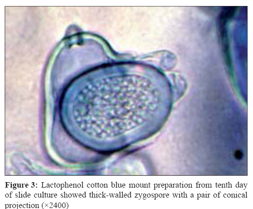
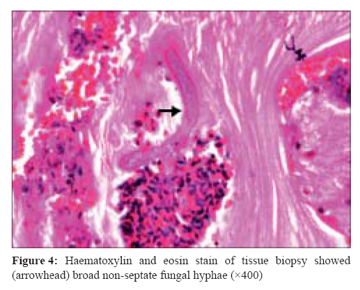
Indian Journal of Medical Microbiology, Vol. 26, No. 3, July-September, 2008, pp. 265-267 Case Report Basidiobolomycosis: A rare case report Singh R, Xess I, Ramavat AS, Arora R Department of Microbiology, All India Institute of Medical Sciences, New Delhi - 110 029 Date of Submission: 30-Dec-2007 Code Number: mb08082 Abstract We report a rare case of basidiobolomycosis seen in an 11-year-old girl from North-Eastern part of India. She presented with complaints of bilateral nasal block and nasal discharge for seven-eight months. CT scan of sinuses revealed polypoidal mass in all the sinuses with extradural extension. The tissue biopsy examined histopathologically and microbiologically, revealed Basidiobolus ranarumKeywords: Basidiobolomycosis, basidiobolus ranarum Basidiobolus species are filamentous fungi belonging to the order Entomophthorales. Unlike other zygomycetes, Basidiobolus species causes subcutaneous zygomycosis in healthy individuals. [1] Basidiobolus ranarum was first described as an isolate from frogs in 1886. It was later cultured from the intestinal contents and ultimately the excreta of frogs. [2] It is commonly found in soil, decaying vegetable matter, and the gastrointestinal tracts of amphibians, reptiles, fish and bats. [3] Basidiobolus is endemic in Uganda and certain other areas of Africa, India, and other parts of Asia. [2] In the past, clinical isolates of Basidiobolus were classified as B ranarum, B. meristosporus and B. haptosporus . But recent taxonomic studies based on antigenic analysis, isoenzyme banding and restriction enzyme analysis of rDNA indicate that all human pathogens belong to B. ranarum. [4] Basidiobolus ranarum was commonly isolated from South India. [4],[5] It was mainly isolated from the extremities, trunk, intestinal tract and rarely other parts of the body. [2],[3],[4],[5],[6] The disease usually occurs in children, less often in adolescents and rarely in adults. [3] Males are much more frequently affected than females. [3] Here, we report a case of submucosal basidiobolomycosis caused by Basidiobolus ranarum , isolated from nasal biopsy specimen of an 11-year-old female patient who was a resident of Agartala, North-East India. To the best of our knowledge, this is the first isolation of Basidiobolus ranarum from north eastern part of India. Case Report An 11-year-old female student presented on 13 th August 2007 to the ENT OPD, All India Institute of Medical Sciences with complaints of bilateral nasal block and nasal discharge for seven-eight months. She was a resident of Agartala, Tripura and from good socioeconomic background. The illness was gradually progressive in nature. Nasal discharge was thick, mucoid and yellow in colour. She gave no history of bleeding from nose, any nasobronchial allergy or trauma in the past. There was no associated headache, vomiting, visual disturbance, aural fullness. She was not a diabetic and had no signs and symptoms suggestive of immunocompromised status. There was no similar illness in the family. Her systemic examination was non-significant. On local examination, there was significant increase in intercanthal distance and broadening of the nose. Her visual acuity and visual fields were normal. Endoscopy examination revealed multiple dirty grayish polypoidal mass in both sides of nasal cavity displacing the middle turbinate with concha bullosa on the right side. Oral cavity was normal but there was postnasal drip and congested post pharyngeal wall. A contrast enhanced CT scan as shown in [Figure - 1] revealed soft tissue density in bilateral nasal cavity, ethmoids, maxillary antrum (with right side more involved than the left) and sphenoid with extradural extension sparing the orbit. Patient underwent endoscopic sinus surgery under general anaesthesia. Polypoidal mass and blackish mucin from all the sinuses were removed with microdebrider and submitted for histopathological and microbiological examination. Tissue material was examined in 10% potassium hydroxide. It showed broad 10-15 μ hyaline, thin walled, aseptate mycelia. Small bits of the tissue were inoculated in two sets of Sabouraud Dextrose Agar (SDA), SDA with gentamicin and SDA with cycloheximide. One set was incubated at 37°C and another at 25°C. Growth was observed within five days at both the temperatures. On seventh day, it showed furrowed cream waxy growth heaped up at the centre and flat at the periphery [Figure - 2]. A lactophenol cotton blue (LCB) mount showed broad, hyaline, aseptate hyphae and conidia of various sizes 25-30 μ x 40-60 μ, globose to pyriform shaped smooth-walled having granules inside. On the tenth day in slide-culture it showed thick-walled zygospores 25-30 μ x 20-25 μ with a pair of conical projection (tubular protuberances), characteristic of Basidiobolus ranarum [Figure - 3]. Histopathological examination of the biopsy also showed dense subepithelial inflammation composed predominantly of eosnophils and plasma cells. A few non-septate fungal hyphae were identified with an eosinophilic sheath (Splendore-Hoeppli phenomenon, [Figure - 4]. Patient could not be treated as she was lost to follow up. Discussion Subcutaneous zygomycosis, the commonest clinical form of basidiobolomycosis, is endemic in South India. [4],[5] In contrast the present case was from North-Eastern part of India. There was no predisposing factor identified in this case, though traumatic implantation is probably the mode of entry. [4] It was isolated from the nasal biopsy specimen which is also an uncommon site. It is mainly isolated from the extremities, trunk and intestinal tract, [2],[3] whereas the present case had nasal and paranasal sinuses involvement. Rhinocerebral presentation is seen in diabetic patients, [2] but this patient was not diabetic. Moreover, the infection occurred in a female patient whereas this infection is reported more in males. [3],[5] The aggressive nature of disease, as shown by the extradural involvement of the mass, is an unusual presentation of basidiobolomycosis. Diagnosis is mainly based on histopathological examination and fungal culture of biopsy specimen. The typical histopathological feature is the presence of thin-walled, broad, often aseptate hyphae or hyphal fragments with an eosinophilic sheath (Splendore-Hoeppli phenomenon), frequently phagocytised within giant cells. [2],[3] On fungal culture, thick-walled zygospore with a pair of conical projection (tubular protuberances) is visualised which is the characteristic feature of Basidiobolus ranarum. [2] An immunodiffusion test has also been developed for specific diagnosis of the disease, [2],[3] but it is not routinely available. This rare variety of fungal disease involving nose and paranasal sinuses should also be kept as a differential diagnosis in similar clinical presentation. The infection may be fatal, however, the outcome in our patient is not known References
Copyright 2008 - Indian Journal of Medical Microbiology The following images related to this document are available:Photo images[mb08082f3.jpg] [mb08082f4.jpg] [mb08082f2.jpg] [mb08082f1.jpg] |
| |||||||||