 |
| About Bioline | All Journals | Testimonials | Membership | News |
|
||||||
|
||||||
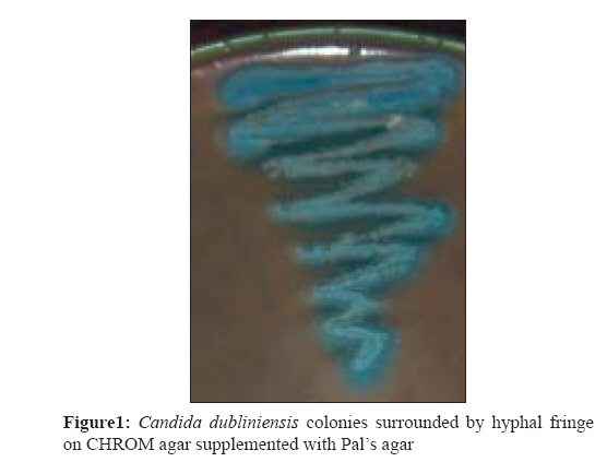
Indian Journal of Medical Microbiology, Vol. 27, No. 1, January-March, 2009, pp. 55-58 Brief Communication Differentiation of Candida dubliniensis on chrom agar and Pal's agar Raut SH, Varaiya A Department of Microbiology, S.L.Raheja Hospital, Mahim, Mumbai-400 016 Date of Submission: 15-Feb-2008 Code Number: mb09013 Abstract Difference in expression of putative virulence factors and in antifungal susceptibility among different Candida species has raised the need for species-level identification. The close relationship of Candida dubliniensis with C. albicans has led to misidentification of C. dubliniensis isolates as C. albicans . Phenotypic tests include ability to produce chlamydospore on casein agar, colony colour development on differential media CHROM agar Candida medium and ability to form hyphal fringe on Pal's agar, have been used to differentiate these two Candida species. Fifty isolates of Candida species were recovered from various specimens (blood, urine, tissue and respiratory secretions) from diabetic and cancer patients between April and July 2007. The isolates were tested for chlamydospore production on casein agar. These were also streaked simultaneously on CHROM agar, Pal's agar and a combination of CHROM agar supplemented with Pal's agar for identification and differentiation of C. dubliniensis from C. albicans . On CHROM agar, 19 isolates were identified as C. dubliniensis , nine as C. albicans , 10 as C. krusei , nine as C. tropicalis and two as C. glabrata . One was indeterminate and later identified as C. dubliniensis . Out of the 20 C. dubliniensis isolates, 19 isolates exhibited hyphal fringe on Pal's agar. On CHROM agar supplemented with Pal's agar, 16 out of the 19 fringe-positive isolates exhibited fringe surrounding the bluish green-coloured colonies of C. dubliniensis . Additional identification tests like growth at 45oC and ability to reduce 2,3,5-triphenyltetrazolium chloride were time efficient, inexpensive and easy-to-use methods for differentiation of C. dubliniensis and C. albicans isolates. CHROM agar when supplemented with Pal's agar gave definitive identification between C. dubliniensis and C. albicans .Keywords: Candida dublinensis, CHROM agar, Pal′s agar Over the past decade, there has been a significant increase in the number of reports of systemic and mucosal Candida infections with non-albicans Candida species that cause a greater proportion of nosocomial infections. Implanted devices, particularly indwelling intravascular catheters, have been a significant risk factor associated with these infections. The potential clinical importance of species-level identification has been recognized as Candida species differ in the expression of putative virulence factors and antifungal susceptibility. [1],[2],[3] Candida dubliniensis was originally isolated from patients with human immunodeficiency virus (HIV) infection and recurrent oral candidiasis. [3] C. dubliniensis was also identified in clinical specimens from diabetics and cancer patients . [4] C. dubliniensis and C. albicans share many morphological and physiological characteristics, such as germ tube positivity, similar biochemical patterns and the ability to form chlamydospores in rice extract agar and cornmeal agar . [5],[6] This close similarity between the two species has led to the misidentification of isolates of C. dubliniensis as C. albicans .[5],[6] The most accurate differentiation between isolates of the two species is performed in reference laboratories with the use of molecular-based techniques such as PCR or DNA fingerprinting with repetitive sequence-containing DNA probes. However, these sophisticated techniques are not readily available in routine clinical microbiology laboratories. [6] CHROM agar Candida medium is widely used for the identification of Candida spp., which develop colonies with distinguishable colours. The two species could be distinguished by culture on Staib agar, Pal′s agar, modified Pal′s agar and casein agar, where C. dubliniensis produces abundant chlamydospores and rough colonies. [3],[7] There is paucity of Indian data on the incidence of C. dublineinsis infections in diabetic foot infections and cancer. In view of this, the aim of the present study was to differentiate various Candida species and to find out the most prevalent species from these patients using casein agar, CHROM agar, Pal′s agar and CHROM agar supplemented with Pal′s agar. Materials and Methods Fifty isolates of Candida were isolated from diabetic and cancer patients over a period of 4 months - April to July 2007. With universal safety precautions, samples were collected from patients in the intensive care unit and transported and processed in the laboratory without delay. Blood cultures were processed using an automated method with Versa Trek (Trivitron, Ohio, USA). Samples were cultured on brain heart infusion blood agar and Sabourauds dextrose agar (SDA). Identification of organisms was done by the standard laboratory technique. [8] Further, these isolates were streaked simultaneously on Casein agar, Pal′s agar and CHROM agar Candida supplemented with Pal′s agar. C. albicans ATCC 10231, C. krusei ATCC 14243 and C. tropicalis ATCC 66029 were used as reference strains. The following media were used for identification and differentiation of different Candida species: CHROM agar Candida (Hi-Media, India) contains enzymatic substrates that are linked to chromogenic substrates, which when acted upon by different enzymes produced by Candida species results in colour variations useful for the presumptive identification of the yeasts. Pal′s agar was freshly prepared with unsalted sunflower seeds (including kernels and shells) and used within 5 days to ensure consistent results. First, an aqueous extract of sunflower seeds was prepared by pulverizing 50 g of seeds in a blender for 5 min and then adding the ground seeds to 1 L of distilled water, followed by boiling for 30 min. Next, the seed extract was cooled and filtered and supplemented with glucose (1 g), KH 2 PO 4 (1 g) and creatinine (1 g). The pH was adjusted to 5.5, the volume was readjusted to 1 L and 15 g of agar (Difco) was added before the mixture was autoclaved at 110°C for 20 min. The cooled medium (45-55°C) was poured into 90-mm-diameter petri dishes. Khan et al . reported that the husk of sunflower seeds can be used as a substitute for whole seeds in the medium without compromising its efficacy. [12] 3. CHROM agar supplemented with Pal′s agar [10] CHROM agar Candida medium supplemented with Pal′s medium was prepared by mixing equal volumes of prepared CHROM agar Candida medium and Pal′s agar. Casein agar has been traditionally used to study the decomposition of casein by aerobic actinomycetes and dematiaceous fungi. Recently, casein agar has been found to be a good medium to induce the production of chlamydospores by C. dubliniensis isolates, a feature that can differentiate C. dubliniensis from C. albicans . Preparation: 10 g of skim milk was dissolved in 90 mL of distilled water and 3 g of agar was dissolved in 97 mL of distilled water. After autoclaving both solutions separately at 121°C for 15 min they were allowed to cool to 45-50°C, after which they were mixed together. After autoclaving, 25 mL of the media was dispensed into each 90 mm-diameter petri dish. 5. Additional tests for identification of C. dubliniensis : (i) Growth at 45oC [5] All C. dubliniensis and C. albicans isolates were isolated on sterile SDA and incubated at 45oC for 48-72 h. Unlike C. albicans, C. dubliniensis lacks the ability to grow at 45ξC. (ii) Ability to reduce 2, 3, 5-triphenyltetrazolium chloride (TTC) [13],[14] Tetrazolium salts possess a reducible chromogenic tetrazolium ring. These compounds are not true dyes but, in the reduced state, the five-membered ring imparts colour to the molecule. TTC is reduced by mitochondrial dehydrogenase into a water-soluble formazan product that is measured spectrophotometrically. 0.5 mL of aqueous solution (0.2%w/v) of TTC dye prepared in phosphate-buffered saline was added to the tube containing 1 mL of 0.1 optical density adjusted Candida cell culture and incubated at 37oC for 6-48 h. Unlike C. dubliniensis , C. albicans lacks the ability to metabolically reduce TTC. Results The results were reported considering CHROM agar Candida as a primary medium for differentiating between various Candida species based on their colony colour and morphology (rough/smooth). Pal′s agar was considered as a secondary medium for differentiation. CHROM agar Candida Based on the colony colour developed on CHROM agar Candida , 50 isolates of Candida were differentiated as: C. dubliniensis (19), C. albicans (9), C. krusei (10), C. tropicalis (9) and C. glabrata (2). One isolate showed an intermittent green-coloured colony on CHROM agar, thereby making it difficult to differentiate between C. dubliniensis and C. albicans . However, this isolate was reported as C. dubliniensis as it failed to grow on SDA and was able to reduce the TTC dye. Maximum C. dubliniensis isolates were from blood cultures followed by respiratory specimens like broncoalveolar lavage from cancer patients whereas C. dubliniensis was isolated from diabetic foot infections followed by C. krusei . Pal′s agar Sixteen of the 19 isolates identified as C. dubliniensis exhibited a hyphal fringe after 48 h of incubation whereas two isolates exhibited a hyphal fringe after 4 days and one isolate failed to exhibit hyphal fringe. All the nine isolates identified as C. albicans failed to exhibit the hyphal fringe. The single Candida isolate that showed an intermittent green-coloured colony on CHROM agar produced fringe on Pal′s agar after 4 days of incubation and thus was reported as C. dubliniensis . Two isolates of C. krusei also exhibited hyphal fringe. CHROM agar supplemented with Pal′s agar Out of the 20 isolates of C. dubliniensis that developed fringe on Pal′s agar, 16 isolates revealed fringe on CHROM agar supplemented with Pal′s agar [Figure - 1] whereas none of the C. albicans developed fringe on the same media. Two C. krusei isolates that revealed fringe on Pal′s agar also developed fringe on this medium. Casein agar All the 20 C. dubliniensis isolates revealed abundant chlamydospores. Addition test for identification (i) Growth at 45oC All C. albicans isolates were found to grow at 45oC whereas all C. dubliniensis isolates failed to do so. (ii) TTC reduction test All C. dubliniensis isolates were able to reduce the TTC dye whereas all the nine C. albicans isolates failed to do so. Discussion C. dubliniensis has been identified in clinical specimens recovered from diabetic and cancer patients with and without symptoms of oral candidiasis . Similarity in morphological and physiological characteristics between C. dubliniensis and C. albicans leads to misidentification of C. dubliniensis as C. albicans . As both these species differ in their antifungal susceptibility, it is essential to identify and differentiate them. In our study, C. dubliniensis was found to be the most prevalent species (38%). When subcultured on Pal′s agar, 18 of these 19 isolates exhibited hyphal fringe surrounding the colonies. In a study by Mosaid et al. , all the C. dublinensis exhibited hyphal fringe on Pal′s agar. [11] Sahand et al. reported that 75% of the C. dublinensis isolates exhibited hyphal fringe. [10] We observed that all the C. dubliniensis isolates, including the single-fringe-negative one, developed rough colonies on Pal′s agar whereas all the C. albicans isolates developed smooth colonies. Sahand et al. in their study reported that 96% of the C. dubliniensis isolates developed rough colonies and all C. albicans isolates developed smooth colony on Pal′s after a 48 h incubation at 30oC. [10] However, in the study conducted by Mosaid et al. , all C. albicans as well as C. dubliniensis grew as smooth colonies on Pal′s agar. [11] Hence, Pal′s agar used alone is not a good differential medium. Among the C. krusei (10) isolates that were identified in our study, two exhibited hyphal fringe on Pal′s agar. Mosaid et al. also reported formation of rough colony with a fringe by C. krusei isolates on Pal′s agar. [11] Among the 20 fringe-positive C. dubliniensis isolated on Pal′s agar, only 16 revealed fringe on CHROM agar supplemented with Pal′s agar whereas none of the C. albicans isolates developed fringe on both media, which correlates well with the study conducted by Sahand et al . [11] Although chlamydospore formation by C. dubliniensis was observed on both the media, there was a reduction in the pseudohyphae production on CHROM agar supplemented with Pal′s agar. CHROM agar helps in preliminary identification of Candida species whereas Pal′s agar, due to the presence of sunflower seed as its major ingredient, facilitates fringe production by C. dubliniensis . Ability to produce hyphal fringe on Pal′s agar is attributed to pseudohyphae production by C. dubliniensis . [10],[12] The volume of Pal′s agar when used in combination being 50% of the total volume, could be a probable reason for the reduction of pseudohyphae in this medium. Unlike C. albicans isolates, all C. dubliniensis isolates failed to grow at 45oC and were able to reduce TTC, which correlates well with the study conducted by Mosca et al. and Giammanco et al . [5],[13] Thus, these additional identification tests were time efficient, inexpensive and easy-to-use methods for differentiation of C. dubliniensis and C. albicans isolates. Thus, in our study, C. dubliniensis was the most prevalent Candida species in both diabetics and cancer patients. CHROM agar Candida medium and Pal′s agar when used singly is time consuming whereas CHROM agar when supplemented with Pal′s agar makes rapid differentiation of both species and can be easily incorporated in routine microbiology laboratories. References
Copyright 2009 - Indian Journal of Medical Microbiology The following images related to this document are available:Photo images[mb09013f1.jpg] |
| |||||||||
