 |
| About Bioline | All Journals | Testimonials | Membership | News |
|
||||||
|
||||||
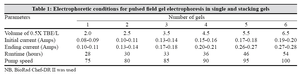
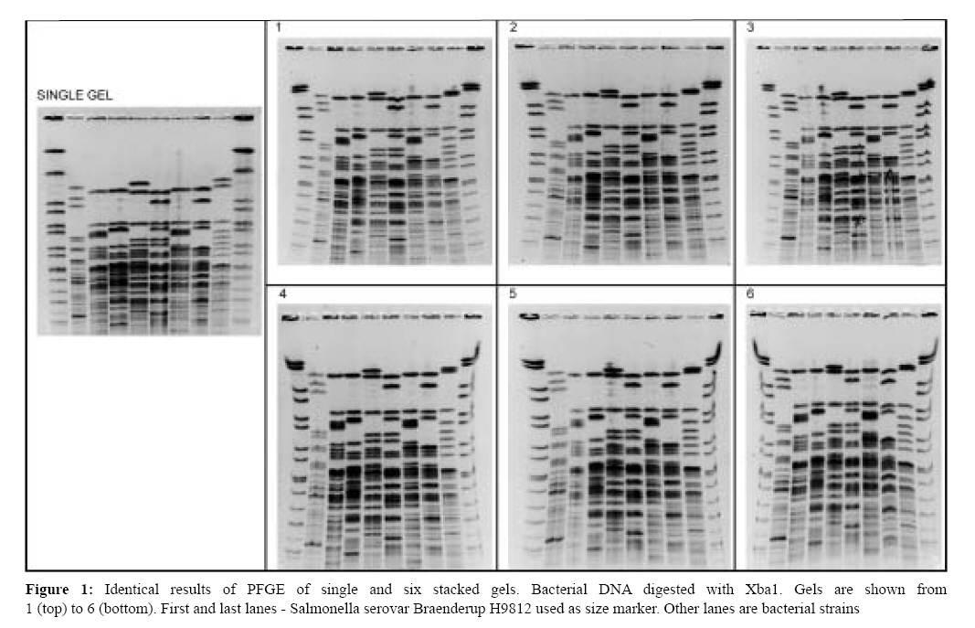
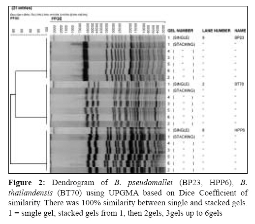
Indian Journal of Medical Microbiology, Vol. 27, No. 2, April-June, 2009, pp. 142-145 Brief Communication Stacking gels: A method for maximising output for pulsed-field gel electrophoresis Heng SeeKah, Heng ChuaKek, Puthucheary SD Department of Medical Microbiology, University of Malaya, 50603 Kuala Lumpur Date of Submission: 26-Jun-2008 Code Number: mb09042 PMID: 19384038 DOI: 10.4103/0255-0857.49428 Abstract Pulsed field gel electrophoresis (PFGE), the gold standard of molecular typing methods, has a major disadvantage of an unusually long electrophoretic time. From the original protocol of 6 days, it was modified to 3 days and subsequently to a single day. We describe the procedure of stacking five to six gels one on top of another in order to increase and maximize the output in a shorter time without compromising the resolution and reproducibility. All the variables that affect pulsed field gels during electrophoresis were taken into consideration. We firstly optimized the parameters to be used and secondly determined whether stacking of five to six gels had any effect on the molecular separation during electrophoresis in comparison with a single gel run. DNA preparation, restriction, electrophoresis, staining and gel documentation was carried out based on previously published methods. Gels were analysed using BioNumerics and dice coefficient and unweighted pair group methods were used to generate dendrograms based on 1.5% tolerance values. Identical band profiles and band resolution-separation were seen in the PFGE patterns with single gel and multiple stacking gels. Cluster analysis further strengthened the fact that results from stacking gels were reproducible and comparable with a single gel run. This method of stacking gels saves time and maximizes the output at the same time. The run time for a single gel was about 28 hours, but with six stacked gels the run time was 54 hours compared with 28 × 6 = 168 hours if they were run separately as single gels thus saving time of 67.86%. Beside the big factor of saving time, stacking gels save resources (electricity, reagents, water, chemicals and working time) by increasing the sample throughput in a shorter time without compromising on quality of data. But optimization of working parameters is vital depending on the PFGE system used.Keywords: Electrophoresis, run time, stacking gels Pulsed field gel electrophoresis (PFGE) allows the separation of very large DNA molecules of up to two million nucleotides and has become a popular and common method for the study of microbial genetics. It is often considered the "gold standard" of molecular typing methods. Apart from being costly and having difficulties with reproducibility, a major disadvantage of the process of PFGE is the unusually long electrophoretic time. From the original protocol of 6 days, Matushek el al . [1] modified the standard PFGE so that it could be completed in 3 days. Subsequently, Gautom [2] developed a rapid PFGE protocol for typing E. coli organisms in a single day. Time savings were achieved by utilizing bacterial cells directly from the culture plates, expediting cell lysis and proteinase K treatment, shortening washing and restriction digestion time and using SeaKem Gold agarose, which allows a more rapid electrophoresis. A single run of one gel, size 14 x 13 cm, with a maximum of 20 samples will take 28 hours using a Chef-DR II machine. This does not include time for the preparation of the plugs. Any methodology to increase the efficiency with regard to the time factor would be very, very useful. Nagy and Choo, [3] using a Chef-DR II apparatus, described a double-decker procedure, which gave identical DNA resolution. More recently, Martins-Wess and Leeb [4] reported the method of stacking up to four agarose gels one on top of another during electrophoresis in order to generate more data in a shorter time. There was no difference between results achieved with a single gel or with multiple stacked gels. These authors suggested that further increases in throughput may be achieved with custom-made combs that allow more samples per gel and with stacking even more gels on top of each other. But there have been no further reports on these methods on reproducibility, electrophoretic parameters necessary nor dendrograms to show evidence of similarities in the results obtained by the stacking gel methodology. Several parameters affect the separation and mobility of DNA molecules during gel electrophoresis. These include the composition and concentration of the gel, the buffer, temperature, pulse time, run time and the voltage gradient of the electric field. Methods to speed up migration of DNA molecules include:
Materials and Methods Bacterial strains Burkholderia pseudomallei ATCC 23343, B. thailandensis ATCC 700388, six clinical strains of B. pseudomallei and Salmonella serotype Braenderup H9812 were grown overnight at 37° C on nutrient agar plates. All cultures were identified using standard laboratory procedures and confirmed by API 20NE (BioMerieux, Marcy l′Etoile, France). Safety precautions were taken in working with these organisms, including the use of a biohazard safety cabinet and gloves. Preparation of genomic DNA DNA for PFGE was prepared by modification of the methods of Gautom et al . [2] and Ribot et al . [6] Bacterial cells in suspension buffer at an optical density of 1.3-1.4 at 610 nm were mixed with 10 uL of proteinase K and an equal volume of molten 1.6% low melting point agarose (InCert agarose; FMC Bioproducts, Rockland, Maine, USA) and pipetted into plug moulds. Plugs were then suspended in cell lysis buffer containing 10 uL of proteinase K and incubated at 50° C overnight in a water bath, then washed twice with distilled water and five times in TEB and stored at 4° C until further use. Digestion with restriction endonuclease Xba1 Plugs were cut to the appropriate size and digested with 10 units of XBa 1 at 37° C with overnight incubation and resuspended in 0.5 x TBE. Salmonella serotype Braenderup H9812, with restriction fragments extending over a major part of the gel, was used as a standard marker. Electrophoresis Plug slices were loaded on to gel combs, aligned in the appropriate order and placed on the casting stand. Molten 1% Sigma agarose was poured over it and allowed to polymerize for approximately 30 min at room temperature. Initially, a single gel was run at 14° C at 200V, angle 120° , pump speed of 75 and a pulse time of 5-65 s for a 28-hour runtime. Subsequently, the same procedure was carried out for two, three, four, five and finally a maximum of six gels. These gels were stacked one on top of another and kept in place by four 10 uL pipette tips at the four corners to prevent the gels from floating away. Optimization of pump speed, electrical current, volume of 0.5 x TBE and runtime was carried out by the "trial and error method" with the different number of stacked gels [Table - 1]. The six stacking gel procedure was carried out three to four times to asses the reproducibility of the results. Image acquisition Following electrophoresis, the gels were stained with 300 mL of ethidium bromide solution and destained. A Gel and Faber Castel ruler was placed on the ultraviolet illuminator and the image was captured using the gel Doc System (Bio-Rad, Hercules, CA, USA). Analysis of TIFF images Gels were analysed using BioNumerics software Version 5 (Applied Maths, Sint-Martens-Latem, Belgium). Dice coefficient and the unweighted pair group method were used to generate dendrograms based on 1.5-2.0% tolerance values. Results Optimization of three parameters was carried out on a "trial and error" basis and there were marked differences in all three of them between single gel and stacked gels. The most significant was seen in the runtime from 28 to 54 hours, pump speed from 75 to 100 and volume of TBE buffer used from 2 to 6.5L [Table - 1]. The runtime for a single gel was about 28 hours but with six stacked gels the runtime was 54 hours compared with 28 x 6 = 168 hours if they were run separately as single gels, thus saving time of 168−54 = 114 hours (67.86%). The increase in the current was mainly due to the increase in the number of gels stacked and more importantly the total volume of buffer used. The concentration of the agarose gel, voltage gradient, running temperature, pulse time and reorientation angle was maintained as for a single gel run. Salmonella serotype Braenderup was chosen as the standard marker as it had "the desired criteria, including coverage of a wide range of DNA fragment sizes, even distribution of bands and stability of the PFGE patterns". This strain has been described as a universal size standard. [7] In our six-gel PFGE, Salmonella Braenderup gave identical banding patterns and DNA size range. Similarly, identical band profiles and band resolution-separation were seen in the PFGE patterns with single gel and multiple stacking gels using the ATCC strains of B. pseudomallei, B. thailandensis and clinical strains of B. pseudomallei [Figure - 1]. Cluster analysis further strengthened the fact that results from stacking gels were reproducible and comparable with a single gel run. We have shown a dendrogram only for three samples instead of 10 in order to make the figure clear and legible [Figure - 2]. Discussion This method of stacking gels saves time and maximizes output at the same time. Besides the big factor of saving time, less consumables were used with six stacked gels compared with six gels run separately. The runtime of 54 hours for the six stacked gels was found to be sufficient and suitable as the molecular weight of the DNA did not exceed 700 kb with the use of Xba 1 as the restriction endonuclease. However, with different restriction endonucleases, the molecular size of the DNA to be separated will be variable and therefore the runtime will also vary. With the stacked gels, the maximum current used was 0.28 Amps and did not exceed the maximum of 0.35 Amps allowed to be used safely with a Chef-DR11 apparatus (personal communication - Bio-Rad). With the stacking gels, some increase in lane distortion (smiling effect) around the edges was observed, but this does not affect the outcome due to normalization of the results during the process of BioNumerics. In agreement with the report by Nagy and Choo, [3] we too did not encounter any cross contamination between the gels. We believe that air bubbles trapped in between the gels during PFGE further prevented cross contamination. In conclusion, stacking gels save resources (electricity, reagents, water, chemicals and working time) and increase the sample throughput in a shorter time without compromising on the quality of data. It is useful and economical particularly in outbreak situations when large number of samples have to be processed in a short time. This method will also be useful in food microbiology where large samples have to be analysed. But, optimization of working parameters is vital depending on the PFGEsystem used. Acknowledgment The authors thank Prof. Tosso Leeb, University of Berne, Dr. Doinita Ispas of Bio-Rad Romania and Dr. Yue Xie of Bio-Rad Laboratories, Hercules, USA, for useful technical advice. SKH is grateful to the Ministry of Health for granting study leave and scholarship for her MSc at the University of Malaya. This study was funded by eSciencefund from the Ministry of Science, Technology and Innovation, Malaysia (no. 12-02-03-2033). References
Copyright 2009 - Indian Journal of Medical Microbiology The following images related to this document are available:Photo images[mb09042t1.jpg] [mb09042f2.jpg] [mb09042f1.jpg] |
| |||||||||