 |
| About Bioline | All Journals | Testimonials | Membership | News |
|
||||||
|
||||||
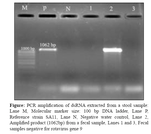
Indian Journal of Medical Microbiology, Vol. 27, No. 2, April-June, 2009, pp. 149-152 Brief Communication Detection of porcine rotavirus from tissue and faecal specimens Prabha Suji, Verghese Susan Department of Microbiology, International Centre for Cardio Thoracic and Vascular Diseases, Frontier Lifeline Pvt Ltd, Dr. K.M. Cherian Heart Foundation, R-30-C, Ambattur Industrial Estate Road, Chennai-600 101 Date of Submission: 21-Feb-2008 Code Number: mb09044 PMID: 19384040 DOI: 10.4103/0255-0857.49430 Abstract Porcine small intestinal sub-mucosa is a cell-free collagen matrix that has demonstrated its ability as a scaffold material. Transplantation poses special hazards because grafted tissues and organs transmit pathogens efficiently, especially viruses. Rotavirus is thought to be confined to the intestine, causing acute diarrhoea. The purpose of this study was to evaluate the porcine intestinal tissue scaffold for Rotavirus and to study the incidence of this virus among pig herds. Only one isolate was successfully adapted to grow in cell line MA 104 from faecal samples. This isolate was further confirmed by reverse transcriptase polymerase chain reaction and sequence analysis.Keywords: MA 104, Rotavirus, reverse transcriptase polymerase chain reaction Xenotransplantation using pig organs or tissues may alleviate the human donor organ shortage. Xenotransplants will enhance the risk of infection because transplanted microorganisms bypass host defenses, recipients lack pre-formed immunity, clinical laboratory assays are often not available, incompatible transplantation antigens may reduce the efficacy of host cellular immune responses and because of unknown effects of genetic or other manipulations used to reduce xenograft rejection. The animals should be screened microbiologically for traditional zoonoses and for pathogens with a broad host range. Animals infected with a certain pathogen should be excluded. Rotaviruses are viruses with a high capacity for recombination. Rotaviruses are characterized by a distinctive morphology, which led Flewett et al. [1] to recognize them as separate from reo- and orbiviruses. In contrast to reo- and orbiviruses, each rotavirus particle is thought to contain 11 segments of double-stranded RNA that form a characteristic pattern on polyacrylamide gel electrophoresis. [2] This virus is widespread in pig populations. It is present in most, if not all, pig herds, with a virtually 100% seroconversion in the adult stock. Rotaviruses are generally species specific, but cross-species transmission is possible, as has been demonstrated experimentally. Several case studies have indicated infection of humans by animal rotaviruses. Rotaviruses causing diarrhoea in pigs were demonstrated by Woode et al . [3] Antibodies to group A rotavirus, whose distribution is worldwide, can be found in 90-100% pigs. Rotaviruses of groups B and C, initially called rotavirus-like agents and para rotaviruses [4] respectively, are also present in swine herds but more detailed data on the evidence of antibodies to these groups are not available. In vitro cultivation of rotaviruses is not easy as they require the presence of proteolytic enzymes both in the sample and in the maintenance medium. The first successful cultivation of human rotaviruses was described in 1981 in cell line MA 104 and this method is also applicable to cultivation of rotaviruses from faecal samples of other animal species. [5] The sub-mucosal layer of the small intestine has been investigated as a source of collagenous tissue with the potential to be used as a biomaterial because of its inherent strength and biocompatibility. The present study was focused on the screening of Rotavirus (one of the potential pathogens in pig) from porcine xenograft tissues and also studying the incidence of Rotavirus in asymptomatic pigs from faecal samples using reverse transcriptase polymerase chain reaction (RT PCR) and the cell culture method. Materials and Methods Cells and viruses MA 104 cells were obtained from the National Centre for Cell Science and grown in a 5% CO 2 atmosphere at 37°C in Eagle′s medium Gibco Laboratories (North andover, MA, USA) that was supplemented to contain 10% foetal bovine serum (FBS) (Sigma, North andover, MA, USA), 2mM L-Glutamine, 500U penicillin/mL and 500µg streptomycin/mL (Gibco Laboratories). Cells were passaged when confluent using 1% trypsin (Sigma, USA). Simian Rotavirus strain SA 11 was kindly provided by Dr. Gagandeep Kang, Christian Medical College, Vellore, as a positive control. Virus stocks were grown in MA 104 cells maintained in Eagle′s medium without FBS. Purified virion preparations were made by infecting MA 104 cells with the virus treated with trypsin (20µg trypsin/mL, incubated at 37°C for 30 min). Cell culture lysates were collected at each passage by two cycles of freezing and thawing (−70°C and 37°C). Sample preparation for cell culture inoculation Intestinal contents obtained from 75 asymptomatic pigs from farm houses were used to prepare 20% suspension in Eagle′s medium MEM (E MEM supplemented with antibiotics: penicillin 20U/mL, streptomycin 20µg/mL and amphotericin B 0.05µg/mL of medium).Cotton swabs were used for swabbing the recta of live pigs and, usually, a small amount of adherent faeces was collected on each swab. The suspension was then centrifuged for 20min at 5000g at 4°C. In the supernatant obtained, rotavirus was demonstrated by RT PCR. The supernatant was used for RNA extraction. For detection of rotavirus from porcine intestinal xenografts, tissues were harvested and transported from animal abattoir in Hanks Balanced Salt Solution Hi Media (Mumbai, India) with antibiotics. The tissues were then dissected, macerated and centrifuged and the suspensions were incubated with trypsin for 45min at 37°C and then inoculated onto an MA 104 cell line. Isolation and propagation of rotaviruses in cell culture Isolation of Rotaviruses from faecal samples and tissue specimens was carried out in MA 104 cells grown in E MEM. The primary virus suspension was pre-incubated for 45 min at 37°C in E MEM with 10µg/mL trypsin. The cell line was cultured for 24-48 h, washed twice in serum-free E MEM, infected with 0.5 mL treated viral suspension and incubated for 90 min at 37°C. Then, the inocula were removed without rinsing. E MEM without serum was used as the maintenance medium. The cytopathic effect (CPE) was monitored for 7 days. Three to five blind passages were carried out in the same manner. Single passages were frozen following a 7-day incubation even in case no CPE was observed. The virus was demonstrated continuously by RT PCR. Rotavirus strain SA 11 was cultured in each passage of virus isolation as a control. RNA isolation Trizol LS reagent was used for RNA isolation according to the manufacturer′s recommendation Invitrogen (Carlsbad, CA, USA). This method can be used to isolate RNA from both tissue and faecal samples and also from tissue culture. Extraction was performed from both faecal samples and cell culture in a passage at the beginning of adaptation and at the end of the adaptation. Extracted RNA was resuspended in RNase-free water and stored at −80°C. Primers Oligonucleotide primers specific for gene segment 9 (or segment 8), which encodes VP7, were synthesized, complementary to the 3′ ends of both viral RNA strands. These primers - beg 9 and end 9, which were 28 and 27 nucleotides long, respectively were selected to produce full-length copies of gene 9 (or gene 8) from any group A rotavirus strain. PCR amplification from dsRNA The pair of primers used for the amplification of full-length sequence was: 5′GGCTTTAAAA GAGAGAATTTCCGTCTGG3′ (beg9) and 5′GGTCACATCATACAATTCTAATCTAAG3′ (end9). The dsRNA extracted directly from the faecal and tissue samples and sample-inoculated tissue culture supernatants were used as templates for RT to synthesize cDNA copies from both the viral strands that were then amplified by the PCR. Various conditions regarding the concentrations of MgCl 2 , dimethylsulphoxide, primers and template were tested. The reverse transcription of dsRNA was carried out using RT MO BIO Laboratories (San Diego, CA, USA) and PCR amplification was carried out with Taq DNA polymerase (Mobio Laboratries, CA, USA). The final concentrations in the RT mixture of 20µL volume were: RT buffer (10x), primer beg9 (2.5µM), primer end9 (2.5µM), RNase inhibitor (40U) and RT enzyme (50U). Reverse transcription was carried out according to the manufacturer′s recommendation. After synthesis of cDNA the RT mixture was brought up to a volume of 100µL of the PCR mixture containing: PCR buffer(1x), MgCl 2 (1.5mM), dNTP mix (200µM),Taq DNA polymerase (5U) and both of the primers (250nM). PCR amplification was accomplished in 30 cycles (94°C for 1 min, 42°C for 2 min, 72°C for 1 min) followed by a final cycle of 7 min at 72°C (Gouvea et al. [6] ). The PCR products (8µL) were analysed in 1.5% agarose in Tris-borate buffer containing 0.5µg of ethidium bromide Bio Gene (Cambridge, UK) per mL. Sequence analysis The PCR product from an isolate was sequenced for further identification. After purification using an ultra clean PCR clean up kit (Mobio Laboratries, CA, USA), the samples underwent sequence analysis with an automated sequencing service Bangalore Genei (Bangalore, India). The sequence was assembled and analysed using the Bioedit sequence alignment editor and NCBI′s analysis tool. The sequence obtained was compared with that of the VP7 gene of the other porcine rotavirus strain nucleotide sequences in gene bank. Results Virus isolation Of the total samples analysed, only one isolate from the pig faecal sample was adapted to growth in the cell line MA 104. Evident CPE was produced after three passages with an incubation period of 4 days. The remaining faecal and intestinal tissue samples from different pigs did not produce CPE even after five passages. Faecal samples were tested to check the incidence of porcine rotavirus in asymptomatic pigs and the porcine intestinal tissues as a xenograft material to check whether the tissues are free of porcine Rotavirus. Faecal and intestinal tissue samples were collected from different pig sources. All the samples were checked with SA11 Rotavirus as a positive control and uninoculated MA 104 cell line as a negative control. RT PCR detection of Rotavirus and sequence analysis The product obtained after RT PCR using primers beg9 and end9 was of the expected length 1062bp. The result is shown in [Figure - 1]. We recognized how important the amount of viral dsRNA could be to the amplification when PCR was applied to stool specimens containing the best range of Rotavirus concentrations. To determine the best amount of viral dsRNA for PCR amplification, we titrated the dsRNA extracted from the control cell culture SA 11 by PCR. The dsRNA suspension extracted, ranging in volume from 0.01 to 10µL, was used as a template for the amplification reaction. The sensitivity of the test was between 20 and 100pg of template dsRNA as 100 pg still produced a visible 1062bp band. Thus, 10µL of this particular RNA preparation containing an estimated 400ng of total RNA produced no visible amplification. It is possible that coprecipitated substances other than viral dsRNA, when present in high quantities, inhibited one of the early steps in the PCR amplification method such as dsRNA denaturing or primer annealing. The primers specific for group A Rotavirus yielded bands of distinct lengths - 1062 bp was demonstrated only in one of the faecal samples, which adapted growth in the MA 104 cell line (1%) of the 75 samples analysed. None of the intestinal tissue or faecal samples showed positivity for Rotavirus even in the RT PCR analysis performed directly from the specimens. The sequence of an isolated strain submitted to gen bank (Gene Accession No: EU445113) showed a 100% homology with the previously published porcine rotavirus strain (ICB2185) (Gene Accession no: AF192267) at the nucleotide level. Discussion Xenografts, bovine or porcine acellular collagen bioprostheses derived from the dermis, pericardium, or small intestine sub-mucosa, were introduced to overcome synthetic mesh-related complications. Small intestines without mucosa act as a growing vascular conduit in children. Clinical pig-to-human xenotransplantation might be associated with the risk of transmission of xenozoonoses. While all kinds of infectious agents such as bacteria, viruses, parasites, protozoa, fungi and others may be transmitted viruses, at the moment, are the major concerns. With good animal husbandry, biotechnology firms ought to be able to produce pigs that are free of most of the infectious agents present under natural conditions. Nevertheless, one must consider and anticipate the potential for xenozoonotic transmission through the human population, constituting a public health concern. Rotaviruses are generally species specific, but cross-species transmission is possible, as has been demonstrated experimentally. Several case studies have indicated infection of humans by animal rotaviruses. Group A is probably common in pigs, but groups B, C and E also occur. Rotaviruses infect intestinal enterocytes, the early events in infection being mediated by virus-epithelial cell interactions. In order to address this concern, the study was carried out for screening of intestinal tissue xenograft and faecal samples of asymptomatic pig herds. Rotaviruses causing diarrhoea in pigs were demonstrated by Woode et al. [3] The clinical form of the disease is common in piglets aged 3-8 weeks; however, all age categories are susceptible to the infections. Group A rotavirus, whose distribution is worldwide can be found in 90-100% of the pigs. Rotaviruses of groups B and C initially called rotavirus-like agents and pararotaviruses, [5] respectively, are also present in swine herds, but more detailed data on the incidence of these groups are not available. The choice of primer pairs for the major outer capsid glycoprotein VP7 allowed amplification of the entire segment. Cell line MA 104 is suitable for rotavirus isolation and, in combination with trypsin treatment, meets the requirements for virus culture in vitro . The trypsin concentration in our experiments increased to 20µg/mL compared with 1µg/mL as mentioned in the literature. [7] Cell culture technique was carried out to identify virus replication capability. We assume that a higher effectiveness of the isolations can be associated with a functional maintenance of VP4 and VP7 in the outer capsid, which are important for penetration and adhesion of the virus in the cell cytoplasm. [8] We have adapted the PCR technique for the amplification of dsRNA of rotavirus gene 9 (or gene 8) directly from both tissue and faecal samples and from tissue culture fluids infected with the virus. The rotavirus-positive sample produced only one segment, 1062 bp long, with the pair of primers beg9-end9. The direct detection of virus by RT PCR from faecal samples resulted in the absence of rotavirus, probably corresponding to the quantity of the viral RNA in the samples. The isolated strain has a 100% homology with the ICB 2185 strain from Brazil, where Maria Lucia et al. demonstrated that the VP7 of ICB2185 exhibited an over 89% amino acid identity with the porcine G4 strain Gottfried, the human G4 strain ST3 and the atypical human rotavirus strain M3014, identified in Australia, whose G type remains undefined. [9] Rotaviral RNA was detected in the stool sample of an asymptomatic fattening pig at a Slovenian pig farm, which was highly identical to the G1 genotype strains. [10] The porcine rotavirus strain HP140 uncovered its close genetic relation to porcine strains, as detected by Ghosh et al. in India in 2006. In conclusion, our experiment confirmed that the occurrence of Rotavirus in both faecal and intestinal samples were very low in the analysed pig herds. RT PCR and sequencing methods can be used for the detection and screening of rotavirus in the xenograft samples. Also, the samples have to be analysed for other groups of rotaviruses (groups C and B) occurring in our country using PCR or other methods. References
Copyright 2009 - Indian Journal of Medical Microbiology The following images related to this document are available:Photo images[mb09044f1.jpg] |
| |||||||||