 |
| About Bioline | All Journals | Testimonials | Membership | News |
|
||||||
|
||||||
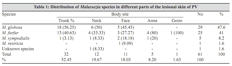
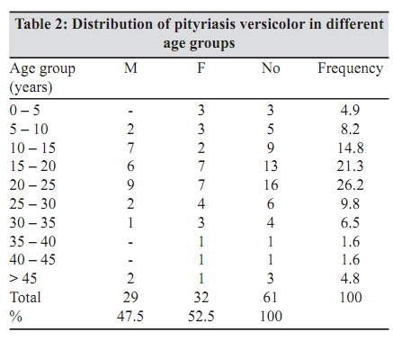
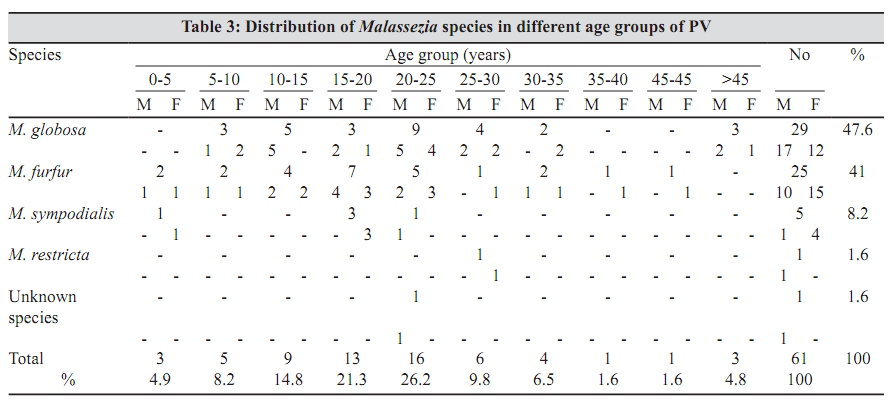
Indian Journal of Medical Microbiology, Vol. 27, No. 4, October-December, 2009, pp. 321-324 Original Article Distribution of Malassezia species in patients with pityriasis versicolor in Northern Iran *T Shokohi, P Afshar, A Barzgar Department of Medical Mycology and Parasitology (TS, AB), Sari Medical School, and Reference Mycology Laboratory (PA), Mazandaran University of Medical Sciences, Sari, Iran Correspondence Address: * Department of Medical Mycology and Parasitology, Sari Medical School, Sari, Iran, shokohi.tahereh@gmail.com Date of Submission: 26-Feb-2008 Code Number: mb09091 PMID: 19736400 DOI: 10.4103/0255-0857.55445 Abstract Purpose : Malassezia yeasts are globally distributed agents of pityriasis versicolor and are implicated in the pathogenesis of seborrhoeic and atopic dermatitis. The aim of this study is to identify the Malassezia species obtained from pityriasis versicolor patients, using morphological, biochemical, physiological as well as Polymerase Chain Reaction-Restriction Fragment Length Polymorphism (PCR-RFLP) methods.Materials and Methods: The identification of Malassezia species is performed according to microscopic features and physiological characteristics, including catalase reaction and Tween assimilation tests. The DNA is extracted from cultured Malassezia using the glass bead, phenol-chloroform method. The internal transcribed spacer 1(ITS1) region is amplified and there is restricted digestion of the PCR products with two enzymes Cfo I and Bst F5I. Results : The most commonly isolated species is M. globosa (47.6%). RFLP analysis of the PCR products of the ITS1 region is in complete agreement with those from the DNA sequences of the internal transcribed spacer (ITS) 1 region and the biochemical tests. Conclusion : Based on the findings of this study, it can be concluded that PCR-RFLP is a relatively simple and quick method, completely comparable to the routine methods used for Malassezia identification. Keywords: Malassezia, pityriasis versicolor, identification, Polymerase Chain Reaction-Restriction Fragment Length Polymorphism Introduction Malassezia yeasts are the causative agents of pityriasis versicolor (PV) with global distribution and are implicated in the pathogenesis of seborrhoeic and atopic dermatitis. Although the taxonomic status of the genus has been recently expanded to include eleven species, namely M. globosa, M. restricta, M. sympodialis, M. furfur, M. obtusa, M. slooffiae, M. pachydermatis, M. dermatis, M. japonica, M. nana and M. yamatoensis , their contribution to skin diseases is under investigation. [1] PV is a chronic superficial fungal disease that is characterized by the appearance of round-to-oval lesions, most commonly found on the trunk and upper aspects of the arms. These lesions vary in color, and can be hypo- or hyperpigmented. Numerous studies have shown the relationship between these species and their role as causative agents and / or triggers of diseases. There is evidence suggesting geographical variations in the distribution of the species. As non-molecular methods are time consuming and the results are sometimes difficult to interpret, molecular typing methods have been developed to evaluate the distribution of different Malassezia subtypes within pityriasis versicolor. There are only a few reports on the molecular identification of Malassezia species in Iran, therefore, this study has been undertaken to identify the Malassezia species isolated from pityriasis versicolor patients using morphological, biochemical and physiological [2] methods in comparison with Polymerase Chain Reaction-Restriction Fragment Length Polymorphism (PCR-RFLP) methods [3] Materials and Methods Subjects A total of 81 patients clinically suspected with pityriasis versicolor, referred to the Mycology Laboratory of the Mazanadaran University of Medical Sciences were recruited. Sixty-nine patients (85.2%) were confirmed with pityriasis versicolor, based on the presence of both hyphae and spores in direct microscopy, however, the Malassezia spp were isolated from 61 patients (88.4%), who were the final enrolled subjects. Collection and culture of sample The samples were collected by scraping with a blunt scalpel blade. The scraped skin was inoculated in modified Leeming and Notman agar (1% w/v peptone, 1% w/v glucose, 0.2% w/v yeast extract, 0.8% desiccated ox bile, 0.1% v/v glycerol, 0.05% w/v glycerol monostearate, 0.5% v/v Tween 60, and 2% v/v oleic acid, 1% w/v agar in distilled water) supplemented with cyclohexamide (0.5%) and chloramphenicol (0.05%), as recommended by Sugita et al ., [1] and was incubated at 32˚C for two weeks. Isolated yeasts were stored at -80˚C for further analysis. Microscopic and physiological characteristics After one week incubation on LNA at 32°C, the yeast cells were examined morphologically by Gram staining. The catalase reaction was performed by the application of a drop of hydrogen peroxide (30% solution) onto a culture smear on a glass slide. Production of gas bubbles was considered as a positive reaction. M. restricta is the only lipid-dependant species lacking catalase. [2],[4] Tween assimilation test The ability to utilize individual Tween compounds (Tween 20, 40, 60 and 80) was tested according to Guillot et al . [2] and Gupta et al . [4] Briefly, the sterile sabouraud's agar (16 ml) was melted and allowed to cool to approximately 50°C . Malassezia yeast suspensions (about 10 5 cfu / ml) were mixed with sabouraud's agar, and the mixtures were plated. Five holes were made in the agar by means of a 2 mm diameter punch and filled with 5 µl Tween 20, 40, 60 and 80. The plates were incubated for one week at 32°C. Utilization of Tween was assessed by the degree of growth and /or reaction (precipitation) of the lipophilic yeasts around individual wells. DNA extraction Colonies of Malassezia were subcultured on LNA to obtain pure culture before DNA extraction. The DNA extraction was done using a glass bead, phenol-chloroform method. [5] Polymerase Chain Reaction Two primers [6] (forward, 18SF1: 5′-AGGTTTCCGTAGG TGAACCT-3′, and reverse, 5.8SR1: 5′-TTCGCTGCGTTC TTCATCGA-3′) were used to amplify the ITS1 region in the rDNA gene in all Malassezia strains. The PCR products were electrophoresed in 1.5% agarose gel and the right PCR products were sequenced. The sequences were aligned and the digestion patterns of the species were checked for the restriction enzymes using CLC Free Workbench software (version_3_0_2). Finally, the enzymes C fo I and B st F5I were selected according to Mirhendi et al . [3] RFLP Digestion was performed by incubating 17 µl aliquot of PCR products with 10 U of the enzyme in a final reaction volume of 20 µl at 37°C for 3 h, followed by 2% agarose gel electrophoresis in a TBE buffer and staining with ethidium bromide. Results Of 81 skin scrapings, 69 (85.2%) were microscopically positive for Malassezia elements. Only 61 (88.4%) of the microscopically positive specimens yielded Malassezia in the culture. The names of the identified species of all cultured Malassezia from PV patients are listed in [Table - 1], along with the body sites from where they were sampled. The most common site was the trunk (52.45%), followed by the neck (19.67%) and the face (18.03%). Chi-square analysis indicated no correlation between the species and the body sites (χ2 = 14.32 df = 12 P = 0.28). The age of the 61 enrolled subjects (29 males (47.5%)) ranged from one to 64 years (average 20.8 years median 20-25 years). The highest prevalence of PV was observed in patients' in the 20 to 25 age group [Table - 2] and [Table - 3]. There was no significant difference between age and gender of patients with PV (χ2 = 29.59 df = 31 P = 0.53). Chi-square analysis indicated that isolated species of Malassezia were not associated with any age group (χ2 = 29.01 df = 27 P = 0.36). Four human-associated lipophilic Malassezia species were isolated from PV lesion scrapings, while normal animal-associated M. pachydermatitis was not obtained. Other species namely M. slooffiae and M. obtusa were not isolated from our population, although the microscopic, morphological and physiological characteristics were mainly used to identify all the strains. Molecular criteria were used to re-confirm the identification of 24 (39.3%) strains. The most common species from all body sites was M. globosa (47.6% of isolates), followed by M. furfur (41%), M. sympodialis (8.2%) and M. restricta (1.6%) with significant statistical differences (χ2 = 24.2 df = 3 P < 0.01). RFLP analysis of PCR products of the ITS1 region with two restriction enzymes C fo I and B st F5I was undertaken on 24 out of 61 isolates. Molecular typing of clinical isolates based on the PCR-RFLP was in complete agreement with those from the DNA sequences of the internal transcribed spacer (ITS) 1 region and the biochemical tests. Discussion Numerous reports have appeared after the taxonomic revisions of the genus Malassezia since 1996, in different geographical areas, including South Eastern Europe, Greece, Spain, Japan and Iran, reporting different frequencies. [7],[8],[9],[10] These studies suggest that M. globosa is the predominant species in normal and / or lesional skin. The same results were found in PV patients examined in our study . M. furfur, the second most common agent, has been isolated more frequently (41%) than by other studies in Japan, Canada, Tunisia and so on, reporting frequencies between 5-30%. [4],[8],[9],[11],[12] M. sympodialis , on the other hand, was isolated less frequently (8.2%) than by many investigators who reported it as the third species [8],[9],[12],[13] or even the first causative agent in Canada. [4] Although M. restricta is a normal skin flora, [2],[4] its lower frequency in our study is attributable, to some extent, to its fastidious nature, as each Malassezia species has a specific ecological niche, as well as specific biochemical and genetic characteristics. Therefore, care must be taken in the interpretation of the culture-based studies. Other species, such as M. slooffiae are less common [9] or extremely rare, [8] hence the absence of this species in the PV patients of our study. M. pachydermatis was also not found in the subjects, because this species is typically associated with animals. Nakabayashi et al . [9] indicated that the pathogenic species involved in PV is M. globosa and it is also detected in 51% of the normal subjects. They showed that PV is caused as a result of an overgrowth of this normal flora. This might be related to the clinical question of whether there is a relationship between this particular Malassezia species and various dermatological disorders, as different authors have debated whether Malassezia yeasts are of primary pathogenic significance or a secondary phenomenon. [14] To the best of our knowledge, the differences between the studies may not only be the result of geographical variation in species prevalence, but also due to ethnic factors, sampling methods (swabbing, scraping and transparent dressing) and different culture media. There is evidence that Malassezia species are common on non-lesional skin of patients with PV and on skin of control subjects, suggesting that the endogenous factors that promote the development of PV in susceptible hosts do not necessarily favour the growth of some species over others. [8],[9],[12] The isolation rate of Malassezia species from PV patients was 88.4%, which was comparable to some studies, [8],[9] but higher than others. [11],[15] The results of the PCR-RFLP analyses of clinical isolates were in complete agreement with those from DNA sequencing and biochemical tests. Alternative laboratory diagnosis, based on molecular identification of each Malassezia species by nested PCR [16] or PCR-restriction enzyme analysis, [6],[17],[18] has been employed using pure cultures, to assist and confirm the conventional laboratory diagnosis [1] of Malassezia -induced [11] or -triggered [8] dermatological conditions. Gaitanis et al . [19] and Gemmer et al . [20] identified the Malassezia species by amplifying targeted DNA extracted directly from skin scales, which could simultaneously reveal potential interactions among Malassezia or other yeast species on human skin. These new methods may resolve the disadvantages of the time-consuming morphological and biochemical techniques and the difficulties in interpretation of some physiological patterns. [12] PCR-RFLP is a sensitive and rapid identification system for Malassezia species identification. However, the technique should be developed further to allow its application in all cases and also on skin scales. Acknowledgements This study was financially supported by the Research Deputy of the Mazandaran University of Medical Sciences. We express our appreciation to Dr. Ahmadali Enayati for his expert advice and critical review of the manuscript. We also thank Prof. Alireza Khalilian for his help with the statistical analysis and Sabah Mayahi for technical assistance. References
Copyright 2009 - Indian Journal of Medical Microbiology The following images related to this document are available:Photo images[mb09091t3.jpg] [mb09091t2.jpg] [mb09091t1.jpg] |
| |||||||||