 |
| About Bioline | All Journals | Testimonials | Membership | News |
|
||||||
|
||||||
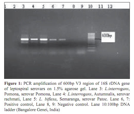
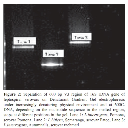
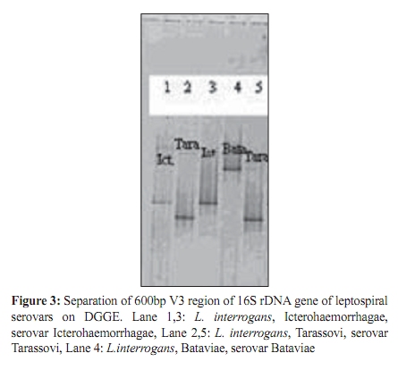
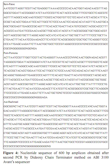
Indian Journal of Medical Microbiology, Vol. 27, No. 4, October-December, 2009, pp. 354-357 Brief Communication Characterization of leptospires using V3 region of 16S rDNA by denaturing gradient gel electrophoresis : A case study *SS Pol, PK Dhakephalkar, RS Bharadwaj Department of Microbiology (SSP, RSB), B.J. Medical College, Sassoon Hospital Campus, Station Road, Pune-411 001, India, Agharkar Research Institute (PKD), G.G.Agharkar Road, Pune-411 004, India Correspondence Address: *Department of Microbiology, B.J. Medical College, Sassoon Hospital Campus, Station Road, Pune-411 001, India, aparnapol@rediffmail.com Date of Submission: 08-Jul-2008 Code Number: mb09098 PMID: 19736407 DOI: 10.4103/0255-0857.55459 Abstract Serological and molecular characterization of Leptospiral isolates helps us to identify serovar, which is useful, for epidemiological study. Serological characterization is tedious and requires a panel of monoclonal antibodies and expertise to read the results. This study is a preliminary work to evaluate the usefulness of Denaturing Gradient Gel Electrophoresis (DGGE) to identify serovars of leptospira. The V3 region of most conserved 16S rDNA of five pathogenic leptospiral serovars and one saprophytic serovar was characterized. DGGE method was employed to separate the amplified V3 region based on the nucleotide sequence. On DGGE, amplified V3 region of leptospiral serovars, under study, showed bands at different positions indicating DGGE as the effective method of characterization in the future. DNA sequencing of V3 region of the three serovars showed great difference in nucleotide sequence supporting the results of DGGE.Keywords: Denaturing gradient gel electrophoresis, polymerase chain reaction, serovars, 16S rDNA, V3 region Introduction Leptospirosis is a zoonotic disease caused by spirochetes of species Leptospira interrogans , gaining public health importance in India due to close association between man and animals. Isolation and characterization of infecting serovar is useful to understand the circulating serovar in the community. Identification of serovars can lead to appropriate control strategies being planned with the development of site-specific vaccine preparation. Characterization of serovars will also help to confirm the source of infection which, in turn, will help to control the epidemic. Serological method of characterization requires a large panel of monoclonal antibodies. [1] Preparation of monoclonal antibodies against more than 250 leptospiral serovars is a time consuming and tedious task. In this situation, molecular methods have come in for characterization of leptospires. DNA-based methods like DNA restriction enzyme analysis (REA), nucleic acid probes, polymerase chain reaction, randomly amplified polymorphic DNA (RAPD) fingerprinting, pulsed-field gel electrophoresis and ribotyping methods have been used for specific detection of leptospires in clinical samples as well as their typing into different serovars. In this study, we have tried the use of small stretch of DNA, 'V3'region, for the characterization of leptospiral serovars by the method of Denaturing Gradient gel Electrophoresis. Materials and Methods Selection of leptospiral serovars Pathogenic serovars selected were Leptospira interrogans , Pomona, serovar Pomona, (Pom-pomona), L.interrogans , Autumnalis, serovar Rachmati, (Aut-rachmati), L.interrogans , Icterohaemorrhagae, serovar Icterohaemorrhagae, L.interrogans , Tarassovi, serovar Tarassovi, L.interrogans , Bataviae, serovar Bataviae, saprophytic serovar L.biflexa , Semaranga, serovar Patoc, (Sem-patoc). The serovars were obtained from National Reference Center for Leptospirosis, Andaman, India. DNA extraction DNA was extracted by the method of Thierry et al . [2] and stored at minus 20 0 C. Polymerase Chain Reaction (PCR) The extracted DNA was subject to first Polymerase Chain Reaction. The primers used were FDD2 (5' CCG GAT CCG TCG ACA GAG TTT GAT CTT GGC TCA G-3') and RPP2 (5' CCA AGC TTC TAG ACG GTA CCT TGT TAC GAC TT-3') (Sigma-Genosys Ltd,Texas,USA). The amplified product obtained from 1 st Polymerase Chain Reaction was subjected to 2 nd Polymerase Chain Reaction using primers 34IFaGC- 5'CCT AGC GGA GGC AGC AG-3'-aGC (aGC is a 40 nucleotide GC rich sequence attached to the 5'end of the primer. The GC sequence is 5'CGC CCG CCG CCG CGC CCC GCG CCC GTC CCG CCG CCC CCG CCC-3') and COM2-5'-CCG TCA ATT CCT TTG AGT TT-3'. The PCR mix and the cycling details were as per Weisburg et al . [3] The amplified product obtained after second Polymerase Chain Reaction was detected in 1.5% agarose gel electrophoresis (Tarsons, India) Result was recorded using Gel Documentation System. (Alpha-innotech, Alpha Imager 2200, India) Denaturing Gradient Gel Electrophoresis (DGGE) The PCR amplified product of 600 bp (obtained after second PCR) of Pom-Pomona, Aut-Rachmati and Sem-Patoc, Ict-Icterohaemorrhagae, Tara-Tarassovi, Bat-Bataviae were subjected to DGGE. (BIO-RAD, Washington D.C., USA) In brief, the principle of DGGE is, the DNA fragments (200-700bp) are allowed to run under increasingly denaturing physical environment (20 to 80% denaturant) and at 60°C in a specially designed electrophoresis apparatus. The denaturant used was a mixture of formamide (Extrapure AR, SRL, Chennai, India) and urea (Extra pure AR, SRL, Chennai, India). As the DNA moves through the gradient, it undergoes partial melting because of which it migrates slowly than the double stranded DNA through polyacrylamide (Extra pure AR, SRL, Chennai, India) matrix. The DNA then stops depending on the nucleotide sequence in the melted region. Therefore, even though DNA fragments are of identical length (600 bp), due to sequence difference; stop at different positions in the gel. DNA sequencing To observe the nucleotide sequence difference, the amplified product of 600 bp from Pom-pomona, Aut-rachmati and Sem-patoc was sequenced using Dideoxy Chain Terminator method, on ABI 3100 Avant's sequencer, as per the manufacturer's instructions. (Big dye terminator kit, version 3.1, Applied Biosystems Inc., Connecticut, USA) The sequence obtained was submitted to National Center for Biotechnology Information (NCBI) for identification through Internet. The identity of the cultures was established on the basis of homology analysis. Results First Polymerase Chain Reaction amplified 16S rDNA gene (1Kb) and second Polymerase Chain Reaction amplified V3 region (600bp) of the gene. After second Polymerase Chain Reaction, we obtained amplified product of 600 bp (V3 region) from Pom-pomona, Aut-rachmati and Sem-patoc. [Figure - 1] On DGGE, every serovar showed separate bands at different positions distinct from each other. [Figure - 2],[Figure - 3] DNA sequencing [Figure - 4] revealed 25% homology between serovar Pom-pomona and Sem-patoc and 20% homology between Aut-rachmati and Sem-patoc. Pom-Pomona and Aut-rachmati showed 25% homology. Discussion In 1989, Masahito Fukunaga and Ichiji Mifuchi revealed some important facts regarding organization of rRNA genes in the leptospiral chromosomal DNA. They found that there are two genes for 16S rRNA and one gene for 5S rRNA in the leptospiral DNA and there was no linkage at all among the rRNA genes and thus are expressed independently in the organism. They found the similar results in Leptospira biflexa. [4] 16S rRNA was selected in this study because it is found in all organisms from bacteria to higher organisms. The16S rRNA gene sequence differs between species. Some sections of the 16S rRNA sequence are highly conserved whereas others differ greatly among organisms showing evolutionary relationships between organisms and 16S rRNA sequences are available on database to use for identification purposes. The 16S rDNA genes include, slowly evolving or conserved regions as well as rapidly evolving or variable regions. There are many variable regions in the structure of 16S rRNA. V3 region, total of 585 bp [positions 341 to 926 Escherichia coli numbering] of the 16s rDNA gene, was amplified using universal primers in the present study. Postic et al . used the first 330bp [positions 46 to 375 Escherichia coli numbering] of the 16s rDNA gene for comparison of leptospira isolates. The study focused on different serovars of L.meyeri species, the classification of which has been controversial,revealed large heterogeities which resulted in suggesting a revised classification of L.meyeri . [5] Woo et al . used the partial 16S rDNA sequences of 8 leptospira like field isolates that reacted weakly or not at all in MAT to identify them. They found that 16S rDNA sequences obtained by them were similar to a nonpathogenic Leptonema illini -type strain 3055. [6] Use of V3 region for the characterization of leptospires was not found to be reported in the past. The amplified V3 region of all the serovars under study was subjected to DGGE,which is the established method to detect mutations in DNA. In normal gel (agarose or acrylamide) electrophoresis, DNA fragments are separated by size. As the size of the DNA fragment increases, its velocity through the gel decreases. In DGGE, DNA fragments of the SAME size are separated by their denaturing profile (how the dsDNA becomes ssDNA). It has been shown to detect differences in the denaturing behavior of small DNA fragments (200-700bp) that differ by as little as one base pair. Gurtler et al . used DGGE to detect mutation in VS2 of the 16S-23S-rDNA-spacer region amplified from Staphylococcus aureus isolates. [7] Pignon et al. analyzed P53 gene coding sequence by DGGE and studied its application in detection of point mutations in acute leukemias. [8] The use of DGGE for the characterization of leptospires has also not been found in the literature. About 600 bp amplicons, in the present study, showed distinct bands on DGGE, which underlined the usefulness of the technique for the characterization of leptospiral serovars. The nucleotide sequence of the V3 region of the three serovars under study supported this result as they showed less2than 70% homology among them. Thus, DGGE will definitely be useful to differentiate closely related serovars of leptospira, especially which are grouped under one genomospecies. Differentiation at the strain level will also be possible, even if there is one or two base pair sequence difference. Along with characterization of leptospires, DGGE will play an important role in the diagnosis and identification of the infecting serovar at the same time. If a ladder of amplified V3 region of prevalent serovars is prepared and run along with the test sample on DGGE, we can match the band of test with the corresponding band in the ladder and identify the infecting serovar. However, if a database using all known serovars is created, it can be utilized for rapid serovar diagnosis in clinical situation. The high cost of the equipment and requirement of the expertise will limit DGGE's use to the reference and research laboratory only. References
Copyright 2009 - Indian Journal of Medical Microbiology The following images related to this document are available:Photo images[mb09098f3.jpg] [mb09098f4.jpg] [mb09098f1.jpg] [mb09098f2.jpg] |
| |||||||||