 |
| About Bioline | All Journals | Testimonials | Membership | News |
|
||||||
|
||||||
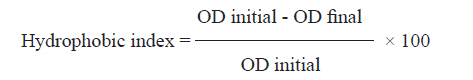
Indian Journal of Medical Microbiology, Vol. 29, No. 2, April-June, 2011, pp. 165-168 Brief Communication Biotypes and virulence factors of Gardnerella vaginalis isolated from cases of bacterial vaginosis J Udayalaxmi1, GK Bhat1, S Kotigadde2 1 Department of Microbiology, Kasturba Medical College, Manipal University, Mangalore - 575 001, India Correspondence Address:G K Bhat Department of Microbiology, Kasturba Medical College, Manipal University, Mangalore - 575 001 India gkbhat61@yahoo.co.in Date of Submission: 02-Sep-2010 Code Number: mb11038 PMID: 21654113 DOI: 10.4103/0255-0857.81798 Abstract The present study was conducted to correlate the biotypes of Gardnerella vaginalis strains isolated from cases of bacterial vaginosis and their virulence factors. Thirty-two strains of G. vaginalis isolated from cases of bacterial vaginosis were biotyped. Adherence to vaginal epithelial cells, biofilm production, surface hydrophobicity, phospholipase C and protease activity were tested on these isolates. Biotype 1 was the most prevalent (8; 25%), followed by biotype 2 (7; 21.9%) and biotypes 5 and 8 (5; 15.6%). We did not find any statistical correlation between G. vaginalis biotypes and its virulence factors. Virulence factors expressed by G. vaginalis were not associated with a single biotype.Keywords: Bacterial vaginosis, biotypes, virulence factors Introduction Bacterial vaginosis represents a unique and complex change in the microflora of the vagina, characterised by a reduction in the prevalence and number of hydrogen peroxide-producing lactobacilli and an increase in the concentration of Gardnerella vaginalis and resident anaerobic bacteria. Although bacterial vaginosis is prevalent, not much progress has occurred in identifying the factors responsible and associated with bacterial vaginosis and its pathophysiology. [1] G. vaginalis is classified into eight distinct biotypes on the basis of lipase and β-galactosidase activity and hippurate hydrolysis.[2],[3],[4] Studies have been conducted to correlate the biotypes with the pathogenicity of G. vaginalis. In previous studies, no significant difference was observed in the distributions of G. vaginalis biotypes in women with or without bacterial vaginosis (BV) 48% of the women acquired a different G. vaginalis biotype after the treatment and a trend toward the acquisition of a new biotype was observed among women who had contact with a new sexual partner. [2],[3],[4] However, limited work has been carried out on correlation between biotypes of G. vaginalis and expression of virulence factors. The objectives of the present study were to identify the biotypes of G. vaginalis, virulence factors and determine any correlation between the biotypes and the virulence factors. Materials and Methods The study population consisted of women attending tertiary care hospitals for antenatal care and intrauterine devices (IUD) insertion or removal, with complaints of discharge and abdominal pain. They belonged to the 21-35 years age group and were non-menstruating at the time of specimen collection and not on any medication up to 1 month prior to specimen collection. The study had the approval of the Institutional Ethics Committee. Diagnosis of bacterial vaginosis was established by Amsel′s criteria and Nugent′s criteria. [5] Vaginal swabs were used for wet mount, Gram stain, whiff test and for determination of pH of the vagina. Vaginal swabs were inoculated on human blood bilayer agar and incubated at 37°C for 48 h. G. vaginalis isolates were identified by standard methods and were preserved in skimmed milk at -70°C for further studies. [6] Thirty-two G. vaginalis isolates were biotyped and virulence factors like adherence to vaginal epithelial cells, biofilm formation, surface hydrophobicity, phospholipase C and protease enzyme were studied. A standard strain of G. vaginalis, ATCC 14018, was included as control with each test. Adherence of G. vaginalis to vaginal epithelial cells was studied by performing an adherence assay, as described previously. [7] In brief, 1 ml of the standard bacterial suspension was mixed with an equal volume of standard vaginal epithelial cell suspension and incubated at 37°C in a shaker water bath at a speed of 35 rotations per minute for 45 min. The epithelial cells were washed free of non-adherent bacteria by passing through a membrane filter of pore size 8 μm. The membrane filter was carefully removed and inverted over a slide, air-dried, alcohol fixed and Gram stained. An average number of bacteria adherent to 30 cells was counted. The method described by Christensen et al. was used for detection of biofilm production. [8] All tests were performed in duplicate. Pseudomonas aeruginosa ATCC 27853 was used as the positive control. Biofilm produced was graded as weak or non-biofilm producer (OD <0.1), moderate (OD 0.1-0.2) and high (OD >0.2). Surface hydrophobicity of the G. vaginalis isolates was determined by a quantitative hydrophobicity assay, as described previously. [9] Standard bacterial suspension in phosphate-buffered saline showing an OD 600 of 0.3 (OD initial) was prepared. Three milliliters of the standard bacterial suspension was mixed with 300 μl xylene and allowed to stand at 25°C for 30 min. The OD 600 of the aqueous phase was determined (OD final). The hydrophobic index was calculated as follows:
Phospholipase C activity of G. vaginalis isolates was detected as described previously. [10] Pseudomonas aeruginosa PA01 strain (MTCC 3541, MTCC, Chandigarh, India) was used as the positive control. The proteolytic activity of G. vaginalis was detected using skim milk agar with 5% horse serum and expressed as diameter of clear zone in mm. [11] G. vaginalis isolates were biotyped using hippurate hydrolysis, ONPG and lipase test, as described in previous studies. [2],[3] Statistical correlation between various biotypes and virulence factors was performed using the Kruskal Wallis test. Statistical correlation of different virulence factors expressed by G. vaginalis strains isolated from asymptomatic women and women with abnormal discharge was performed by the Chi square test. Statistical relevance for frequency of isolation of different biotypes from asymptomatic women and women with abnormal discharge was determined by the Chi square test. Result Of 527 women screened for bacterial vaginosis, 97 were diagnosed as having bacterial vaginosis by Amsel′s criteria and 100 by Nugent′s criteria. Of these 100 women, 45 yielded G. vaginalis. Of these 45 women, 32 yielded a heavy growth of G. vaginalis and hence these 32 isolates were preserved and used for the biotyping and virulence factor study. Out of the 32 isolates studied, 22 (68.8%) showed a relatively better adherence (an average of 26.79 bacterial cells per vaginal epithelial cell). Of the 32 strains of G. vaginalis under study, 23 (71.8%) produced biofilm. Out of the 32 isolates studied, 24 (75%) showed a relatively high surface hydrophobicity index (>50%). Out of the 32 isolates tested, 28 (87.5%) produced phospholipase C and 14 (14.82%) were protease positive. Out of the 32 isolates of G. vaginalis, eight were from asymptomatic women and 24 were from women with vaginal discharge. There was no statistical correlation between the different virulence factors expressed by the strains of G. vaginalis isolated from asymptomatic women and women with abnormal discharge [Table - 1]. Biotype 1 (8; 25%) was the predominant isolate, followed by biotypes 2 (7; 21.9%) and 5 and 8 (5; 15.6%). There was no statistical correlation between the different G. vaginalis biotypes and expression of virulence factors [Table - 2]. The frequently isolated biotypes 1, 2, 5 and 8 were more common in women with abnormal discharge than in asymptomatic women. However, the difference was not statistically significant [Table - 3]. Discussion An attempt was made to determine the biotypes and the virulence factors of G. vaginalis isolated from cases of bacterial vaginosis. Presence of clue cells in the vaginal discharge of cases of bacterial vaginosis indicates the role of adherence exhibited by G. vaginalis in the pathogenesis of bacterial vaginosis. Adherence and colonization of G. vaginalis could be considered as the initial stages in the pathogenesis of bacterial vaginosis. A previous study showed that G. vaginalis biofilms were more resistant to H 2 O 2 and lactic acid than planktonic cells. [12] Therefore, the formation of a biofilm in the vagina could increase the bacterial resistance to H 2 O 2 and lactic acid and lead to colonization, even in the presence of lactobacilli. Bacterial phospholipase C may damage the reproductive tract tissue by both direct and indirect mechanisms. [10] Phospholipase C-mediated degradation of placental tissues by microorganisms triggers the onset of premature labor. This may be the reason for complications like pre-term birth, low-birth weight of infants, post-partum endometritis and post-operative cellulites in women with bacterial vaginosis. [10] There is hardly any study showing protease production by G. vaginalis. Protease breaks down the tissue proteins, resulting in the release of amino acids that may support the growth of G. vaginalis and other bacteria in the vagina. The frequency of isolation of various biotypes from cases of bacterial vaginosis and normal women show varying results. [2],[3],[4] A past study showed that G. vaginalis isolates from cases of bacterial vaginosis expressed better adherence and biofilm formation than isolates from normal women. [13] A previous study positively correlated sialidase activity with biotypes 5 and 8. [3] Biotyping is a simple marker system that may be useful to study the epidemiology of bacterial vaginosis. A study of the association of virulence factors with symptoms of bacterial vaginosis helps in the study of pathogenesis of bacterial vaginosis. Study of the frequency of isolation of various biotypes of G. vaginalis from cases of bacterial vaginosis or from symptomatic women may help to develop better treatment options for bacterial vaginosis. Development of probiotics that are active against the biotypes more frequently isolated from cases of bacterial vaginosis or symptomatic women may help in treatment. The present study showed that virulence factors expressed by G. vaginalis are not associated with a single biotype. Studies involving a bigger sample size may be required to explore any correlation between biotypes and virulence factors of G. vaginalis. Acknowledgemen The authors thank the staff and students of of Department of Obstetrics and Gynecology, for Lady Goshen Hospital and Kasturba Medical College, Hospital, Attavar, for facilitating the collection of clinical material for the study. References
Copyright 2011 - Indian Journal of Medical Microbiology The following images related to this document are available:Photo images[mb11038t2.jpg] [mb11038t3.jpg] [mb11038t1.jpg] |
| |||||||||

![[Table - 1]](/showimage?mb/photo/mb11039t2.jpg)
![[Table - 2]](/showimage?mb/photo/mb11038t2.jpg)
![[Table - 3]](/showimage?mb/photo/mb11038t3.jpg)