 |
| About Bioline | All Journals | Testimonials | Membership | News |
|
||||||
|
||||||
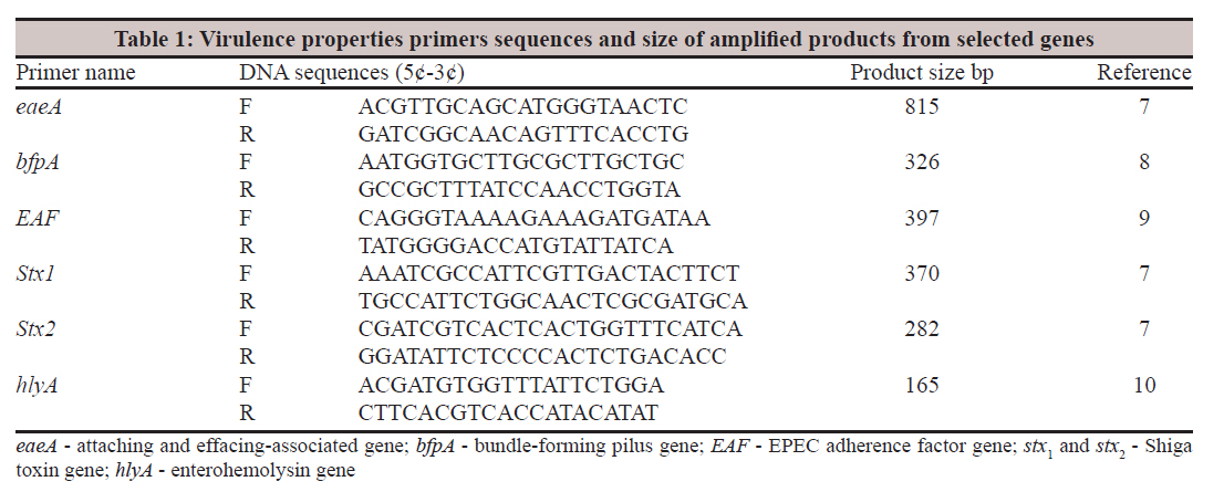
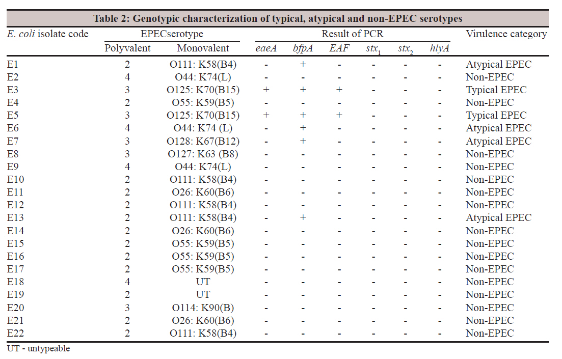
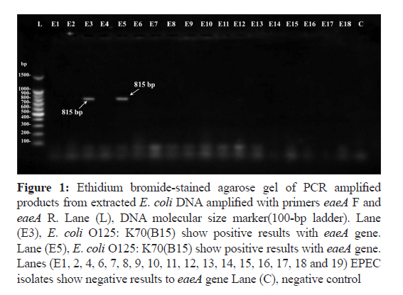
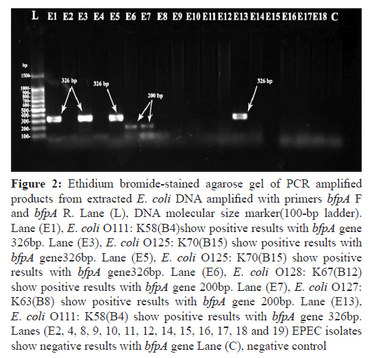
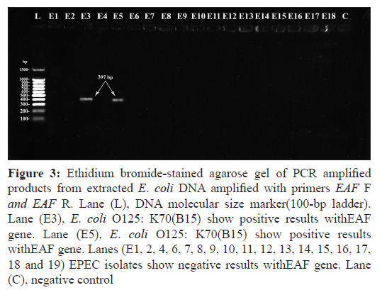
Indian Journal of Medical Microbiology, Vol. 29, No. 4, October-December, 2011, pp. 383-388 Original Article Occurrence and molecular characterization of enteropathogenic Escherichia coli serotypes isolated from children with diarrhoea in Najaf, Iraq Samer A Al Hilali, Ali M Almohana Department of Microbiology, College of Medicine, Kufa University. Iraq Najaf Kufa p.o. Box(18), Iraq Date of Submission: 05-Jun-2011 Code Number: mb11093 PMID: 22120799 Abstract Purpose: Enteropathogenic Escherichia coli (EPEC) are among the most important pathogens infecting children worldwide and are one of the main causes of diarrhoea. The study was carried out to investigate the occurrence of EPEC as a cause of infectious diarrhoea in children younger than 2 years of age and characterize their virulence genes. Keywords: Atypical EPEC, bundle-forming pilus, enteropathogenic Escherichia coli, intimin, typical Enteropathogenic Escherichia coli Introduction Diarrhoeal disease continues to be a health problem worldwide. The causes of diarrhoea include a wide range of viruses, bacteria and parasites. Among the bacterial pathogens, E. coli plays an important role. EPEC is an important category of diarrhoeagenic E. coli which has been linked to infant diarrhoea in the developing world. As with other diarrhoeagenic E. coli strains, transmission of EPEC is faecal-oral, with contaminated hands, contaminated foods or contaminated fomites serving as vehicles. EPEC adhere to the mucosal cells of the small bowel. The result of EPEC infection is watery diarrhoea, which is usually self-limited, but can be chronic. [1] EPEC strains are noninvasive, infecting their hosts by attaching to intestinal epithelial cells (IEC), effacing the epithelial microvilli and producing pedestal-like structures. The formation of attaching and effacing (A/E) lesions is required for these microbes to cause diarrhoeal disease. [2] EPEC possess both the bfpA and eae for E. coli-attaching and effacing. It is a well-recognized pathogen in developing countries as class I EPEC or typical EPEC. However, atypical EPEC organisms possessing eae alone have been reported to be more prevalent in both developing and developed countries, and animals can be reservoirs of atypical EPEC, in contrast to typical EPEC, in which humans are the sole reservoir. [3] In Iraq, till now, little information was available on the prevalence of EPEC infection. However, in Najaf, there is no molecular identification and categorization of EPEC associated with diarrhoea in children younger than 2 years old. The study is aimed to determine the occurrence of EPEC serotypes in children with diarrhoea in Najaf and detecting their virulence properties. Materials and Methods Patients and stool collection This study was conducted in Najaf. Two hospitals (Alzahraa Teaching Hospital and Alhakeem Teaching Hospital) were included in this study. A total of 656 stool specimens were collected from children younger than 2 years old suffering from watery diarrhoea characterized in Nguyen et al., [4] and 54 apparently healthy control. The specimens collected during the period from September to November, 2009. Isolation and identification of bacterial isolates All stool specimens were cultured on MacConkey agar (Himedia M081; Himedia, Mumbai, India) and were incubated for 24 h at 37°C. Colonies morphologically resembling E. coli were identified by standard laboratory techniques. [5] Escherichia coli serotyping The E. coli-positive cultures were set for further serological test. Isolates biochemically identified as E. coli were serologically examined by slide agglutination test (remel Escherichia coli Agglutinating Sera; Remel Europe Ltd., UK) according to kit procedures, using polyvalent O antisera for EPEC, (2, 3, and 4), separately. From the colony which showed a positive reaction with polyvalent antisera a subculture was prepared and the isolates were retested by using the following EPEC monovalent O: K antisera: (O26: K60(B6), O55: K59(B5), O111: K58(B4), O119: K69(B14), O126: K71(B16), O86: K61(B7), O114: K90(B), O125: K70(B15), O127: K63(B8), O128: K67(B12), O44: K74(L), O112: K66(B11), O124: K72(B17), O142: K86(B), O18c: K77(B21). DNA isolation and polymerase chain reaction assay Enteropathogenic E. coli DNA extraction was done according to the Pospiech and Neumann [6] method. DNA templates were subjected to PCR using six sets (F and R) of primers targeting virulence properties genes listed in [Table - 1]. The reaction mixture moreover contained GoTaq® Green Master Mix, X2 (Promega M 7122; Promega Corporation, USA), which is premixed ready-to-use solution containing Taq DNA polymerase dNTP, MgCl 2 and according to Promega procedure, the reaction mixtures were prepared in 0.2 ml eppendorf tube with 25 ml reaction volumes. The PCR were performed with PCR system (GeneAmp PCR system 9700; Applied Biosystem, Singapore) at 94°C for 2 min. For 1 cycle followed by 30 cycles of 94°C for 1 min, 55°C for 1 min and 72°C for 1 min. For the eaeA gene, 94°C for 2 min for 1 cycle followed by 30 cycles of 94°C for 1 min, 60°C for 1 min and 72°C for 2 min for the bfpA gene, 94°C for 5 min. For 1 cycle followed by 30 cycles of 94°C for 1 min, 57°C for 45 s and 72°C for 1 min for the EAF gene, 94°C for 5 min for 1 cycle followed by 35 cycles of 95°C for 40 s, 65°C for 20 s and 72°C for 45 s for stx1 and stx2 genes and 95°C for 5 min for 1 cycle followed by 35 cycles of 95°C for 1 min, 58°C for 1.30 min and 72°C for 1.30 min for the hlyA gene. The amplified PCR products were detected by agarose gel electrophoresis and visualized by staining with ethidium bromide using gel documentation system (BioDocAnalyze Live; Biometra biomedizinische Analytik GmbH, Germany). Statistical analysis The Chi-square test was used to determine the statistical significance of the data. A P value of <0.5 was considered significant. Results It was observed that the number of E. coli isolates from 656 stool specimens were 535 (81.6%) (Basically, 1 E. coli isolate from each patient). A total of 52 (96.3%) E. coli isolates were obtained from 54 healthy individuals. Of the total of E. coli isolates recovered from 656 children with diarrhoea, only 22 (3.4%) agglutinated with EPEC polyvalent antisera. By contrast, no EPEC isolate belonging to classical EPEC polyvalent serotypes was detected in stool of healthy individuals. However, the highest number of the EPEC isolated belonging to polyvalent 2 sero-group with following serotypes, O55: K59(B5) (22.7%), O111: K58(B4) (18.18%), O26: K60(B6) (13.63%), followed by polyvalent 3 sero-group with the following serotypes, O125: K70(B15) (9.09%), O128: K67(B12) (4.54%), O127: K63(B8) (4.54%), O114: K90(B) (4.45%), followed by polyvalent 4 sero-group with serotype O44: K74(L) (13.63%). From the total isolates, 6 (27.3%) isolates were PCR-positive for at least one of the targeted virulent genes [Table - 2]. Only two (9.1%) isolates gave positive results with the eaeA gene belonging to serotype O125: K70(B15) [Figure - 1]. Six (27.3%) isolates gave positive result with bfp, of these, four isolates gave amplified product size equal to 326 bp, belonging to O125: K70(B15) (two isolates), O111: K58(B4) (two isolates), while, two isolates gave amplified product size equal to 200 bp, belonging to O44: K74(L) and O128: K67(B12) [Figure - 2]. Only two isolates (9.1%) that belonged to serotype O125: K70(B115) was found to harbour the EAF plasmid gene [Figure - 3]. Based on these criteria [Table - 2], two isolates (33.3%) were classified as typical EPEC, these isolates belonged to O125: K70(B15) serotype, obtained from infants younger than 6 months. Four (66.7%) isolates were classified as atypical EPEC. The atypical EPEC isolates belonged to O111: K58(B4) (two isolates), O44: K74(L) (one isolate), O128: K67(B12) (one isolate) serotypes, obtained from children with diarrhoea aged from 3 months to 17 months. Discussion Enteropathogenic E. coli has been identified as important cause of infantile diarrhoea in all the developing countries where it has been looked for, but the incidence has varied greatly in different studies. The present study revealed that EPEC was higher than been demonstrated previously by Almohana [11] who reported that 37 (2.1%) E. coli isolates recovered from 1798 patients suffering from diarrhoea were assigned as EPEC. The relative frequency has been demonstrated in Tehran, Iran, [12] that EPEC represent (8.4%) of diarrhoeal causes. A high frequency of EPEC was observed in Korea (56%) and Brazil (34.0%). [13] In another study, EPEC serogroups were isolated as the sole pathogen from 44.9% of the Iranian children with diarrhoea. [14] The WHO has considered isolates in the 12 O serogroups to be EPEC strains; [15] O26, O55, O86, O111, O114, O119, O125, O126, O127, O128, O142 and O158. However, the serological investigation revealed 8 E. coli serogroups of the 12 E. coli serogroups described by WHO [15] as EPEC strains. We found that O55: K59(B5) was the most common EPEC isolates that represented 0.8% of cases of diarrhoea in Najaf. In agreement with this study, the O55 serogroup was the most prevalent (12.6%) in Iran. [14] While, in Romania the O55 serogroup represent 1.2% diarrhoeal causes in children. [16] Although serogrouping proposed by Levine et al. [17] has been carried out to define the EPEC strains, it is now recognized that the serogrouping is not well correlated with the presence of pathogenic factors. Therefore, at present, the detection of pathogenic genes by PCR may be the best way to identify the diarrhoeagenic E. coil. [18] Only 2 (9.1%) isolates belonging to serotype O125: K70(B15) gave positive results with eaeA gene. Since there is no data recorded for the presence of the eaeA gene in the EPEC isolates in Najaf, we cannot identify the difference in the incidence of this virulence factor. However, the isolation frequency of EPEC having the eaeA gene was not as high as expected in Najaf. The isolation frequency having this gene may vary tending on the district, season, child age and so on. Notably, a high incidence of EPEC defined on the basis of the eaeA gene was reported by Nguyen et al. [4] in Hanoi, Vietnam, who found that all tested EPEC isolates were eaeA PCR positive. In an Iranian study, the eaeA gene was detected in 40.5% of the 111 EPEC examined strains. [14] A lower incidence of 10.9% has been reported in South Africa. [19] On the other hand, Nishikawa et al. [20] reported that all EPEC strains isolated from children with diarrhoea did not react with eaeA-specific primer in Japan. However, the results of this study appeared that EPEC isolates possess the eaeA gene is an uncommon cause of diarrhoea in Najaf. Only 18.2% isolates gave positive results with bfpA primers that equal to target product size (326 bp). The ethidium bromide-stained agarose gel of PCR-amplified products show other two products (200 bp), which may be considered as positive result depending on a previous study conducted by Carneiro et al., [21] who reported the presence of 200 bp amplicons with bfpA primers, suggesting that a deletion on this gene could contribute for the lower efficiency with which these strains attached to cells, producing a localized adherence like type phenotype. The occurrence of bfpA in the present study was analogous to that recorded by other studies such as, Alikhani et al. [14] who found that 31.5% strains in Iran gave positive results for the bfpA gene. In South Africa, Obi et al. [15] showed that the bfpA gene coding for EPEC was most frequently detected (22.7%) among E. coli isolates. The most regular matter in EPEC is the presence of eaeA alone (atypical EPEC) or with bfpA (typical). Unusual in this study was the detection of bfpA without the eaeA gene [Table - 2]. This observation was also documented by Kobayashi et al. [2] who found that two isolates were positive to the bfpA gene but negative to the eaeA gene. Unusual in this study is the detection of the bfpA gene in four isolates but not the EAF gene [Table - 2]. It′s questionable whether isolates positive for the bfpA gene but not for EAF plasmid should be classified as EPEC and whether they actually are pathogenic. The reason for these phenomena is currently unknown. However, this may be due to the absence of the EAF probe from the EAF plasmid without any deleterious effects on localized adherence expression. [22] The EAF probe used to detect EPEC not recognized in some typical EPEC isolates, and the frequency of these organisms in infection may have been underestimated. [23] There are no report in Iraq that can be compared with the results of the present study and concerning detection of the EAF probe and plasmid in EPEC. However, the absence of the EAF plasmid in most bfpA-positive EPEC isolates in the present investigation was in agreement with the observation of Trabulsi et al. [15] Based on this study observation, the EAF PCR assay proved to be a nonspecific and inefficient method for the detection of EPEC isolates carrying the EAF plasmid. In this study, two isolates were classified as typical EPEC (eaeA +, bfpA +). Typical EPEC strains remain an important cause of potentially foetal infant diarrhoea in developing countries. [14] In Brazil, up to the 1990s, typical EPEC was the main cause of acute diarrhoea in children younger than one year old, of low socio-economic status. [23] In Iran, Alikhani et al. [14] reported that the typical EPEC strains continue to be an important cause of diarrhoea in children, mostly encountered among the strains belonging to serogroups O55 and O86. In a meeting on EPEC held in 1995, a consensus definition of atypical EPEC was established, namely that they are EAF- (bfpA- ), eaeA+ strains that promote attaching and effacing lesions. [24] The present study demonstrated unusual detection of four isolates carrying the bfpA gene without the eaeA gene [Table - 2]. Hardegen et al. [25] detected five isolates which were positive for EAF plasmid only and reported this question whether E. coli strains positive for EAF plasmid but not for eaeA should be classified as EPEC. Alikhani et al. [14] reported that the presence of only one gene (eaeA or bfpA) used to identify EPEC as atypical. Therefore, these four isolates recovered in the present study were classified as atypical EPEC. According to the results of the present study, we concluded that the serotyping of E. coli is not likely corresponding to the pathogenic factors. Significant differences were found between the serotyping method and molecular techniques in detection of EPEC (P<0.05). The molecular methods was more specific but slower than the serotyping method. The possession of EPEC-related O and K antigens is no longer deemed an essential characteristic of true pathogenic EPEC strains. References
Copyright 2011 - Indian Journal of Medical Microbiology The following images related to this document are available:Photo images[mb11093t2.jpg] [mb11093f3.jpg] [mb11093t1.jpg] [mb11093f1.jpg] [mb11093f2.jpg] |
| |||||||||