 |
| About Bioline | All Journals | Testimonials | Membership | News |
|
||||||
|
||||||
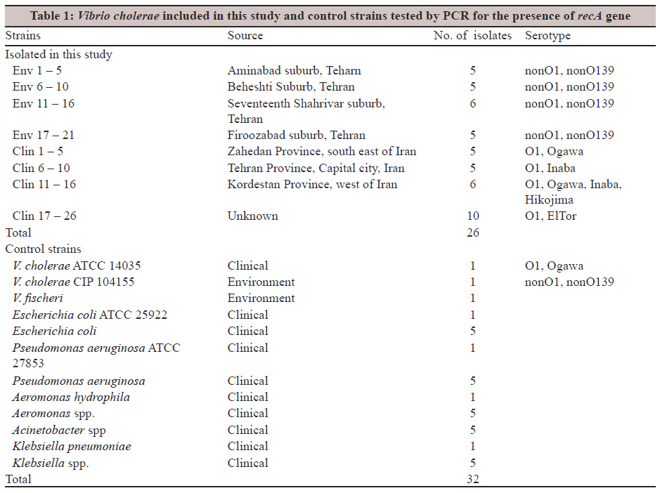
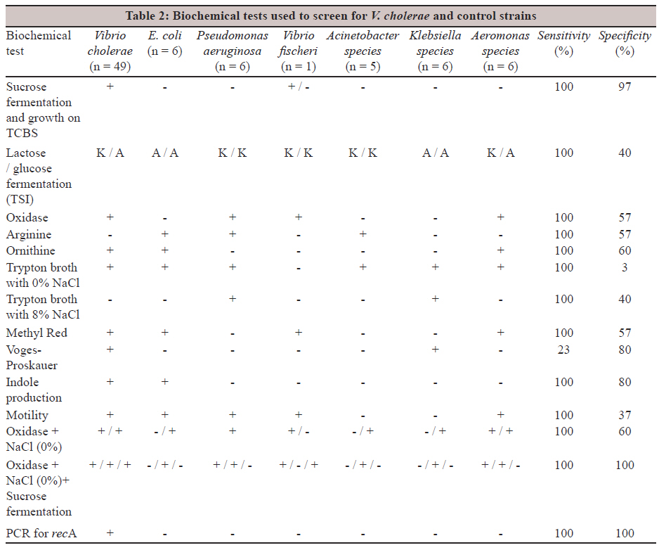
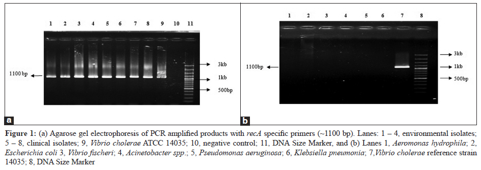
Indian Journal of Medical Microbiology, Vol. 30, No. 1, January-March, 2012, pp. 39-43 Original Article A rapid and reliable species-specific identification of clinical and environmental isolates of Vibrio cholerae using a three-test procedure and recA polymerase chain reaction AD Roozbehani1, B Bakhshi2, MR Pourshafie1, M Katouli3 1 Department of Bacteriology, Pasteur Institute of Iran, Correspondence Address: Date of Acceptance: 19-Dec-2011 Code Number: mb12008 PMID: 22361759 DOI: 10.4103/0255-0857.93027 Abstract Purpose: Vibrio cholerae, the cause of cholera, is one of the leading causes of morbidity and mortality in many developing countries. Most laboratories initially rely on biochemical tests for a presumptive identification of these strains, followed by a polymerase chain reaction (PCR)-based method to confirm their identification. The aim of this study is to establish a rapid and reliable identification scheme for V. cholerae using a minimal, but highly specific number of biochemical tests and a PCR assay. Keywords: Housekeeping gene, identification, polymerase chain reaction, recA, Vibrio cholera Introduction Cholera is a life-threatening diarrhoeal disease with high mortality among human populations. At least eight pandemics of cholera have been recorded, with the first one occurring in 1817. Of these, all but one originated in the Indian subcontinent, with the fifth, sixth, and seventh pandemics being caused by the classical strain of serogroup O1, although the last one introduced a new O1 strain with different phage susceptibility and biochemical properties. [1] The most recent pandemic was caused by a non-O1 serogroup, O139, in the 1990s. The aetiological agent of the disease, V. cholerae, is a water-borne pathogen for which 209 O serogroups have been recognised. Of these serogroups O1 and O139 have so far, been associated with human diseases. The other serogroups, collectively referred to as non-O1 non-O139 serogroups, have not been associated with epidemics, but can cause sporadic diarrhoea [2] and are ubiquitously distributed in the aquatic environment. Rapid and accurate identification of V. cholerae O1 in potable water, aquatic environment, and fecal samples is important for disease management and the public health system. Identification of V. cholerae is usually achieved by conventional bacteriological methods, such as, culturing stool or surface water samples on a selective medium, Thiosulfate Citrate Bile Salts Sucrose (TCBS) agar, followed by biochemical testing of colonies and confirmation by a slide agglutination test, using specific antisera. [3] The TCBS agar eliminates most non-target bacteria in the clinical samples, but it is not satisfactory for surface water samples, because some bacteria present in the water sources can produce yellow colonies resembling those of V. cholerae. [4] Furthermore, the series of biochemical tests commonly used to identify V. cholerae[5],[6],[7] are originally selected and used for clinical samples in order to specifically detect pathogenic Vibrios. In addition, the close relation between V. cholerae and certain other members of the Vibrionaceae or Aeromonas spp., with respect to their biochemical properties, has often made unambiguous identification of the organism quite difficult. Although the commercial availability of O1 and / or O139 antisera has helped the identification of the epidemic strains of V. cholerae considerably, it may not be true for the non-O1 / non-O139 strains, which until recently, have been known to range from serogroups O2 to O138 and from the O140 to O193. [8] The problem has become prominent recently, as the non-O1 / non-O139 strains of diverse serogroups have been implicated as the causative agents of a large number of diarrhoeal cases in many developing countries. [9] The use of a 16S rRNA sequence for the identification of V. cholerae strains has not been successful so far due to the lack of appreciable differences between V. cholerae and other members of the Vibrionaceae family. [6] While different molecular-based methods have been developed for the rapid identification of V.cholerae, most laboratories use the biochemical methods for the initial and / or presumptive identification of these bacteria. In this study, we have evaluated the use of a triple biochemical test procedure and a highly specific recA PCR for the rapid identification of V. cholerae strains, using a collection of 49 clinical and environmental strains of V. cholerae, isolated from different parts of Iran against 29 control strains belonging to different species, and have shown the specificity and sensitivity of the method. Materials and Methods Bacterial isolates Environmental samples were collected from different surface water sources in the north, west, and central parts of Tehran over a three-month period. The processing of water samples has been described previously. [10] Briefly, 500 ml from each water sample was concentrated using a vacuum pressure of 15 to 20 lb / in2 on filter water samples, through a 0.45-μm pore size membrane, after pre-filtration through filter paper (Wattman No.1; Maidston, UK). After enrichment of the filter papers in phosphate-buffered saline (PBS) and alkaline peptone water (APW), each sample was streaked on TCBS agar plates and incubated overnight at 37°C. [11] All the yellow colonies on the TCBS agar plates suspected of being V. cholerae were further examined by colony morphology, oxidase test, motility, sucrose, and lactose fermentation, growth in trypton broth containing 0% NaCl, arginine dehydrolase, ornithin decarboxylase, Voges-Proskauer, and indole tests. [4] Alkaline peptone water was used for the enrichment of V. cholerae in clinical samples, which was then streaked on TCBS agar plates and incubated at 37°C for 18 to 24 hours. Suspected V. cholerae colonies were confirmed by using the 12 biochemical assays as described earlier. [11] The V. cholerae colonies were further examined for their serogroups by the slide agglutination test using the polyvalent V. cholerae O1 antiserum. All isolates that agglutinated with the polyvalent V. cholerae O1 or O139 antiserum were then serotyped by specific monovalent antisera against Inaba- and Ogawa-specific antigens. [12] Selection of the biochemical tests The effectiveness of each biochemical test was evaluated for its sensitivity and specificity, and 100% sensitivity was sought, in order to eliminate false negatives. These were calculated as follows: sensitivity = [(number of positive isolates, as determined by the biochemical tests and PCR) / (total number of positive isolates, as determined by PCR)] Χ 100; and specificity = [(number of negative isolates, as determined by biochemical tests and PCR) / (total number of negative isolates, as determined by PCR)] Χ 100. [13] Confirmation of presumptive V. cholerae isolates Presumptive V. cholerae isolates were confirmed by PCR using species-specific primers as described by Chun et al. [7] The PCR primer sets were, prVC-F (5'-TTAAGCSTTTTCRCTGA GAATG-3') and prVCM-R (5'- GTCACTTAACCATACAACCCG-3'), which amplified a portion of the intergenic spacer region between the 16S and 23S RNA genes specific for V. cholerae. Polymerase chain reaction assay for recA gene Amplification of the target gene was carried out by a PCR assay, using bacterial cell lysate as the source of template DNA. Bacterial isolates were initially grown overnight at 37°C on brain-heart infusion (BHI) agar plates. From each plate isolated colonies were picked up and suspended in 100 ml of double-distilled water and boiled for 10 minutes. The cell debris was removed by centrifugation at 1000 rpm for five minutes, and the supernatant containing the template DNA was used for the PCR assay. The PCR amplified a fragment of approximately 1100 bp from the housekeeping gene recA, using primers recA-F (5΄- TGGACGAGAATAAACAGAAGGC-3΄) and recA-R (5΄- AACCTCTTTGCATTCAGCCC-3΄). [14] PCR amplification of the target DNA was carried out in a thermal cycler using 200-μl PCR tubes, with a reaction mixture volume of 25 ml. Each of the reaction mixtures contained 20 µL sterile double distilled water, 2.5 µL 10x Taq polymerase buffer, 0.3 µL dNTPs (10 mM), 0.5 U Taq DNA polymerase, and 25 pmol of each primer. The cycling conditions were as follows: preincubation at 94°C for five minutes, 30 cycles of one minute at 94°C, for denaturation, one minute at 64°C for annealing, two minutes at 72°C for elongation, and incubation at 72°C for three minutes for final elongation. [3] Results Bacterial isolates In all, 26 clinical and 21 environmental isolates were included in this study. All the clinical isolates belonged to serogroup O1 of different sero- and biotypes, while environmental isolates belonged to the non-O1, non-O139 serogroups [Table - 1]. Two culture collection strains of V. cholerae isolated from the clinical cases (i.e., ATCC 14035) and the environment (i.e., CIP 104155) were also included in this study as the control. Other control strains (n = 30) belonged to different Gram-negative species, identified and characterised in different clinical settings [Table - 1]. Evaluation of the biochemical tests and polymerase chain reaction of recA The results of the biochemical tests for V. choleae strains isolated in this study and each of the control strains are shown in [Table - 2]. As depicted, a combination of oxidase test, sucrose fermentation on TCBS, plus growth in trypton broth containing 0% NaCl, were the selected tests, with 100% sensitivity and specificity for V. cholerae strains. All V. cholerae strains positive for these biochemical tests also showed positive PCR results for the recA gene [Table - 2] and [Figure - 1]a. To determine the specificity of the primers for detection of V. cholerae strains, DNA obtained from Aeromonas spp., Escherichia coli., V. fischeri, Acinetobacter spp., Pseudomonas spp. and Klebsiella spp. were included in the study and showed no gene amplification by PCR [Figure - 1]b, confirming the specificity of the primers for detecting V. cholerae O1. Discussion As V. cholerae is autochthonous to the aquatic environment, [6],[10] monitoring of this bacterium in water sources is important for the control of cholera. The simple triple set of tests introduced in this study, provided 100% sensitivity and specificity for the identification and confirmation of the V. cholerae isolates against the control strains that showed distinct biochemical test patterns, different from those of the V. cholerae strains. To verify the application of this set of tests for the successful isolation and detection of V.cholerae in field studies, we used this procedure to isolate and detect V.cholerae from the clinical and water samples. All suspected bacterial colonies, with a biochemical profile of + / + / + from sucrose fermentation on TCBS agar, positive oxidase test, and growth of alkaline peptone water with 0% NaCl, were subjected to tests with PCR, for the presence of recA gene. The results were in complete agreement with those obtained by the procedure, as described earlier. [13] One shortcoming of this study, however, might be that the sensitivity and specificity of the proposed combined tests was not tested for other members of Vibrionaceae that might grow on TCBS medium and partially ferment sucrose. In our study V. fischeri, a species closely related to V.cholerae was found to grow weakly on TCBS. However, this strain showed a characteristic pattern against this triple set of biochemical tests that was different from those obtained from the V. cholerae strains isolated from the clinical and environmental sources. Several molecular methods for the identification of V. cholerae have been reported previously, which include 16S rRNA [15] and 16S-23S rRNA intergenic sequence analysis. [7] The inability of 16S rRNA sequences to unambiguously distinguish between the different Vibrio species, however, is well-recognised. [6] In this study, we used an assay specific to the V. cholerae species that was developed in our laboratory to confirm the specificity of the triple biochemical tests, for detection of V.cholerae isolates. Amplification of the recA gene by the polymerase chain reaction technique was not reported in the identification of the V. cholerae strains in pure cultures and our results indicated that the presence of the recA gene was consistent with the selected biochemical tests in all these isolates. It should be noted, however, that the primers used were just for the detection of V. cholerae and not for differentiation of the pathogenic and nonpathogenic strains. Thus, according to this study, PCR of the recA gene can be used as a confirmatory alternative test for the accurate identification of the species V.cholerae. In conclusion, our results indicate that the clinical and aquatic samples can be screened rapidly for the presence of V. cholerae by enriching the samples with alkaline peptone water without NaCl, followed by selecting the yellow colonies on the TCBS agar, and performing an oxidase test. Isolates showing positive reaction in these three tests can then be confirmed by PCR amplification of the recA gene. This protocol can be adopted in cholera surveillance programs for the efficient monitoring of V. cholerae in surface water, and fecal samples as well. Acknowledgment The study was funded by the Iranian National Science Foundation grant No. 88002358. References
Copyright 2012 - Indian Journal of Medical Microbiology The following images related to this document are available:Photo images[mb12008t1.jpg] [mb12008t2.jpg] [mb12008f1.jpg] |
| |||||||||