 |
| About Bioline | All Journals | Testimonials | Membership | News |
|
||||||
|
||||||
African Journal of Biomedical Research, Vol. 3, No. 2, May, 2000, pp. 109 - 115 Original article HAEMATOLOGICAL VALUES OF APPARARENTLY HEALTHY SHEEP AND GOATS AS INFLUENCED BY AGE AND SEX IN ARID ZONE OF NIGERIA EGBE-NWIYI T.N*1, NWAOSU, S.C.2 AND SALAMI, H.A3 Departments of
Veterinary Pathology1, Mathematics /statistics2 and Human
Physiology3, University of Maiduguri, P.M.B. 1069, Maiduguri, BornoState.
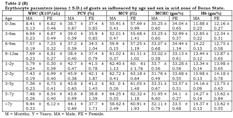
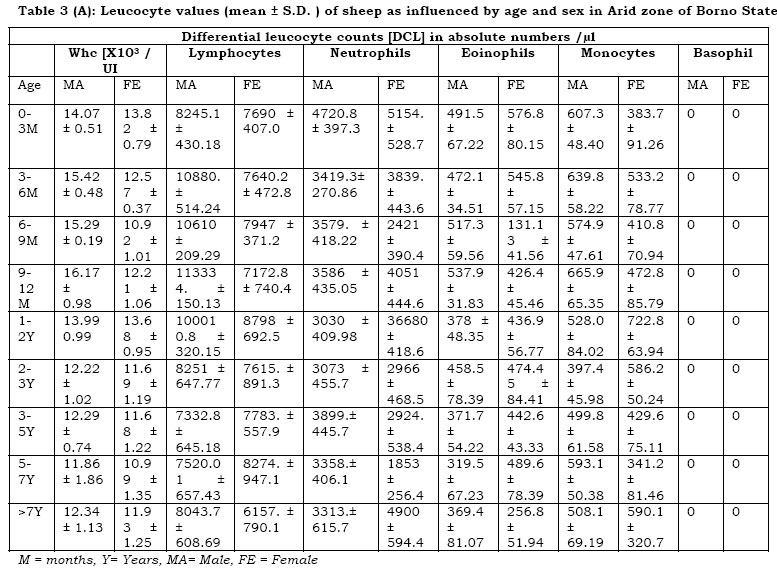
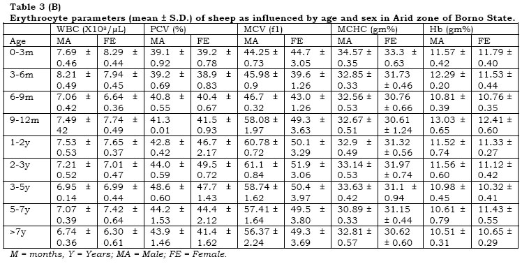
Received: July 1998 Code Number: md00031 SUMMARY The influence of age and sex on the heamatological values of goats and sheep studied in the arid zone of BornoState of Nigeria. Age and sex remarkable influence (P<0.05) on the red blood cell (RBC) counts of goats. Age influenced (P<0.05) the haemolglobin (HB) and the packed cell volume (PCV) values. Age and sex greatly influenced (P<0.01) the mean corpuscular volume (MCV) values. Mean corpuscular haemoglobin concentration (MCHC) was influenced by age. Lymphocytes constituted more than 60% of the total white blood cell (WBC) counts in male and female goats. Neutrophill and eosinophil counts were influenced by sex and age. Sex influenced (P<0.05) monocyte and lymphocyte values in goats. Sex and age influenced (P<0.05) the RBC values in sheep. PCV and MCHC values of sheep were influenced (P<0.05) by both age sex. The MCV was influenced (P<0.05) by age. Sex significantly influenced (P<0.05) the total WBC and monocyte counts. Keywords:- Goats, Sheep, Haematology, Age, Sex, Arid zone. RESUME L’ influence de l’age et du sexe sur les valeurs Hematologiques des chivres et des moutons etudie’s dans la region. Semi-aude de l’etat de Borno du Nigeria. L’age et le sexe influencent remarquablement (P<0.005) sur le taux de globules rouges des chevres. L’age influencait les valurs d’ Hemoglobine et d’Hematocrite. L’age et Le sexe influencaient grandement (P<0.01) les valeurs du volume corpusculaire moyenne des cellules. La concentration corpusculaire moyenne de l’Hemoglobine etait influencee par l’age. les lymphocytes constituaient plus de 60% du taux de globules blancs chez les chevres males et femelles. Les taux de neutrophiles et d’oesonophiles etaient influence’s par le sexe et l’age. Le sexe influencait (p<0.05) les valeurs en Lymphocytes et monocytes chez les chevres. Le sexe et L’ age influencaient (P<0.05) le taux de globules louges chez les moutons. L’es moutons. L’es valeurs d’ Hematocuite et La concentration corpusculaire moyenne d’Hemoglobine chez les moutons etaient influence’es (p<0.05) par le sexe et L’ age. Le volume corpusculaire moyen etait influence’ (P<0.05) par L’ age. Le sexe influencait significamment (p<0.05) les taux de monocytes et de globules blancs. mols cle: Chevres, moutons, Hematologique, age, sexe zone arude. Small ruminants (Sheep and goats) are one of the domestic animals of they inhabitants of Borno state and they provide meat, milk hides and skin. It has been reported that regardless of age, sex and climate, sheep and goats reared under traditional husbandry system have low haematological values compared to those reared under modern husbandry (Coles, 1980; Schalm et al, 1975). Low nutritional grassland pasture, stress, purturition and climatic factors greatly alter the blood values of goats and sheep (Anosa and Isoun, 1978, Radostits et al 1994). Blood is an important and reliable medium for assessing the health status of individual animals (Oduye, 1976). Much work has not been done on haematological values of goats and sheep in the arid zone of Borno States especially as it applies to sex and age. Therefore, this paper focused on the haematological values of apparently healthy sheep (Balami and Ouda breeds) and goats (Red Sokoto and Borno White breads) as influenced by age and sex in Borno State, of Nigeria. MATERIALS AND METHODS The goats and sheep used in this study were obtained from the University of Maiduguri animal farm and various quarters in Maiduguri metropolis especially Kasuan shanu area. The animals were apparently healthy. Faecal sample from each animal was collected and examined for the presence of helminth ova using floatation method (Basu, et al, 1994, Uqurhart et al, 1992). 5 mls of blood was collected from each animal from the external jugular vein following proper restraint by the owners or attendants and with minimal excitement. The blood was collected in ethylene diamine tetracetate (EDTA) vacutainer tubes and transported to the laboratory for analysis. The samples were analyzed within two hours collection. The blood samples were screened for the presence of haemoparasites using standard laboratory techniques (Schaim et al, 1975, Coles, 1980). The red blood cell (RBC), white blood cell (WBC), packed cell volume (PCV), haemoglobin concentration (Hb), differential leucocyte counts (DLC) mean corpuscular volume (MCV) and mean corpuscluar heamoglbin concentration (MCHC) were determined as described by Schaim et al (1975). All animals with lesions, gastrointestinal or haemoparasites were excluded from the study. The animals were grouped as shown in Table 1. RESULTS The haematological (mean ± SD) of the goats are presented in tables 2A and B. The RBC counts in the males range between .79 ± 0.24 to 9.44 ± 0.33. The RBC was high at birth (0-3 months) and decreased as the animal grew and later increased especially when the animal was above 7 years, particularly more so in the males. In the female, on the other hand, the value was low 0-6 months and increased between 6 to 9 months and later fluctuated with age. The value ranged from 6.12 ± 0.62 to 7.25 ± 0.32. Age and sex had significant influence (P<0.05) on the RBC values of goats. The Hb concentration increased with age in both sexes and later fluctuated. The highest value was recorded in both sexes and later fluctuated. The highest females at 2 to 3 years of age. Age had influence (P.05) but sex did not have significant influence on the HB contraction of goats. The PVC fluctuated at early age (0-12 month) and gradually increased with age up to 2 to 3 years in the females on the other hand the value fluctuated with age. Highest value was recorded in both sexes at 2 to 3 years of age. Sex had no significant influence but age did influence [ p<. 05] , the PVC values of goats. The MCV and MCHC values fluctuated with age in both sexes. Both age and sex showed significant (P< 0.05) influence on the MCV value of goats. Similarly, age had remarkable influence (p<. 05) on the value. The total WBS counts range was 10.45 ± 0.26 and 10.86 ± 0.35 to 14.36 ± 0.97 in males and females respectively. The highest value in males and females was observed at 3 to 5 years and 6 to 9 months respectively. In the males, the relative differential leucocyte counts (DLC) showed 62% lymphocyte, 24.8% neutrophil, 7.8% eosinophil and monocyte 5.1%, and 21.8% neutrophil, 7.8% eosinophil and monocyte 5.4%. In the females on the other hand, iit was eosinophil 7.1%, monocyte 5.1%, and 21.8% and 66.0% for neutrophils and lymphocytes respectively. Age showed significant (P < 0.05) influence on the WBC values of goats. The absolute lymphoocyte counts increased with age in the males. Similarly, absolute neutrophil counts followed the same trend and reached the peak at 1 to 2 years of age. Eosinophil and monocyte absolute counts showed gradual decrease with age to some extent and later fluctuated. There was sharp increase in the absolute lymphocyte counts in the females from 6 to 9 months and later the value fluctuated. Absolute neutrophil count fluctuated with age and the highest value was recorded at 3 to 5 years, while the lowest value was seen at birth (0-3 months). Eosinophil counts gradually decreased with age and the highest and lowest values were obtained at 0 to 3 months and 5 to 7 years respectively. On the other hand, absolute monocyte counts increase from 0 to 9 months and later decreased. The highest value was witnessed at 6 to 9 months while the lowest value was seen at 2 to 3 years. Sex had influence (P < 0.05) but age had no influence on the absolute neutrophil and eosinophil counts in goats. Age had no influence on monocyte counts but sex had (p < 0. 05). Sheep The heamatological values (mean ± SD) are presented in table 3A and 3B. In the males, the RBC counts fluctuated from 6 months to 7 years and the highest value was recorded at 3 to 6 months. The females showed highest value at 0 to 3 months of age and from 6 months and above. The value decreased remarkably (P < 0.05). The value at birth (0-3 months) in females is higher than the male value. Age and sex showed significant influence (P < 0.05) on the RBC counts of sheep. The Hb concentration increased from 0 to 6 months in males and later fluctuated. The highest value was witnessed at 9 to 12 months while the lowest value was seen as from 7 years and above. Females did not show consistency in the value and at 9 to 12 months of age, the highest value was recorded from 7 years and above. The Hb was significantly (p<0.01) influenced by age. The PCV in males showed gradual increase with age and this increase became significant at 6 to 12 months. The value reached the peak at 3 to 5 years of age and later, decreased gradually and fluctuated. Lowest value was seen at birth (0-3 months) similarly, the value increased with age in females up to 3 years of age. Highest value was seen at 2 to 3 years and the lowest value was obtained at 7 years and above. In the sheep, age and sex exhibited remarkable influence (P < 0.05) on the PCV value. The MCV value in males increased remarkably with age up to 2 to 3 years. The value in the females fluctuated. The highest value in both males and females was seen at 2 to 3 years of age. On the other hand, lowest values were observed in males and females from 0 to 3 months and 3 to 6 months respectively. In sheep, age had a significant influence (P < 0.05) on the MCV values. The MCHC values fluctuated with age in both sexes. Highest value was recorded from 0 to 3 months in both sexes. Both age and sex had remarkable influence (p < 0.005) on the MCHC value of sheep. The total WBS counts range in males and females is between 11.86 ± 1.86 to 16.17 ± 0.98 and 10.92 ± 1.01 to 13.82 ± 0.79 respectively. In the males, the highest value was obtained at 9 to 12 months of age while the lowest value was witnessed a 5 to 7 years of age. In the females on the other hand, the lowest and highest values were obtained at 6 to 9 months and 0 to 3 months of age respectively. Age showed no significant influence but sex had an influence (P < 0.05) on the total WBC counts. The relative DLC indicated in the males, 58.62% lymphocyte, neurtrophil 33.5%, eosinophil 3.8% and moncyte 4.3%. In the females, it was lymphocyte 55.0%, neutrophil 37.4%, eosinophil 4.7% and monocyte 4.3%. In the females, it was lymphocyte 55.0%, neutrophil 37.4%, eosinophil 4.7% and monocyte 2.9%. In the absolute DLC there was gradual increase with age in lymphocyte counts in males at early age (0 – 12 months) and as the animals grew in age the values fluctuated. Highest and lowest absolute lymophocyte counts were seen at 9 to 12 months and 3 to 5 years of age respectively. In the females, the highest count was seen when the animals were between the ages of 1 and 2 years. Lowest count was obtained when they were more than 7 years. Age showed no significant influence but sex had an influence (P<0.05) on the total WBC counts. The relative DLC indicated in the males, 58.62% lymphocyte, neutrophil 33.5%, eosinophill 3.8% and monocyte 4.3%. In the females, it was lymphocyte 55.0%, netrophil; 37.4%, eosinophil 4.7% and monocyte 2.9%. In the absolute DLC, there was gradual increase with age in lymphocyte counts in males at early age (0 –12 months) and as the animals grew in age the values fluctuated. Highest and lowest absolute lymphocytes counts were seen at 9 to 12 months and 3 to 5 years of age respectively. In the females, the highest count was seen when the animals were between the ages of 1 and 2 years. Lowest count was obtained when they were more than 7 years. Age had no influence (P < 0.05) but sex showed an influence (P < 0.05) on the lymphocyte counts. The absolute neutrophil counts in both sexes fluctuated with age. Highest value was recorded at birth (0 –3 months) in both sexes. Lowest value in males and females were obtained at 1 to 2 years and 5 to 7 years of age respectively. Both sex and age did not have influence (P <0.05) on the neutrophil value. Absolute eosinophil and monocyte counts fluctuated. In the males and females, the highest eosinophil count was obtained at 9 to 12 months and 0 to 3 months of age respectively, while the lowest count in the males and females was witnessed at 5 to 7 years and 6 to 9 months of age respectively. Both age and sex exhibited remarkable influence (P > 0.05) on the eosinophil counts. The monocyte count in males was highest at 9 to 12 months and lowest at 2 to 3 months. In the females on the other hand, the lowest and highest values were witnessed at 5 to 7 years and 1 to 2 years of age respectively. In sheep, sex had influence (P < 0.05) on the monocyte values but age did not. In both species of animals, the Hb content was very high (P <0.01) at 2 to 3 years. The Hb concentration of goats was higher than that of sheep (P < 0.05). The male values are generally higher (P < 0.05) than the female values. In WBC values, males recorded in both species. Goats showed lower PCV values than sheep (P < 0.05). Male animals generally exhibited higher (P < 0.05) PCV values than female animals. Goats showed higher MCV values (P <0.05) than sheep in both species, males recorded higher (P <0.05) MCV values than females. The MCHC value of goats was higher (P< 0.05) than that of sheep. Males exhibited higher lymphocyte counts than females in both species (P< 0.05). DISCUSSION The RBC, Hb, PCV, MCV, MCHC and WBC values obtained in this study in both sexes in goats and sheep were comparable to those previosly reported (Sarror and Schil, 1977; Anosa and Todd et al, 1952; Oduye, 1976; Holman; 1944a; Holman and Dew, 1965a; Schalm et al, 1975). In both species, high RBC counts was recorded in early life ( 0- 6 months) of the animals when compared to the adult values and this was in agreement with the observations of Holman and Dew (1964) and Schalm et al., (1975). The high RBC values in the young ruminants in this study may among other things be due to excitement or strenuous exercise during handling (Gartner et al., 1969). This leads to the release of adrenaline and hence spleen contracts and this causes the release of more RBC into circulation. It is only psychological tranquillisation that can reduce the splenic influence (Schalm et al., 1975) and this can only be achieved by holding the animal for 15-20 minutes by the owner or sympathetic attendant before sexes of goats and increased in the first few with the observations of other workers (Schalm et al., 1975), who reported that the Hb and PCV values decreased during the first week of life; then stablished with a slight downward trend, during the first few months and later increased as age increased. It might be pertinent to note that the difference in the present workers may be due to breed difference, management and environmental factors. The MCV and MCHC values in both species fluctuated and their values are dependent upon RBC, Hb and PCV values. Goats showed higher neutrophil values than sheep and in both species, males exhibited higher neutrophil counts than female (P <0.05). Goats had higher eosinophil counts than sheep (P <0.05). It was observed in this study that both sexes of both species of animals showed high number of WBC in the first few months of life, and this agrees with previous work (Schalm et al., 1975). Tropical environments are known to be havens of parasites and high WBC counts in the young ruminants may probably be due to environmental disposition. The white blood cells (WBC) are the soldiers of the body and their high counts may also be due to increase or complement the immune systems o the animals at the early stage of life which they may not obtain from the colostrum of the dam (Coles, 1980; Schalm et a;., 1975). It may also be attributed to physiological phenomenon i.e. excitement or strenuous exercise during handling (Coles, 1980). The lymphocytes constituted majority of the WBC counts and the cells increased with age in early life in both sexes of both species of animals. The high lymphocyte counts in the animals in this study are favoured by the findings of Milson et al., (1960) and Wilkins and Hodges (1962) and it might be attributed to stress and immune response to the environment (Cole, 1980) which harbours various detectable and undetectable parasitic and or bacterial organisms. The total mean WBC counts in male and female sheep in this study ranged between 11.8 ± 1.86 to 16.17 ± 0.98 and 10.92 ± 1.01 to 13.82 ± 0.79 respectively. The total WBC in sheep in this study in some ge groups are higher than the value (4,000 – 12,000) reported by Holman (1944b), who recorded 13,000 as slight leucocytes. Similarly, the total mean WBC counts range in male and female goats is 10.45 ± 0.19 to 15.91 ± 0.26 and 10.86 ± 0.35 to 14.36 ± 0.97 respectively. The range agrees with the one (6,000 – 16,000) reported by Schalm et al., (1975) but was lower than the value (19,180) reported by Holman and Dew (1965b) in individual animals by the 4th month. Since the animals are apparently healthy, any value beyond the upper limit in any of the sex in both species may be regarded as leucocytosis and any vale below the lower limit may be termed leucopaenia. In conclusion, age group and sex showed remarkable influence on the haematological values of small ruminants in the arid zone of BornoState. The values obtained are comparable to values recorded elsewhere. There was fluctuation in all the hematological parameters of both species of animals. What cause the fluctuation in various parameters may be undetected minor infections, weather extremities and poor management in this area, since the economic hardship is on the advanced stage and most of the farmers are very poor. They have to attend to themselves first ‘feeding wise’ before attending to their animals. ACKNOWLEDGEMENT The authors are grateful to Mr. Benson Okeh, Ismaila A. Gadaka for their technical assistance; Dr. I.O. Igbokwe for his useful criticism and University of Maiduguri farm manager for his cooperation. REFERENCES
© 2000 - Ibadan Biomedical Communications Group
The following images related to this document are available:Photo images[md00031t1.jpg] [md00031t3b.jpg] [md00031t2b.jpg] [md00031t3a.jpg] [md00031t2a.jpg] |
| |||||||||
