 |
| About Bioline | All Journals | Testimonials | Membership | News |
|
||||||
|
||||||
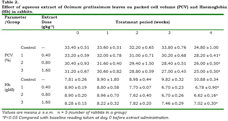
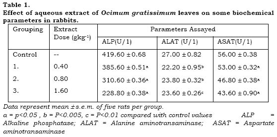
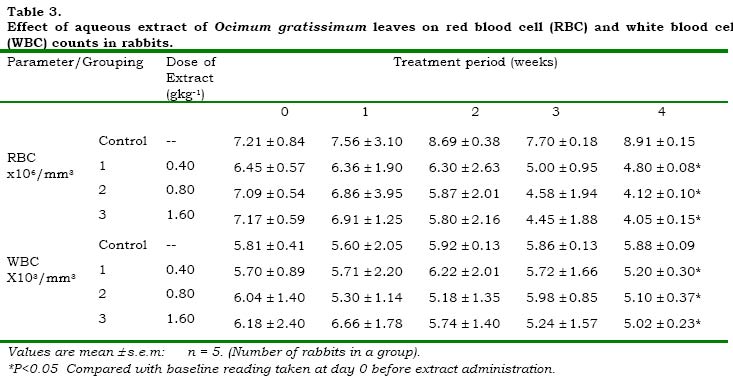
African Journal of Biomedical Research, Vol. 3, No. 3, 2000, pp. 175 -179 THE EFFECT OF AQUEOUS LEAF EXTRACT OF OCIMUM GRATISSIUM ON HAEMATOLOGICAL AND BIOCHEMICAL PARAMETERS IN RABBITS. **EFFRAIM, K.D.1., SALAMI, H.A2. AND OSEWA, T.S3. Departments Of Pharmacology1, Physiology2 and Biochemistry3, College of Medical Sciences, University of Maiduguri, Maiduguri.
Received: September 1999
Code Number: md00049 The effect of an aqueous leaf extract of Ocimum gratissimum (Linn), on haematological and biochemical indices in rabbits was studied. Increasing doses (0.4, 0.8 and 1.6 gkg-1 body weight) of the extract were administered orally to different groups of rabbits for a period of 4 weeks. Significant (P<0.05) decreases in the level of haemoglobin (Hb), packed cell volume (PCV), red blood cells (RBC) and white blood cells (WBC) were observed. The extract significantly (P<0.05, 0.005 and 0.01) reduced the activities of the liver enzymes, alanine aminotransamine (ALT), aspartate aminotransaminase (ASAT) and alkaline phosphatase (ALP). The decreases were dose dependent. There were clinical signs of loss of appetite and general malaise. Ocimum gratissimum (Linn) of the family Labiaceae is a herbaceous plant commonly found in tropical Asia especially India, where it is used for aromatic baths of fumigations in the treatment of rheumatism and paralysis (Uphotz, 1968). The plant is also found in West Africa. In Nigeria, it is found in the savannah and coastal areas. In the coastal areas of Nigeria, the plant is used in the treatment of epilepsy (personal communication), high fever (Oliver, 1960) and diarrhoea (Sofowora, 1995), whilst in the savannah areas, decoctions of the leaves are used to treat mental illness (Abdurahman, 1992). The leaves of the plant are used as a condiment in cooking. In the Southern part of Nigeria, the plant is called “effirin-nla” by the Yoruba speaking rtribe. It is called “Ahuji” by the Igbos while in the Northern part of Nigeria, the Hausas call it “Daidoya”. In view of its many uses, especially in Nigeria and the fact that traditional medicine practitioners prescribe and administer decoctions of the leaves to patients without regard to its possible adverse effects, the present investigation was undertaken to assess the effect of the crude leaf extract of this plant on haematological and biochemical parameters in rabbits. This is of important because there is no such information available in literature. MATERIALS AND METHODS Collection and Identification of Plant Materials: The fresh leaves (800g) of the shrub were collected from the University of Maiduguri campus. The leaves were identified and authenticated as Ocimum leaves, by Dr. S.S. Sanusi, a plant taxonomist in the department of Biology, Faculty of Science, University of Maiduguri. Experimental Animals. Male healthy rabbits bred in the department of Pharmacology were used for the study. Their average weight was 1.2kg. They were fed with groundnut leaves and tap water throughout the period of the experiment. Preparation of Extract. Fresh leaves of Ocimum gratissimum were kept in the oven at 800C for 10 min to stop any enzyme activity and then at 600C for 30 min. They were then air dried and ground into coarse powder. Fifty grammes (50g) of the powdered leaves was stirred into 450ml of boiling distilled water. Boiling was allowed to continue for 5 min. The mixture was kept aside, off the hot plate, for 30 min to allow it to infuse. It was then filtered through cheese cloth. The filtrate was concentrated to 200 ml (1ml of the extract being equivalent to 0.25g of the starting material). The extract was kept in a referigerator at 40C until used. Administration of the Extract Twenty male rabbits were randomly distributed into four groups of five. Group 1 served as the control and received distilled water. Groups 2, 3 and 4 received the aqueous extract of Ocimum gratissimum at doses of 0.4, 0.8 and 1.6 gkg-1 respectively. Animals in the control group received a quantity of distilled water equivalent to the dose in the 1.6 gkg-1 group. Administration of the extract was done orally, by means of a polythene cannula. Animals received their doses twice a week for four weeks. They were observed daily for clinical signs of toxicity or pharmacological signs, throughout the period of study. Collection of Blood Haematological Parameters: Blood samples were collected from the ear vein of the rabbits into heparinized sample bottles, using sterile surgical blade. Blood so collected was used for the various estimations. Sampling was done at four days interval. Biochemical Parameters.: At the end of the fourth week of extract administration, 2ml of blood was collected from each rabbit. They were allowed to clot and then centrifuged at 150g for 10min. Serum so obtained was used for the assay of alkaline phosphatase (ALP), alanine aminotransaminase (ALAT) and aspartate aminotransaminase (ASAT). Blood Analysis: The haematological examinations performed were according to standard methods. Haematocrit was determined by the micro-haemotacrit method described by McGown,et al., (1955). Erythrocytes and total leucocytes were counted using the improved Neubauer haemacytometer. The packed cell volume of each sample was determined by using a Hawksley microhaematocrit centrifuge at 1200 g for 5 min (Dacie and Lewis, 1984). Biochemical analysis of the serum enzymes for ALAT and ASAT was by the method of Reitman and Frankel, (1957). ALP was assayed according to the method of REC (1972). RESULTS Haematological Parameters The mean PCV and Hb values of the rabbits treated with the extracts of O. gratissimum are shown in table 2. The mean PCV values in the treated animals were significantly (p<0.05) reduced at the end of the treatment period as compared with the baseline measurement taken at day 0. The decrease was dose dependent. But all the untreated rabbits demonstrated an increase in the PCV values. The mean Hb also decreased significantly (p<0.05) as compared with the baseline reading (day 0). The rabbits in the control group that did not received any extract demonstrated progressive increase in Hb concentration. Thus showing that the extracts produced effects in the rabbits (Table 1). The mean RBC count decrease significantly (P,0.05) at the end of the treatment period as compared with the initial reading on day 0 (table 3). The percentage decrease were 25.3, 41.2 and 43.5 percent in groups 1,2 and 3 respectively. The total WBC count also decreased significantly (P<0.05) as compared with the baseline measurement at day 0 prior to extract administration. But with the untreated rats in the control group, the WBC count increased as compared with its baseline reading (Table 3). Biochemical Parameters The activities of the three most prominent maker enzymes, alkaline phosphatase (ALP), alanine aminotransaminase, (ALAT) and aspartate aminotransaminase, (ASAT) were markedly affected after pre-treatment of the animals with extract doses of 0.4, 0.8 and 1.6 gkg-1 body weight. The activity of alkaline phosphatase significantly (P<0.05) decreased, at a dose of 0.4 gkg-1 with activity level of 385.60 ± 0.51 u/l as compared with a control value of 419.60 ± 0.68 u/l. As the dose was increased to 0.8 gkg-1 the activity further decreased (310.60 ± 0.36 u/l). At 1.6 gkg-1 body weight of the extract, the activity of ALP significantly (P<0.05) decreased to 228.80 ± 0.38 u/l as compared to the control value (table 1). Alanine transaminase activity decreased in the rats pretreated with various doses of the extract. At a dose of 0.4 gkg-1 the activity was significantly (P0.005) reduced to 22.2 ± 0.95 u/l as compared with a control value of 27.0 ±0.82 u/l. The activity further and significantly reduced to 23.8 ± 0.32 u/l (P<0.005) when the dose was increased to 0.8 gkg-1. A significant (P<0.01) decrease (23.60 ± 0.26 u/l) was observed when the dose was further increased to 1.6 gkg-1 (table 1). There was a significant (P<0.05) decrease in the activities (53.0 ± 0.32, 46.8 ± 0.38 and 43.60 ± 0.90 u/l) of aspartate amintransaminase in all the rats pretreated with various doses (0.4, 0.8 and 1.6 gkg-1 respectively) of the extract as compared with control value. The effect of the extract was observed to be dose dependent. DISCUSSION The results of the study show that, the leaf extract of O. gratissimum administered at the dosages used and for the duration of the experiment appear to suppress the haemopoietic system. There was reduction in the haematological parameters, in the treated animals, as compared with the control and also with the baseline measurement taken before extract treatment. The reduction may have occurred due to lysis of blood cells and probably suppression of blood cell synthesis by saponins found in the leaf extract (Irvine, 1961). Saponins are known to be toxic to body systems (Watt and Breyer-Brandwijk, 1962). The effect of saponins may have contributed to the clinical signs of loss of appetite (as deduced by reduction in feed intake) and loss of weight observed in all rabbits administered with the extract. The extract in the dosage range used also reduced/suppressed the activity of the liver enzymes in the treated animals compared with the controls. The effect was dose dependent. Liver enzymes (ALAT and ASAT) are liberated into the blood whenever liver cells are damaged and enzyme activity in the plasma is increased (Edwards, et al., 1995). The fact that the enzyme activity was reduced indicated that the extract did not have necrotic effect on the liver. The extract was found to contain flavonoids. Flavonoids are reported to exhibit antioxidant activity (Ramanathan, et al., 1989) and are effective scavengers of superoxide anions (Robak and Gryglewski, 1988). The extract may have exhibited hepatoprotective activity due to its antioxidant property attributable to flavonoids. Interestingly, saponins especially terpene glycosides are reported to enhance natural resistance and recuperative powers of the body (Singh, et al. 1991). Saponins were found in the extract. Ir was not possible to identify the type of saponins present. But there is the possibility that terpene glycosides might have contributed to the lowering of the levels of enzyme activity, despite the fact the blood they affected parameters. In conclusion, inspite of the popularity of the plant as a condiment and herbal medicine, the extract has been observed to suppress the haemopoietic system. On the other hand, it has possible hepotoprotective activity in rabbits because of the presence of flavonoids and saponins (terpene glycosides). It is therefore suggested that chronic usage of the leaf is not advisable. Further researches on phytochemical analysis and identification of the components responsible for the suppression of the blood parameters and enzymes activity is yet needed. ACKNOWLEDGEMENT The authors gratefully acknowledge the technical assistance of Mr. Justus Jibrin, and the secretarial work of Mrs. Gladys Oguocha. REFERENCES
© 2000 - Ibadan Biomedical Communications Group
|
