 |
| About Bioline | All Journals | Testimonials | Membership | News |
|
||||||
|
||||||
African Journal of Biomedical Research, Vol. 4, No. 3, Sept, 2001, pp. 143- 145 Original article LIPOLYTIC EFFECT OF CALOTROPIS PROCERA IN THE SKIN OF WISTAR RATS. AKINLOYE A.K1*, ABATAN M.O2, ONWUKA S.K1, ALAKA O.O3 & OKE B.O1 Departments of Veterinary Anatomy,
Veterinary Physiology/Pharmacology znd Veterinary pathology, University of
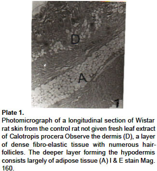
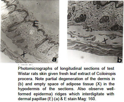
Ibadan. Ibadan, Nigeria Code Number: md01069 Histopathological studies of skin samples from Wistar rats treated once daily for four weeks with 20mg/gm body weight of the fresh leaf extract of Calotropis procera by the oral route showed significant degeneration of adipose tissue in the hypodermis and partial degeneration of the dermis at the site of merging with the hypodermis. No significant histological changes were, however, observed in the stratified squamous epithelium of the epidermis. Hair follicles and sebaceous glands were not affected. It was concluded from these observations that the subcutaneous layer was the one mainly affected by the treatment with the leaf extract. Keywords: Calotropis procera, lipolysis, skin, rat INTRODUCTION Calotropis procera is of the family Asclepiadaceae. It is perennial shrubby treelet with thick cottony tomentose leaves when young and frequently glabrescent when fully developed (Huber, 1985). It is distributed widely in the tropics, especially in the and waste places (Hussein et. al., 1994). It is a common plant in Nigeria but it is more abundant in the northern part of the country (Sofowora, 1984). Jam et al (1996) reported that Calotropis procera was used in traditional medicine as a purgative, anthelmintic, anticoagulant, anticancer as well as. tntipyretic, analgesic arid antimicrobial. Fleurentin and Pelt (1982) also observed that -;-he plant was used as an antiseptic for skin infection. Several studies have been carried out on the effects of various extracts of Calotropis procera on different organs of animals (Al-Robai et al, 1993a, 1993b; Jam et al, 1996; Basu et al, 1997). On the contrar), there is little or no information on the effect of the plant extract on the skin. Anatomically, the skin is the largest organ in the body. It is also a kind of biological window, which not only "mirrors" the internal condition of the animal but serves, in addition, as an inter-face between the animal and the external environment (Onwuka et al 1998). The series of studies of which this is an initial report was designed to determine the effect of fresh leaf extract of Calotropis procera on the histology of Wistar rats skin. MATERIALS AND METHODS Animals 20 white albino Wistar rats weighing between 100- I 50gm were used. They were fed ad libitum on rat cubes (Ladokun and Sons Livestock Feeds, Nigeria Limited) and allowed free access to fresh clean water in their rat cages. IO rats served as test animals while-the remaining 10 rats served as control animals. Preparation of extract Fresh leaves of Calotropis procera plant were collected after identification at the Department of Botany and Microbiology, University of lbadan, lbadan. An extract was prepared everyday by macerating 20gm of the fresh leaves with 10ml of distill water and later squeezed and filtered. The filtrate served as the stock solution. Administration of the extract The stock solution was administered orally at a dose rate of 2mg/gm body weight once daily for 30 days using an oral cannula. The control animals received only distilled water for the same number of days. Histopathology Skin samples were collected from different sites on the animals (viz: back, trunk and neck) immediately after exsanguination. The samples were histologically prepared as earlier described by Ahmed and Onwuka (1996). RESULTS No gross lesion was observed in all the skin samples collected. Fig. 1 is a photomicrograph of a longitudinal section of rat skin from a control rat. It is typical of the observations made on samples from the control animals. The epidermis and dermis showed normal inter-digitation of epidermal ridges and dermal papillae. The dermis was observed to merge with the loose areolar connective tissue of the hypodermis which consisted largely of adipose tissue (A). Fig.2a &b show sections from the test animals. As seen in photomicrograph (a), there was an empty space where the subcutaneous adipose tissue had been in the controls. Additionally, there was partial degeneration of the dermis (fig. 2b) at the site of merging with the hypodermis. However, the layers of the epidermis were normal. Well-formed epidermal ridges which interdigitated with dermal papillae as well as hair follicles and sebaceous glands were observed. DISCUSSION Although the literature is replete with information on the numerous medicinal and economic importance of Calotropi sprocera (Hilal and Youngken, 1983; Nsekuye, 1994; Basu et al, 1997), there is, however, paucity of basic data on the effect of the plant on the skin. The results of this study showed that fresh leaf extract of Calotropis procera had no observable histopathological effect on the epidermal and dermal layers of the skin apart from partial degeneration of the dermis at the site where it merged with the hypodermis. However, significant histological changes were observed in the hypodermis where there was degeneration of adipose tissue. The reason for this observation may however not be unconnected with the effect of the cardiac glycoside chemical components of Calotropis procera which was reported to cause cytolysis in kidney tubules of Wistar rats (Al-Robai et al, 1993a) The observation of lipolysis of adipose tissue in the hypodermis is medical significance since prolonged exposure of livestock to the leaves of Calotropis procera may lead to threatening loss of fat in the body and thus weight loss. Secondly, baring other histopathological effects of Calotropis procera, this finding could be of importance in reducing fat levels in both obese animals and humans. For livestock farmers who engage in fattening of their animals for maximum profit, chronic ingestion of this plant by the animals may have negative economic impact However, in man where obesity predisposes to cardiovascular disease, diabetes and shortening of life (Keele and Neil, 1978), the plant might be desirable. Fleurentin and Pelt (1982) reported the antiseptic effect on the plant extract on the skin. The observed lipolytic effect on the subcutaneous tissue may be the histological basis for that. Further studies need to be undertaken to identify the chemical components of the plant which are responsible for these observations and determine their safety margins REFERENCES
The following images related to this document are available:Photo images[md01069f1.jpg] [md01069f2.jpg] |
| |||||||||
