 |
| About Bioline | All Journals | Testimonials | Membership | News |
|
||||||
|
||||||
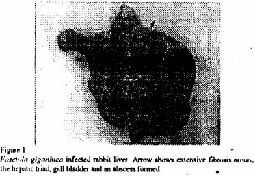
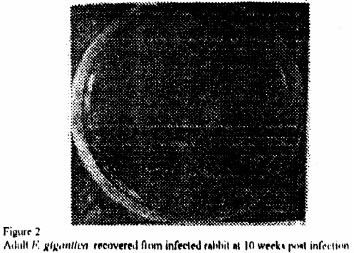
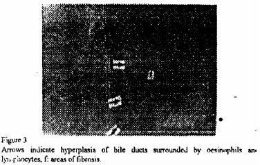
African Journal of Biomedical Research, Vol. 4, No. 3, Sept, 2001, pp. 161-163 Short communications THE ASSESSMENT OF FASCIOLA GIGANTICA INFECTION IN THE RABBIT (ORYCTOLAGUS CUNICULUS) AS A LABORATORY MODEL PARASITE DEVELOPMENT - CLINICAL SYMPTOMS AND LIVER PATHOLOGY ADEDOKUN, O. A and FAGBEMI, B. 0. Department of Veterinary
Microbiology and Parasitology, University of Ibadan, Nigeria Code Number: md01074 In this study, the rabbit was assessed as a laboratory host for the helminthes parasite, Fasciola gigantica. Three groups of rabbits were Infected experimentally with 5, 10 end 15 metacercariae of F. gigantica respectively. Clinical signs found included pale mucous membrane, progressive emaciation and rough hair coat. The post mortem findings Included haemorrhage and scattered calcified nodules on the omentum. Echymosis. fibrosis and necrosis were prominent on (he Ever in addition to fibrosis and thickening of the gall bladder and bife ducts. Histopathology of the Ever revealed congestion. haemorrhage, ceffular Infiltration end necrosis, BE. ducts were hyper plastic and fibrotic with sever Inflammatory reactions. The ova of gigantica were recovered from the faeces of some infected rabbits at 10 weeks post infection. Mean number of flukes recovered from the rabbits was 0 in the 5 metacercariae group, 2 in the 10 metacercariae group and 5 in the group given 15 metacercariae. Keywords: Fasciola gigantica , fascioliasis, rabbit, laboratory model. Fascioliasis in rabbits INTRODUCTION Fasciouasis, the disease caused by Fasciola iganrica and Fasciola hepatica constitutes one of the most Important helminthic diseases of livestock, especially the natural ruminant hosts. The disease is of great economic important due to the mortality which occurs in acute cases and the morbidity encountered in chronic cases Graber, 1971 and Cawdery, 1978. The recognized importance of fasclolosis has stimulated continued research and the veterinary literature is almost replete with studies on various aspects of the disease. F. hepatica, the temperate species is the better known of the two. It has been extensively studied in the natural ruminant hosts and various laboratory models su as rabbit, guinea pig, and rats have been developed for this trematode infection. However this is not true for F. gigantica which is the much more important species in tropical Africa and Asia. Previous studies in laboratory hosts seem to reveal that laboratory animals are susceptible to F. hepitica but refractory to F. gigantic, Gerber et a!., 1974; Strivasta and Singh, 1974. Furthermore, it was observed that when the development of F. gigantica occurred in laboratory animals’ growth did not progress to sexual maturity, Ogunrinade, 1978. - The availability o{ cheap and easy-to handle, laboratory hosts facilitates rapid advances in the understanding of the biology and immunology of parasitic infections especially at the cellular and molecular level. For Instance an inventory of the immunology of schistosomiasis in the last two decades re” the great Importance of laboratory models of Infection UNDO/World Bank/WHO 1987. In view of this fact, this study was designed to re-assess the rabbit as a laboratory host for F. gigantica infection.. MATERIALS AND METHODS Experimental Animals: The experimental animals consisted of seventeen 9-week — old rabbits divided into four groups of 4, 5, 6 and 2 animals each. They were treated with anthelmintic (levainisole) and stabilized over a period of 8 weeks. The first group were infected with 10 and 15 metacercariae respectively. The last group of 2 rabbits served as uninfected control. The parasite materials consist of metacercariae obtained from Lymnaeea naf&ensis 33 days after infection with F. gigantica miracidia. Faecal examination was carried out using the sedimentation method ofThlenpont eta!. 1979. Histopathology was carried out on tissue samples obtained from the liver of the rabbits and preserved in 10% formnlin, RESULTS Clinical Symptoms: No clinical signs were observed during the first 6 weeks after mi Paleness of the mucous membranes was noticed by the seventh week and this progressed with the duration of the infection. Anorexia, dullness, progressive emaciation and rough hair coat were observed by the eighth week. The first mortality was recorded at 10 weeks in the group infected with 15 metacercariae. The last surviving rabbits were slaughtered 16 weeks in the group infected with 15 metacercariae. The last surviving rabbits were slaughtered 16 weeks after infection. Post Mortem Findings: The post mortem findings included haemorrhage and scattered calcified nodules on the omentum, liver, diaphragm and stomach walls. Severe adhesions between the organs occurred in few cases. Bile ducts and gall bladder were fibrotic and thickened. Echymosis, necrosis and severe fibrosis were observed on the liver. Abscesses were found on the organ in two cases (Plate 1). The adult and immature flukes were recovered from the bile duct and gall bladder at post mortem. (Plate 2). Histopathology revealed hepatic congestion heemorrhage, lymphocytic infiltration, pseudolóbulation and necrosis f liver cells were observed as prominent features. Bile duct hyperplasla with severe inflammatory reactions characterised by zones of necrosis surrounded by infiltration of oesinophils and lymphocytes with extensive fibrosis also occurred in the liver. (Plate 3). The ova of gigantica were recovered from the faeces of infected rabbits as from 10 weeks after infection (Fig 4). Number of flukes recovered at post mortem ranged from 0 to 1 in the 5 metacercanal group, 0 to 4 in those infected with 10 metacercariae and 2 to 8 in the group infected with 15 metacercariae. DISCUSSION in this study rabbits were infected experimentally with F. gigantica in order to assess the parasite development, clinical symptoms and liver pathology with the aim of developing these animals Into a suitable model as laboratory host for fasciollasis. The gross and histo-pathological findings In the Infected rabbits especially as these related to the degree of liver destruction were found to be directly related to the number of flukes recovered at post mortem. The works of Batakana et aL, 1976, Lizuokwu and Ikerne, 1978 and Schilihorn Van Veen, 1980, corroborates this. The result of this study though will seem to negate the findings of some previous author such as Gether et a!., 1974, Ogunrlnade, 1978 who reported rabbit as being refractory to F. gigantica infection. However it was ouserved that the greater the number of surviving juvenile flukes that reaches the liver (in excess of 3), the lower their growth rate and in some, failure to attain sexual maturity was observed, This is probably -as a result of competition for nutrient and space — which in the light of size of the rabbit liver may nol permit development to adult stage Rabbit liver may be up to 11cm at its widest point and adult flukes recovered measured between 30mm — 40mm in length and 6— 7mm in breadth. In the natural ruminant hosts this may reach up to 25— 75mm by 12mm. The main features o6served in rabbits which Included anorexia, dullness, weakness, pale mucous membrane, are similar to the findings of Batakana et a 1976 in the natural ruminant host. However Berry and Dargie, 1977 concluded that symptoms in any host depends on the dose and viability of metacercariae, type of host, age, plane of nutrition and absence or presence of inter-current infections. These factors could be further examined in the rabbit. The death recordød at 10 weeks in the 15 metacercariae group of rabbits may not be strange as Bitakaramino and Bwangamoi, 1969 also recorded death at 12 weeks In the cattle infected with F, gigantica metacercariae in execs of 1000. REFERENCES
© 2001 - Ibadan Biomedical Communications Group
The following images related to this document are available:Photo images[md01074f3.jpg] [md01074f2.jpg] [md01074f1.jpg] |
| |||||||||
