 |
| About Bioline | All Journals | Testimonials | Membership | News |
|
||||||
|
||||||
African Journal of Biomedical Research, Vol. 5, No. 1-2, Jan & May, 2002, pp. 57-61 HISTOMORPHOMETRIC AND HISTOPATHOLOGICAL STUDIES ON THE EFFECTOF CALOTROPIS PROCERA (GIANT MILKWEED) ON THE MALE REPRODUCTIVE ORGANS OF WISTAR RATS AKINLOYE, A. K1* ABATAN, M.O2 ALAKA O. O AND 'OKE, B. O1 Departments of 1Veterinary
Anatomy, 2Veterinary
Physiology/ Pharmacology and 3Veterinary Pathology, University of
Ibadan. Ibadan, Nigeria. Received: February,
2001 Code Number: md02011 Histomorphometric and histopathological evaluations of the effects of fresh leaf extract of Calotropis procera on the reproductive organs of male wistar rats given 20mg\gm body weight of the extract once daily, orally, for varying number of days showed varying degrees of desquamation of seminiferous epithelial cells, degeneration of seminiferous tubules and presence of large-sized multinucleated cells as well as significant reduction (P < 0.05) in the seminiferous tubular diameter. The epididymis of the test rats showed cell debris, numerous immature round cells and pinkish homogenous material in the lumens while the epithelia appeared normal. There was a general reduction in the mean ductular and luminal diameter while fluctuating changes were observed in the epithelial height of the epididymis of treated rats. The accessory glands of test rats showed pinkish homogenous fluid as well as inflammatory cells in the lumen and glandular degeneration of the seminal vesicles. The result from this study revealed that Calotropis procera has a potentially deleterious effect on the testes and accessory sex org an s. Key words: Calotropis procera, testes, epididymis, accessory sex glands histomorphometric INTRODUCTION Calotropis procera (giant milkweed) is a perennial, greyish-green, woody shrub with broad obovate fleshy leaves that grows wild in the tropics and in warm temperate regions (Huber, 1985; Hussein et al, 1994). The plant is found in almost all parts of Nigeria but more abundant in the northern part of the country (Sofowora, 1984). Jain et al (1996) reported that Calotropis procera was used in traditional medicine as a purgative, anthelmintic, anticancer as well as to treat leucoderma, ulcers, piles and disease of the spleen. Saha et a/ (1961) described the plant to be an abortifacient while Malhi and Trivedi (1972) observed it to be an antifertility agent. Hilal and Youngken (1983) observed that calotropis procera has uterine stimulating effect while Prakash et al (1978) showed that it has embryotoxic effects. Edman (1983), Al-Robai et al (1993a) and Hussein et al (1994) reported the presence of alkaloids, flavonoids, cardiac glycosides as well as sterols and uscharin in the entire part of the plant, Calotropis procera. Studies on the effects of the plant extract on the ultrastructure of kidney (AlRobai et al, 1993a), cardiac muscle and blood serum composition (Al- Robai, et al, 1993b) have been carried out. This study was carried out to provide an insight into its activities on the histology of the testis, epididymis, and the accessory sex glands. MATERIALS AND METHODS Animals, Grouping and Experimental Design: Fifty (3 weeks old) sexually mature male albino Wistar rats, bred and maintained at the Experimental Animal unit, Faculty of Veterinary Medicine, University of lbadan, were used for this study. There were 5 groups (of 10 rats each) designated as GA, GB, GC, GD and GE; 5 rats served as control and 5 rats served as test animals. Preparation of Calotropis procera extract: Fresh leaves of Calotropis procera plant were collected within the campus of the University of lbadan and were identified at the Department of Botany and Microbiology, University of lbadan, lbadan. A leaf extract of Calotropis procera was prepared everyday by macerating 20gm of the fresh leaves with 10ml of distilled water and later squeezed and filtered. The filtrate served as the stock solution. Administration of Calotropis procera: The stock solution was administered orally at a dosage of 2mg/gm body weight once daily using a 5ml oral cannula for 7 days, 14 days, 21datys, 28days and 35 days to the experimental animals in groups GA, GB, GC, GD and GE respectively. The control animals in each group received only distilled water for the same number of days. Sample collection: The animals were dissected and the testes, epididymis, seminal vesicles and prostate glands were collected as described by Oke (1988) immediately after exsanguination. Histological and Histopathological procedures: The samples collected were fixed in Bouin's fluid and processed by the usual method for paraffin embedment and stained with Hematoxylin and Eosin (H & E) as described by Akinloye et al (2000). The slides of testes, epididymis, seminal vesicle and prosrate gland were evaluated for pathological changes under light microscope. Histomorphometry: The slides were examined under the microscope and the following measurements were taken; seminiferous tubular diameter, epididymal tubular diameter, epididymal luminal diameter and epididymal epithelial height. For each parameter, ten measurements were made per section using a calibrated eye-piece micrometer (Graticules Ltd. Toubridge Kent). The means of the measurements of parameter in each section were recorded for each animal. Statistical Analysis: All data obtained were expressed as means with the standard errors. The data were subjected to the pooled variance "t" test for comparison and Duncan multiple range test as described by Steel and Torrie (1986). RESULTS Histomorphometry: Table 1 shows the mean seminiferous tubular diameter of the testis and percentage change in the mean seminiferous tubular diameter of control and Calotropis procera treated rats. The result shows general reduction in the mean seminiferous tubular diameter in the test rats in comparison to the control. However, significant reduction (P<0.05) of 49.9% and 31.20% were observed in groups GD and GE respectively. Table 2 shows that reduction observed in seminiferous tubular diameter in the test animals in group GA was significantly different (P < 0.05) from reduction observed in group GB whereas there was no significant difference in reduction observed in seminiferous tubular diameter in groups GB and GC. Similarly, reductions observed in diameter of seminiferous tubules in groups GD and GE were not significantly different (P < 0.05). Table 1 Mean seminiferous tubular diameter (microns) and percentage change in testis of control and Calotropis procerafresh leaf extract treated rats in groups GA-GE.
STD= Seminiferous tubular diameter, *P<0.05; Table 2 Means of groups for seminiferous tubular diameter of testes for rats in groups GA- GE (in microns).
STD= Seminiferous tubular diameter Means with same superscripts within the row are not significantly different at P < 0.05 (Duncan Multiple Range Test) Table 3 shows mean ductular diameter, luminal diameter and epithelial height of epididymis of control and treated rats. The result shows general reduction in the mean ductular and luminal diameters of epididymis of test rats in comparison to the control group. Table 3 Mean ductular diameter, luminal diameter and epithelial height of epididy-mis of control and Calotropis procera fresh leaf extract treated rats in groups GA-GE (in microns).
*P < 0.05; DD=Ductular diameter, LD=Luminal diameter, EH=Epithelial height. Table 4 Percentage change in the mean ductular diameter, luminal diameter and epithelial height of epididymis of Calotrops procera fresh leaf extract treated rats in groups GA-GE
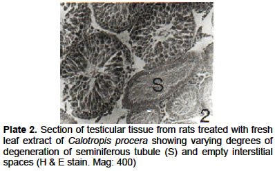
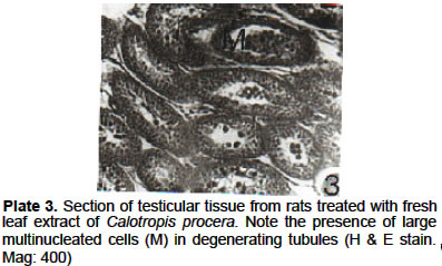
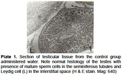
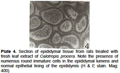
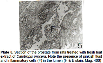
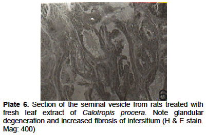
Significant reductions (P < 0.05) of 31.07% and 50.55% were observed in ductular diameter in groups GC and GD respectively while significant reduction of 36.07% and 68.35% were recorded in groups GC and GD respectively for luminal diameter of the epididymis (Table 4). Though not statistically significant, mean epithelial height of epididymis of treated rats were observed to be 2.05%, 3.64%, 15.18% and 1.55% higher than the control rats in groups GA, GC, GD and GE respectively. Only treated rats in groups GB had a lower mean epithelial height of 12.42% of the epididymis when compared to the control rats. Histopatholoqy: Rats treated with fresh leaf extract of Calotropis procera showed varying degrees of degeneration of seminiferous epithelium as well as presence of large-sized multinucleated cells in the tubules and empty interstitial spaces (Plates 2 and 3) when compared with testicular tissue from the control rats (Plate 1). Sections of epididymal tissue from the treated rats showed presence of cell debris and numerous immature round cells in the epididymal lumens as well as normal epithelial lining of the epididymis (Plate 4). Fresh leaf Calotropis procera extract treated rats showed pinkish fluid and inflammatory cells in the lumen of the prostate gland (Plate 5) while glandular degeneration and increase fibrosis of interstitium were noticed in the seminal vesicles (Plate. 6). DISCUSSION Calotropis procera (giant milkweed) has been reported to have numerous medicinal arid economic importance (Hussein et al, 1994; Jain.et al, 1996; Khurma et al, 1997) but was observed to be potentially injurious to the body especially after prolonged or chronic use (Hilal and Yungen, 1983; Sofowora, 1984; Nsekuye, 1994). In this study, apart from the group given the extract for 7 days that had no significant testicular lesions, histopathological observations showed that all other groups had varying degrees of testicular lesions which were more severe in the last two groups that received the extract for longer periods. In addition significant reductions were observed in the seminiferous tubular diameter of the testes of treated rats in comparison to the control rats. The pattern of cellular damage observed in this study is consistent with the effects of phoxim (Atef et al, 1995), oestradiol valerate (Kohter-samouilidis et al, 1998) and Curcuma comosa extract (Piyacchaturawat et al, 1998) on the male reproductive organs. The histological changes observed in the testis of the treated rats in this study may be due to the of cardiac glycosides found in the latex extract of Calotropis procera which was incriminated to be responsible for pathological and ultrastructural changes in the kidney tubules of Wistar rats (Al-Robai et al, 1997). As the interstitium was observed to be devoid of Leydig cells in most cases, the histological changes observed may also be due to decreased production of testosterone known to be responsible for normal testicular architecture (Eik-Nes, 1970). The general reduction observed in this study in the mean ductular and luminal diameters of the epididymis as well as the apparent increase in the mean epithelial heights of the epididymis of the treated rats in comparison to the control suggest that the luminal epididymal volume would be lower in the treated rats than in the control rats. Although the effect of the extract of Calotropis procera on the sperm volume was not examined in this study, it may be inferred from this work that Calotropis procera extract may affect sperm volume negatively. These observations may partially explain why Calotropis procera is reportedly being used as an anti-fertility agent (Malhi and Trivedi, 1972). Histo-pathological examinations of the epididymis showed desquamated immature and degenerated germinal cells in epididymal lumen, and presence of unidentified cell debris in the lumen of the epididymis of test rats. Despite these observations in the epididymal lumen, epithelial height and pseudostrafied features of the epithelium were relatively normal. The presence of the various debris in the epididymal lumen is probably a reflection of degenerate testicular lesion observed in the treated rats which was passed to the epididymis via the ductuli efferentes and probably not as a result of the effect of Calotropis procera extract on the epididymis itself. Therefore, it may be suggested from this study that Calotropis procera extract has destructive effect on the germ cells which are actively dividing. This observation may also proffer partial explanation for the reason why Calotropis procera was reported to be used in the treatment of tumours (Jain et al, 1996). The plausible reason for the observation in accessory sex gland is probably the extension of the effect of destructive activity of the extract on the testicular tissue that has probably affected testosterone production. If decreased testosterone production holds true to be responsible for these observations on the accessory sex glands, then this study lends credence to earlier findings that male accessory sex glands are highly dependent on the male sex hormone for development and secretory activity (Mann, 1964) Acknowledgement The authors wish to acknowledge the senate research grant No. SRG/FVM/94-95/13A of the University of Ibadan which was used to carry out the greater part of this work. REFERENCES
©
2002 - Ibadan Biomedical Communications Group
The following images related to this document are available:Photo images[md02011p1.jpg] [md02011p4.jpg] [md02011p6.jpg] [md02011p5.jpg] [md02011p2.jpg] [md02011p3.jpg] | |||||||||||||||||||||||||||||||||||||||||||||||||||||||||||||||||||||||||||||||||||||||||||||||||||||||||||||||||||||||||
| |||||||||
