 |
| About Bioline | All Journals | Testimonials | Membership | News |
|
||||||
|
||||||
African Journal of Biomedical Research, Vol. 5, No. 1-2, Jan & May, 2002, pp. 93-95 Case Report ASCARIDOSIS IN CAPTIVE RETICULATED PYTHON (PYTHON RETICULATUS) V.O. TAIWO1, O.O. ALAKA1, N.A. SADIQ2 and J.O. ADEJINMI2 Departments of 1Veterinary
Pathology and 2Veterinary Microbiology & Parasitology University
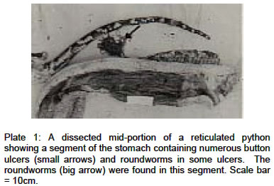
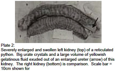
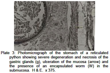
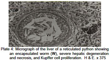
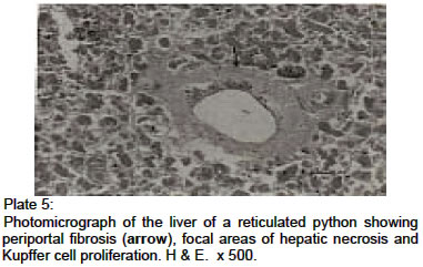
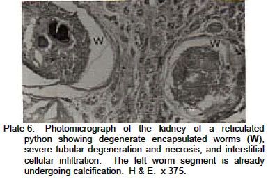
of Ibadan, Ibadan, Nigeria. Received: March 2001 Code Number: md02020 Two captive reticulated pythons, Python reticulatus, in the Zoological Gardens, University of Ibadan, Ibadan, Nigeria died and were submitted for necropsy at the Diagnostic Laboratory of the Department of Veterinary Pathology, University of Ibadan. Both pythons had been infected with Ascaridia galli for a long period of time during which there was no record of anthelminthic medication to the pythons. Organ lesions associated with these worms included severe ulcerative gastroenteritis, necrotic hepatitis and nephritis. It is advised that chickens meant for feeding captive-reared pythons or other wildlife must be certified worm-free to avoid such economic and wildlife resource loss in future. Key words: Python, chicken, Ascaridia galli, worm infestation, wildlife preservation. INTRODUCTIONThe management, propagation and preservation of wildlife are very important, not only to the maintenance of the world’s floral and faunal biodiversity for future generations, but to encourage and sustain tourism and recreation. Game parks and zoological gardens or animal orphanages offer formidable sources of recreation and revenue generation for nations that have them; Notable examples are found in Eastern and Southern Africa. The need to maintain the health of these animals should be of utmost importance to their keepers. We report on two pythons that died from Ascaridia galli infection and the pathology associated with this parasite. CLINICAL HISTORY AND NECROPSY REPORT The two reticulated pythons, Python reticulatus (Family Boidae; Superfamily Phythominae, one >20 years old and the other 7 years old, were kept in captivity in the same pen at the Zoological Gardens, University of Ibadan, Ibadan, Nigeria. The younger python was brought into the zoo when it was about one year old. The older python had previously undergone a surgical operation for the removal of papillomatous growths on the oral mucosa in 1983. Both pythons had been maintained on two or three live or dead chickens once every 3 days. The pythons died 1 year apart after several days of exhibiting dullness, anorexia and lethargy. There was no record of anthelminthic therapy for the pythons in the last 5 years. Both pythons were presented fresh for necropsy. The older python was in an advanced stage of molting when presented and had numerous firm papillomatous outgrowths on the mucosa of the left hard palate, Both carcasses had variable amounts of thick, light greenish mucopurulent exudate in their bronchi and both anterior and posterior airsacs. A total of 269 roundworms, identified by routine methods (Soulsby, 1968; Long 1979) as Ascaridia galli Schrank, consisting of 99 smaller and thinner (larvae) and 170 large and robust (adult) forms were found along the entire length of the digestive tract of both pythons. Both specimens have been deposited at the Natural History Museum, Obafemi Awolowo University, Ile-Ife, Nigeria with Accession No. UNIFEM IV 961 (larvae) and Accession No. UNIFEM IV 962 (adults). A segment of the digestive tract of the older python, measuring about 45cm in length, from the distal end of the stomach and the anterior portion of the duodenum was markedly thickened and had rough, leathery and severely haemorrhagic mucosa. Within this segment was found a large number of raised button ulcers, between 5 to 15mm in diameter. Some of these ulcers contained embedded roundworms (Plate 1). This segment was adherent to the underlying peritoneum by a thick fibrous connective tissue. The peritoneum was cloudy and grayish around this region. Numerous smaller scattered ulcers were also found along the remaining portions of the small and large intestines. The stomach of the younger python also contained many ulcers, but these were not as deep and numerous as those of the older python, and there was no adhesive peritonitis. Thick blood-stained mucoid fecal material was found in the rectum of both pythons. The liver of the older python had numerous nodules, up to 2cm in diameter, protruding from the surface and also deep into the parenchyma. Upon incision, a thick creamy fluid exuded from these nodules. The left kidney of this python was markedly enlarged, swollen, and the anterior portion and ureter contained lumps of gritty and whitish urate crystals and stones (Plate 2). About 250 milliliters of yellowish gelatinous fluid was found in the ureter, which had its distal end blocked by the urate stones. The lungs, hearts, spleens and brains of both pythons appeared normal grossly. Samples were collected from each organ, fixed in phosphate-buffered formalin, embedded in paraffin, sectioned at 5µm, and stained with haematoxylin and eosin (H&E). Histopathological changes include severe ulcerative gastritis and enteritis with hemorrhages into the mucosa, and areolar tissue surrounding the submucosal glands. There was degeneration and necrosis of gastric mucosal glands (Plate 3), clubbing of villi and moderately severe cellular infiltration, mostly by neutrophils, lymphocytes, plasma cells and a few eosinophils. Cross-sections of roundworms were found in the submucosa of the stomach as well as the various segments of the small and large intestines. These sections were associated with mild lymphocytic and eosiniphilic infiltration and in some areas with granulation tissue. The liver had multiple foci of encapsulated sections of roundworms (Plate 4). There was multifocal hepatocellular degeneration, especially around the worm segments, Kupffer cell prolifereation and priportal fibrosis (Plate 5). In the kidney, there was severe degeneration of renal tubular epithelial cells and presence of pinkish homogenous casts in the lumen of some tubules. A large number of encapsulated worm segments were found in the interstitium. Some of these segments were in various degrees of calcification (Plate 6). Deeply staining basophilic crystals were found in some of the dilated tubules. Numerous lymphocytes, macrophages, eosinophils and few plasma cells diffusely infiltrated the renal interstitium. The cellular infiltration was more severe in the younger python. DISCUSSION Pythons are generally omnivorous (Grzimek, 1975), but could be entirely carnivorous depending on their environment (Bogart, 1974). They are reported to feed indiscriminately on reptiles, birds and mammals such as leopards (Leo pardus), domestic and wild pigs (Sus scrofa) and antelopes (Hippotragus equines) (Bogart, 1974; Grzimek, 1975). When around human habitations, pythons prey on cats, dogs and, on rare occasions, humans (Bogart, 1974). The pythons in this report have been held in captivity for more than five years and have been fed exclusively on domestic chickens. The nematode, Ascaridia galli, responsible for the gastrointestinal, hepatic and renal lesions described in these pythons are strictly avian parasites (Soulsby, 1968; Long, 1979). They may have been inadvatently transmitted to the pythons through consumption of worm-infested chickens. Thus, these pythons are aberrant hosts to this worm species. The known ascarid nematodes of pythons are Ophiascarid spp., Polydelphus anoura and Amphicaecum robertsi (Sprent, 1970). Ascaridia galli infection of these pythons might have been aided by favorable humidity and the tropical body temperature of these poikilotherms (Grainger, 1959). In their natural hosts, A. galli, along with Anisakis spp., have direct life cycles (Burke and Rodgers, 1982) wherein the adults live in the gut, while larval development takes place in the gut epithelium (Soulsby, 1968; Long 1979). The larval development was what Tugwell and Ackert (1952) described as the tissue phase of the life cycle of this nematode. Severe infestations of birds by these nematodes usually result in loss or reduction in performance or productivity such as unthriftiness in chicks and growers, and reduced egg production in layers, while clinical infections and deaths are reportedly rare (Long, 1979). The presence of more severe lesions in the organs of the older python suggests either a more chronic infection or a more active anti-parasite immune reaction in the younger python. The cellular inflammatory response, especially of lymphocytes, macrophages and plasma cells, was evidence for an immunological reaction by the host (Unanue, 1980). The presence in the younger python of a more severe immunologic reaction supports this idea. However, both eosinophilic and lymphocytic cellular reactions have been suggested to lead to ulceration and appetite depression in reptiles, and a compromise on the already narrowed digestive tract (Burke and Rodgers, 1982), blocked in this case by numerous roundworms. The nematode parasites were found at various stages of development, both in the gut, liver, and the kidneys, suggesting possible cycles of re-infection within the python or a continuous re-infection through feeding with worm-infested chicken. The ulceration, peritonitis, hepatic and renal lesions, which are severe enough to cause the death of the pythons, may be due to the effects of migratory larvae through the intestinal mucosa and submucosa to the peritoneum and from there to other body organs. It may also suggest an aberrant tissue tropism by the parasite. It is noteworthy that reticulated pythons are endangered species (Bogart, 1974), thus when kept in captivity, conscious efforts should be made to feed them with parasite-free food. This can be ensured by routine certification of the chickens by qualified veterinary personnel. Pythons in captivity should be routinely given anthelminthic therapy, especially with drugs like CitarinR and ConcuratR both of which have been found to be non-toxic to reptiles (Lehmann, 1971). REFERENCES
© 2002 - Ibadan Biomedical Communications Group The following images related to this document are available:Photo images[md02020p5.jpg] [md02020p6.jpg] [md02020p4.jpg] [md02020p1.jpg] [md02020p3.jpg] [md02020p2.jpg] |
| |||||||||
