 |
| About Bioline | All Journals | Testimonials | Membership | News |
|
||||||
|
||||||
MEMBRANE POTENTIAL CHANGE EFFECTS ON CATIONIC AND NEUTRAL DRUG - INDUCED ERYTHROCYTE SHAPE CHANGE AND CELLULAR UPTAKE OF DRUGS. NWAFOR A AND COAKLEY W. T 1 Department of Human Physiology, College of Health Sciences University
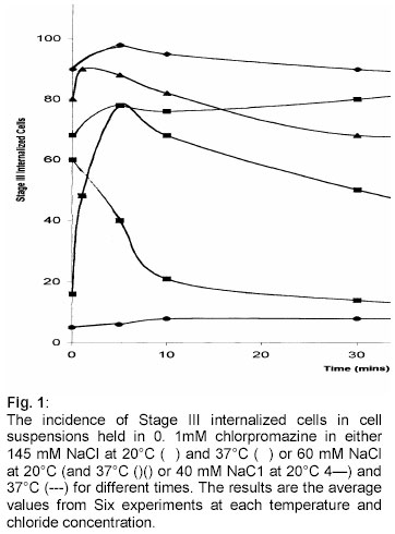
Of Port Harcourt , Nigeria Received: September 2001 Code Number: md03002 ABSTRACT The effect of membrane potential change of the human erythrocytes on cationic drugs tetracaine and chlorpromazine and neutral drug benzyl alcohol induced cell shape change and red cell uptake of drug has been quantitated using light microscopy and spectrophotometry respectively. At the drug concentration necessary to cause cell membrane cell shape change membrane potential change from -7.1 mV to 1 6.4mV let to the reversal of the cup-forming property of chlorpromazine and tetracaine to that of a crenetor at both 20°C and 37°C. The effect of altering the membrane potential from -7. lmV to 16.4mV also led to the decrease of cellular uptake of drug with increasing membrane diffusion potential. The membrane potential dependent drug induced cell shape change with also reversible on reversing the membrane potential. The results therefore suggest that the cellular uptake of drug and drug induced cell shape change in human erythrocytes was dependent on change in extracellular chloride concentration Key words: Membrane potential, cationic drugs, drug uptake, blood, human. INTRODUCTION Glaser (1979, 1982) found some correlation between the membrane potential of human erythrocytes and the discocyte -echionocyte and the discocyte - stomatocyte transformation. The membrane potential was changed by alteration in pH of the suspending phase or by ionophore treatment. Nwafor and Coakley (1985, 1991) pointed out that there was some relationship between membrane potential of human erythrocytes and drug induced cell shape change. In the study membrane potential was changed by reducing the extracellular chloride concentration significantly while maintaining the ionic strength and the osmolarity of the cell suspending solution constant. Cell volume changes are in principle undersirable in systems in which cell morphology is studied (Nwafor and Cockley 1991). The influence of transmembrane potential on cellular uptake of drug has not been properly quantitated. Mohandas and Feo (1975) studied the uptake of anionic and cationic derivatives of phenothiazine by the red blood cells and pointed out that there was some correlation between cellular uptake and red cell morphology Sheetz and Singer (1974), Kanaho et al (1981) observed that ghosts and intact erythrocytes undergo very similar drug - induced shape changes. Their studies led to the suggestion that membrane potential of human erythrocytes was not of primary importance to the observed shape changes. However, the studies were carried out at extracellular chloride contraction around l30mV - 145mV NaC1 (-4.2mV to -7. lmV). In the present study we report spme observation on the contribution that membrane potential make on cellular uptake of cationic drugs chlopromajine and tetraceine (Deuticke 1968 Fuji et al 1976) and neutral drug benzyl alcohol (Deuticke, 1968) with their effects on the morphology of human erythrocytes at E= -7. lmV (145mM NaC1) to E = 16 .4mV (60mM NaC1) (Nwafor and Cockley 1989, 1991). MATERIALS AND METHODS Composition of erythrocytes suspending solutions: The compositions of the erythrocyte suspending solutions of sodium chloride with sorbitol/sodium buffered with 5mM Hepes at pH calculated to keep the intracellular chloride ion concentrations and cell volume constant has been described elsewhere (Nwafor and Coakley, 1991). Briefly, buffered NaCl/sorbitol NaCI/Na gluconate solutions were prepared with 5mM Hepes as follows: 145mM NaC1 with 5mM Hepes, pH 7.32, diffusion potential E,-7. l4mV and 60mM NaC1 with 170mM sorbitol (or 85mM sodium gluconate with 5mM Hepes, pH 6.94, diffusion potential E = 16.43mV. Preparation of buffered solutions of drugs: Cationic drugs (chlorpromazine hydrochloride, tetracaine hydrochloride and neutral drug benzyl alcohol (BDH chemicals) were used for the study. The chemical to be tested was dissolved in the buffer and the pH of the drug solution was adjusted to the value determined for the buffer by addition of 1% NaOH. The concentration of the drug solutions was assessed spectrphotometrically using an S.P 1805 Unicam double beam spectrophotometer (Unicam Ltd. England) and their Ultraviolet absorption wavelength (mm) were obtained as follows. benzyl acohol 259, chlopromasine HCL 254 and tetracenic HCL 310. Light Microscopy: Cell suspension (5 x 10 cells/ml) in drug solution and glularaldehyde in buffer (0.5%, vtv) were maintained at the same temperature of eithet or 37°C. Following exposure of the cells to drug for a known time, 0.2m1 of the glutaraldehyde solution was added to 1 .Oml of cell suspension. The cell suspension was then allowed to stand at the desired temperature of at least 3 minutes. A sample of the fixed cell suspension was draw by surface tension unto 5.0cm long glass microcapillaries of rectangular cross-section 0.2mm pathlength and 1.2mm width and observed using Normaski differential interference contrast with a x 100 oil immersion objective on a Nachet 400 microscope (Nwafor and Coakley 1985, 1986). The initial erythrocyte shape change (0 mm) was determined as described elsewhere (Nwafor and Coakley 1986), the erythrocyte shape were characterized according to the criteria proposed by Fujii et al (1979), Deuticke (1968). Measurement of cellular uptake of drugs and preparation of blood suspension: Various blood was collected into acid! citrate !dextrose. 1.0 to 5.Oml of the cell suspension media (145mM NaCl 5mM Hepes, pH 7.32 or 60mM NaCl 170mM sorbitol 5mM H pH 6.94). The cell suspension was washed and the washed cells were resuspended in 1 Oml (V) of the same buffer containing a known concentration of drug or without drug (control). The percentage of the collected whole cells to the total volume of the cell suspension was estimated by the relationship: Haematocrit H (%) = PCV + N X 100 (1.0) PCV + (N+V) Where PCV, packed cell volume, was taken to be 0.45% Cell pellets washed as described below were resuspended in buffer containing a known drug concentration and maintained at 37°C for 10min in a thermostat controlled water bath. After 10 mm the suspension was centrifuged at 3,5 OOg for five minutes in a bench centrifuge at 37°C and the supernatant was carefully collected. The absorbance of the initial drug solution (OD') was measured against a buffer blank at the appropriate ultraviolet absorption wavelength of the drug tested. The absorbance of the supernatant (0D was measured against the supernatant of the control cell in order to reduce any interference of haemoglobin with the results. The absorbance measurements were obtained with S.P. 1805 Unican double beam spectrophotometer. The percentage drug uptake by erythrocytes was calculated as the difference in absorbance 0D and 0D expressed as a percentage of the initial drug solution (OD 1 ) i.e Change in absorbance (%) = OD1– OD2 X 100 The absorption of the drugs reached equilibrium in less than two minutes and no difference in the absorbance value was detected with increasing time up to 60 minutes. RESULTS The influence of transmembrane potential on cellular uptake of drugs: Table la shows the effect of different extracellular chloride concentration on cellular uptake of tetracaine, chlorpromazine and benzyl alcohol. Erythrocytes exposed to cationic drugs at membrane diffusion potential, E, = 16.4 mV had low cellular uptake of drug compared with cells at E - 7. lmV. There was only a small difference in uptake of benzyl alcohol by erythrocytes in the two isotonic solutions. Generally, the partitioning of the positively charged drugs - tetracaine and chlorpromazine and neutral drug benzyl alcohol across the membrane decreased with increasing membrane potential. The ratio of cellular uptake of tetracaine, chlorpromazine and benzyl alcohol for a constant extracellular drug concentration by erythrocytes at 145 NaC1 to 60 mM NaCl for the different drugs and haematocrits, has the values given by column 5 of table la. The ratio of the values for the distribution of the drugs across the membrane calculated at 145 mM NaC1 and 60 mM NaCl at constant haematocrit gave the relative change in intracellular concentatration of drug for constant extracellular chloride concentration. Drug induced erythrocyte shape change: Chlorpromazine: The percentage stage III internalized cells scored for cells maintained with 0.1mM chlorpromazine in 60mM NaCl at 37°C was highest immediately and then fell gradually with time. The initial peak following exposure of cell, to drug in 145 mM NaC1 at 37°C was not found for erythrocytes exposed to the drug in 60mM NaCl (Table 2). Generally cells maintained at 20°C with 0. 1mM chlorpromazine had a higher incidence of stage III internalized shapes compared with erythrocytes at 37°C over the same exposure time (Fig. 1). Table 1: The Influence Of Membrane Potential On Drug Membrane Association a. The influence of membrane potential on cellular uptake of drugs
? = not relevant Table 2: The Influence Of Membrane Potential On Drug Membrane Association b: Shape changes of human erythrocytes induced by drugs in buffered 60mM NaCl 5mM Hepes 85mM Na gluconate, pH 6.94 at 20°C and 37°C
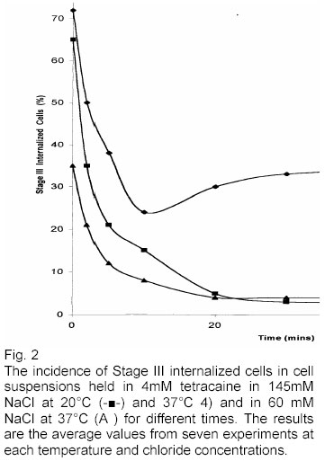
Tetracaine: About 35% of the cells internalized membrane (Stage III stomatocytes) immediately on exposure to 4mM tetracaine in 6 mM NaCI at 37°C in constant to 70% in the situation for cells in 145 mN NaCI with the drug. Cells in both 60 mM NaCI and 145mM NaCl with the drug rapidly decreased their shapes towards a lower incidence of stage III internalized form with time (Fig. 2). After 10 minutes cells exposed to 4mM tetracaine in 60mM NaCI at 20°C gradually became externalized. 10% of the cells showed stage III externalized shape after 60 minutes. Benzyl alcohol: Fig. 3 shows the incidence of stage III crenation of cells in cell suspensions maintained in 50 mM benzyl alcohol. The initial (0 to 2 minutes) high incidence of stage III externalized shapes as w observed when first exposing cells to changed drugs were not found with benzyl alcohol at either 20°C or 3 7°C. the erythrocytes maintained with the drug in 60 mM NaC1 showed low incidence of stage Ill externalized form at 20°C and 37°C (Fig. 3). DISCUSSION In the present study, a strong influence of extracellular chloride ion concentration (E = -7.1mV to E = 1 6.4mV) on cationic drugs tetracaine and chlorpromazine (Fuji et a l 1976) and neural drug benzyl alcohol (Deuticke 1968) induced human erythrocytes shape change and on cellular uptake of drug has been established. Decreasing the extracellular chloride ion concentration (Nwafor and Coakley 1991) led, in the case for tetracaine and chlorpromazine to reduce their cup forming property The direction of shape change was reversible through changes in the extracellular chloride ion concentrations. The study also revealed that unlike charged drugs, NaCl concentration did not influence the shape changes of human erythrocytes induced by benzyl alcohol at 20°C. Unlike charged drugs, benzyl alcohol in 145mM NaCI at 37°C did not show immediate externalization. At membrane diffusion potential E, - 7mV the cells became increasingly echinocytic (Fuji et al 1979) with time. The slow development of externalization in cells in 145mM NaC1 with benzyl alcohol at 37°C suggest that the development of the echnocytic forms in cells may be due to a slowly developing secondary modification of the bilayer which occurs when the membrane potential is approximately - 7mV and not when the membrane potential is 16.4mV. The cells in 6OmV NaCl (E 16.4 rnV) at 37°C were not initially, and did not become echinocytic. These results are consistent with the views that drugs do not penetrate the hydrophobic region of the lipid bilaryer of the cell membrane (Seeman 1972, Kanaho et al 1981). The bilayer couple hypothesis (Sheetz and Singer 1974) was not affected by the above results since it requires only that a differential drug- induced change in the surface free energy at the two faces of the membrane gives rise to a bending couple. Thus the observation that changes in the extracellular chloride concentration which alter the potential across the erythrocyte membrane can modify the morphological consequences of exposure to drug is consistent with the bilayer couple hypothesis (Sheetz and Singer 1974). Table lb shows the values of the ratios for the partitioning of tetracaine, chlorpromazine and benzyl alcohol across the membrane when we would expect from the left hand side of equation 3 (Nwafor and Coakley; in press) the ratio to be 145/60 = 2.43. The results support the view that membrane potential alters the partitioning of drug across the erythrocyte membrane in a manner which is strongly dependent on the chloride ion distribution The differences in drug membrane interaction at 37°C for cells in 60mM NaCl or in 145mM NaC1 with 50mM benzyl alcohol (Fig. 3) may explain the small differences in drug uptake for different membrane potential (Table 1b). The partitioning of a neutral drug across the membrane would be expected to be independent of membrane potential so that the expected ratio would also be expected to be independent of membrane potential in that the expected ratio would be 1.0. The ratio 1.22 above (table lb) may reflect potential dependent change in the extent of the interaction of the neutral drug with the membrane at 37°C. However, study on the possible influence of membrane potential change effects on anionic drugs induced cells shape change and a cellular uptake of drug is needed to reach a definite conclusion. REFERENCES
© Ibadan Biomedical Communications Group The following images related to this document are available:Photo images[md03002f3.jpg] [md03002f1.jpg] [md03002f2.jpg] | ||||||||||||||||||||||||||
| |||||||||
