 |
| About Bioline | All Journals | Testimonials | Membership | News |
|
||||||
|
||||||
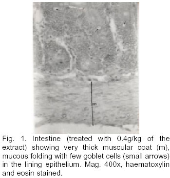
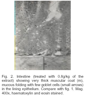
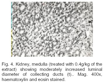
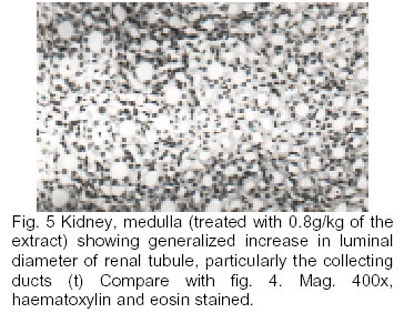
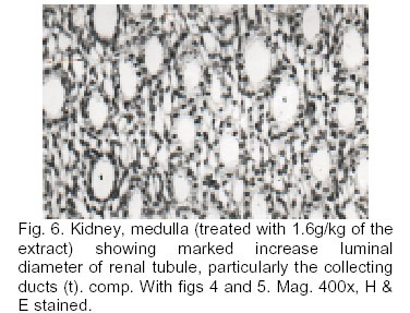
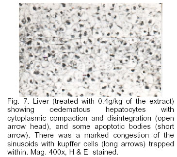
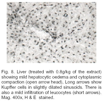
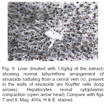
African Journal of Biomedical Research, Vol. 6, No. 1, Jan, 2003, pp. 21-25 HISTOPATHOLOGICAL STUDIES ON THE TOXICITY OF OCIMUM GRATISSIMUM LEAVE EXTRACT ON SOME ORGANS OF RABBIT EFFRAIM K. D 1 , JACKS T. W 2 AND SODIPO O. A 3 Departments of Pharmacology, Biochemistry and Anatomy, College of Medical Sciences, University of Maiduguri . Borno State , Nigeria Received: December 2000 Code Number: md03004 ABSTRACT Aqueous extract of leaves of Ocimum gratissimum (OG) at doses of 0.4, 0.8 and 1.6 g/kg was experimentally tested through oral administration twice weekly to rabbits, for four weeks. At the end of the fourth week, the animals were sacrificed after ether anesthesia and transection of the jugular vein. The livers, kidneys and the intestine (jejunum) were removed and fixed In 10% formal saline. Paraffin sections were prepared from these organs and stained with haematoxylin and eosin for histopathological assessment. The extract of OG at all doses showed changes In the intestine which were indicative of Its effects on bowel discharge and mucous secretion. The effect on the kidney was indicative of diuretic activity at all the tested doses. In the liver the extract showed higher necrotic changes at a low dose (0.4g/kg'). The laxative and hepatoprotective potentials of aqueous extract of leaves of Ocimum gratissimum have been discussed. Keywords: Ocimum gratissimum, neem, toxicity, hepatoprotection INTRODUCTlON Ocimum gratissimum (Linn) family Labiaceae Is a herbaceous plant commonly found In tropical Asia especially India , where It is used for aromatic baths of fumigations in the treatment of rheumatism and paralysis. The plant is also found in West Africa . In NIgeria , it is found In the Savannah and coastal areas. In the coastal areas of Nigeria , the plant Is used In the treatment of epilepsy, (Osifo, 1992), hIgh fever (Oliver, 1980) and diarrhea (OlIver, 1980, and Sofowora, 1993), whilst in the Savannah areas decoctions of the leaves are used to treat mental illness (Abdulrahman, 1992). The leaves of the plant are used as a condiment In cooking. In the southern part of Nigeria , the plant Is called "effinrin-nla" by the Yoruba speaking tribe. It is called "Ahuji" by the Igbos, while In the Northern part of Nigeria , the Hausas call it "Daidoya". In view of its many uses especially In Nigeria and the fact that traditional medicine practitioners prescribe and administer decoctions of the leaves to patients without regard to its possible adverse effects, the present investigation was undertaken to assess Its potential toxic effects on some organs (kidney, Intestine and the liver) of localization In rabbits. MATERIALS AND METHODS Collection and Identification of Plant Materials The fresh leaves (800 g) of the shrub were collected from the University of Malduguri . The leaves were Identified and authenticated as Ocimum leaves, by Dr. S.S. Sanusi, a plant taxonomist in the Department of Biology, Faculty of Science, University of Maiduguri. Experimental Animals Male healthy rabbits bred in the Department of Pharmacology were used for the study. Their average weight was 1.2 kg. They were fed with ground leaves and tap water throughout the period of the experiment. Preparation of Extract Fresh leaves of Ocimum gratissimum were kept in the oven at 80°C for 10 mm to stop enzyme activity and then 60°C for 30 minutes to dry. They were then air dried and ground into coarse powder. Fifty grams of the powdered leaves was stirred into 450 nil of boiling distilled water. Boiling was allowed to continue for 5 mm. The mixture was then kept aside, for 30 mm to allow it to infuse. It was then * filtered through cheese cloth. The filtrate was concentrated to 200 ml (i ml of the extract being equivalent to 0.25g of the starting material). The extract was kept in a refrigerator at 4°C until used. Administration of the Extract Twenty male rabbits were randomly distributed into four groups of five. Group I served as the control and received distilled water. Groups 2, 3 and 4 received the aqueous extract of Ocimum gratissirnum at doses of 0.4, 0.8 and 1.6gkg respectively. Animals in the control group received a quantity of distilled water equivalent to the dose given to the 1.6 g/kg group. Administration of the extract was carried out orally, by means of a polvthene cannula. Animals received their doses twice a week for four weeks. The animals were observed daily for clinical signs of toxicity or pharmacological signs, throughout the period of study. At the end of the fourth week, the animals were sacrificed after ether anaesthesia while transection of the Jugular vein! The kidneys, liver and a length of intestine were removed and observed grossly. These were then fixed in 10% formal saline for histological studies. Paraffin sections were cut at 8u and stained with Haematoxylin and Eosin. RESULTS During the bi-weekly oral administration of the aqueous leaf extract ( 0.4, 0.8 and 1.6 gkg" body weight) of Ocimum there was no mortality in all the groups treated. Gross examination of the organs harvested did not reveal any pathological changes due to the treatment with the extract General histo pathologic evaluation of the organs, revealed a dose-dependent effect of the extract on the structure of the organs, with the effect increasing with dose Histopathologic changes In the intestine of animals treated with 0.4gkg dose of extract showed very thick muscular coat (m), mucous folding with few goblet cells in the lining epithelium (fig. 1). Increasing the dose of the extract to 0.8gkg" body weight, produced thick muscular coat, mucous folds with many goblet cells in the lining epithelium (fig.2) when compared with fig.1. At an extract dose of 1.6gkg" body weight, numerous goblet cells in mucous folds were observed in the lining epithelium. The glandular tissue showed some apoptotic bodies (fig. 3). The effect of the extract on the intestine seemed to be dose-dependent. The structure of the kidney exposed to an extract dose of 0.4gkg body weight revealed moderately increased luminal diameter of the collecting ducts (fig. 4). Increasing the dose of the extract to 0.8g/kg body weight caused further increase In the luminal diameter of the renal tubules particularly the collecting ducts (fig. 5). Marked Increase in the luminal diameter of the renal tubules was observed when the extract dose was increased to 1.6gkg body weight (fig. 6). Thus showing a dose response effect of the extract on the structure of the kidney. The structure of the liver also showed dose-dependent changes when exposed to various doses of the extract. At a dose of 0.4gkg' body weight of the extract, there was a generalized edema/hypertrophy of the hepatocytes resulting In a marked widespread, sinusoidal congestion. About 80% of the hepatocytes showed cytoplasmic compaction and disintegration, with some apoptotic bodies as well as nuclear piknosis. Kupfer cells were many and were trapped within the sinusoids. These indicate a degenerative/necrotic process (fig. 7). Increasing the dose of extract to 0.8gkg body weight produced a result similar to fig. 7. There was a reduction in all the parameters observed. There was less hepatocytic edema/hypertrophy resulting in slightly widened sinusoidal spaces (fig. 8). Hepatocytes showed reduced cytoplasmic compaction and disintegration with less prominent apoptotic bodies. In addition there was mild leukocyte infiltration. compaction was observed in the hepatocvtes. These indicate a mild tissue lesion or damage as compared with the 0.4g/kg treated group. The group of animals treated with 1.6 gkg' of the extract depicted a re establishment of the normal structure of the liver. Hepatocytes showed no sign of oedema hvpertrophv resulting in the i of sinusoids with larger (normal) diameter. Only a mild cytoplasmic DISCUSSION The study of toxic or adverse effects of crude drugs of plant origin is essential in order to provide a guide to their safe usage and eventual standardization. This is especially pertinent, as traditional medicine practitioners often administer such preparations without regard to their possible adverse effects. Ocimum gratissimum (O.G) extract has been reported to have antipyretic and anti- diarrhea activities (Oliver, 1960 and Sofowora, 1993). Besides, the extract has been used in the treatment of mental illness (Abdulrahman, 1992). The OG extract has also been shown to have sedative activity (Effrairn et cii unpublished) and to have therapeutic benefit in patients with inflammatory joint disease (Tanira et al., 1988). However, there is a paucity of information concerning the toxic or adverse effects of repeated oral administration of aqueous preparation of 0G. The present study shows that the OG extract can exert itself to cause some functional damages to critical organs such as the liver and kidneys when applied in varying doses (0.4, 0.8 and 1 6 g/kg body weight) of the extract. Changes observed in the muscular coat (both inner and circular longitudinal layers) of the intestine indicate that 0.G may be implicated in the enhancement of bowel discharge. The increase in the number of goblet cells in the epithelial lining suggests increased mucus secretion for lubrication of intestinal val1 and hence enhanced bowel discharge. However, the presence of numerous goblet cells in the intestine, particularly, the small intestine is suggestive of necrotic change. Enhancement of bowel discharge seem to contradict the report of Oliver (1980) and Sofowora (1970) about the alleged usale of OG as an anti-dlarrhoeal agent The method of preparation of the aqueous extract may have reduced the eugenol content of the extract which is implicated in the anti-diarrhoeal property of the extract (Sofowora, 1993). The extract also produced dose-dependent increase in luminal of all the renal tubules both in the the medulla the medullary collecting ducts. This suggested its implication in renal clearance and hence diuresis. The finding Is consistent with the report of Dhawan et al (1977), despite the use of rats as the animal model. The effect of the extract of O.G on the liver showed an interesting pattern. At low doses (0.4 g/kg') there was characteristic degeneration or necrosis of the liver. But as the dose was increased, the degenerative effect diminished. In fact at a high dose of 1.6 g/kg body weight of extract, only a mild cytoplasmic compaction of the hepatocytes was observed. The results of this study were supported by the biochemical data obtained recently (unpublished data). The extract may possess hepatoprotective activity in higher doses. This may explain the lack of deleterious effects in humans despite the fact that the leaves are consumed in large quantities as vegetable and also as condiment In Nigeria. The present study has shown that, the repeated oral administration of the aqueous leaf extract of OG may bring about changes In the liver, kidneys and the intestines. Such changes may be beneficial (in the case of the liver) especially as the dose is increased. This finding thus provides an experimental support for the use of decoctions of OG as a diuretic, laxative and a possible hepatoprotective agent. Further researches on the laxative and possible hepatoprotective activities are In progress. ACKNOWLEDGEMENT The authors gratefully acknowledge the technical assistance of Mr. Justus JIbrin and the secretarial work of Miss Gladys Ogoucha REFERENCES
© Ibadan Biomedical Communications Group The following images related to this document are available:Photo images[md03004f5.jpg] [md03004f3.jpg] [md03004f2.jpg] [md03004f8.jpg] [md03004f6.jpg] [md03004f4.jpg] [md03004f1.jpg] [md03004f9.jpg] [md03004f7.jpg] |
| |||||||||
