 |
| About Bioline | All Journals | Testimonials | Membership | News |
|
||||||
|
||||||
African Journal of Biomedical Research, Vol. 6, No. 1, Jan, 2003, pp. 27-31 ENHANCEMENT OF CUTANEOUS WOUND HEALING BY METHANOLIC EXTRACTS OF AGERATUM CONYZOIDES IN THE WISTAR RAT. OLADEJO O.W 1 ., IMOSEMI I.O 1* ., OSUAGWU F.C 1 ., OLUWADARA O. O 1 ., AIKU A 3 ADEWOYIN O 2 . EKPO O.E 1 , OYEDELE O.O 1 , & AKANG E.E.U 4 . Departments of 1 Anatomy, 2 Pharmacognosy, 3 Physiology
and 4 Pathology, College of Medicine , University of Ibadan
. Ibadan , Nigeria . Received: May 2002 Code Number: md03005 ABSTRACT In a bid to test the wound healing effect of a crude methanolic extract of Ageratum conyzoides (Linn.), 20 animals were divided into two groups of ten animals each representing control and experimental groups. Each animal had a 2cm x 2cm area of skin on the right dorsolateral flank area marked and excised. The resulting area of skin wound in the experimental group was dressed with crude methanolic extract of Ageratum conyzoides at a five daily interval while the animals in the control group were dressed with normal saline at the same interval. The wound area was measured at the tenth post-operative day for animals in both groups and the percentage wound contraction calculated. Sample of granulation tissues and end scar obtained from these animals and prepared by routine paraffin wax method. Fibroblast and blood vessel counts were determined in both groups. The result showed a significant increase in the percentage wound contraction at day 10 in the experimental group compared with the control (82.3 ± 1.6 % vs 55.0 ± 4.2 %), P < 0.05. The wound of animals in both groups showed excellent granulation tissue formation and minimal signs of wound infection. There was a significant reduction in the mean fibroblast count in the experimental group compared with the control (44.2 ± 5.8/ high power field vs 90.2 ± 17. 4 / high power field),P < 0.05. The exact significance of this cannot however be determined. There was no significance difference in the vessel count. It was concluded that extract of Ageratum conyzoides has a better wound healing enhancing action compared with normal saline treated controls. This effect may be due to the antimicrobial properties of Ageratum conyzoides. Key Words: Wound healing, Ageratum conyzoides, Effect of. INTRODUCTION Wounds arise from injury by various agents (Bailey and Love, 1998)]. Healing of wounds is an important part of the reparative process. Like the alchemist's dream of turning base metal into gold, efforts aimed at achieving a perfect wound healing has pushed many researchers into trying various therapeutic options which were thought to aid or accelerate the wound healing process. The cheaper and more effective the agent, the better for the patient. The aim of this study was to compare the wound healing effect of a crude methanolic extract of Ageratum conyzoides with that of Normal saline acting as control. Ageratum conyzoides has long been known in herbal or folk medicine as a remedy for various ailments in Africa (Almagboul et al , 1985), Asia , and South America [Ekundayo et al , 1987; Borthakur and Baruah, 1987]. Amongst the Yorubas of Southwestern Nigeria, it is known as Imí esú. The plant is used by the Fipa in South Africa as application to fresh wounds and in central Africa , the leaf is used to aid the healing of wound especially those caused by burns (Watt et al , 1962). The fresh leaves are rubbed between both palms until well macerated; the juice is squeezed into the wound and covered by a bruised but intact leaf. Dressing this is generally done once a day and the process of healing is said to be enhanced. Durodola (1977) demonstrated the effectiveness of crude extract of this plant in inhibiting the growth of Staphylococcus aureus a major wound pathogen in in-vitro cultures of the organism. MATERIALS AND METHODS Animals: Twenty rats in all were used in the study. They were divided into two groups containing ten animals each as follows: Group 1 : The control group. The animals in this group had their wounds treated with normal saline (CG). Group 2: Ageratum group. Animals in this group had their wounds treated with crude methanolic extracts of Ageratum conyzoides (Linn), AG. All the animals were acclimatized in the lab for one week before the commencement of the experiment. They were fed ad libitum with a rat pellet diet and water. Methanol extraction of the plant was carried out in the research laboratory of the pharmacognosy department of the University of Ibadan using the following procedure: Plant Material: Two hundred and fifty grams of dried leaves of Ageratum conyzoides collected from the herbarium of Forestry Research Institute of Nigeria (FRIN) on the 10 / 9 / 2001 with Voucher Specimen Number FHI 106480 was pulverized and soaked for 72hrs in methanol at room temperature. The resulting extract was concentrated and dried to a weight of 8grams.This was then made into a 1.5% suspension in water. Wound induction : The animals were anaesthesized using a combination of diazepam and chloroform anaesthesia. The animals were weighed individually and the skin on the dorsolateral flank area shaved. After wound preparation with 70% alcohol, 2cm x 2cm area of skin was measured and cut on the right dorsolateral flank area of the animal. The resulting area of skin wound was measured using a transparent white plastic which had previously been sterilized using a sodium hypochlorite solution. Hemeostasis was secured by application of direct pressure. The wounds were then packed with sterile gauze soaked in the dressing agent. A further layer of dry sterile gauze was placed on top of this and then secured with adhesive zinc oxide plaster. Change of wound dressing was done at 5 days interval until complete wound re-epithelization had taken place. Measurement of wound size: This was carried out at the 10 th day post wounding using transparent plastic sheets sterilized with sodium hypochlorite solution. The area of the traced outline was then determined by using a graph paper. A random sample of the wound in each group was taken at day ten post wounding. Another random sample of the end scar tissue was also taken following complete wound re-epithelization. Following a 48hr fixation in 10% formalin the tissues were then processed routinely using paraffin wax technique and stained using haematoxylin and eosin stains. The day 10 granulation tissue was studied as well as the blood vessel and fibroblast density per area of end scar tissue harvested. The result obtained were statistically analyzed using student – t test. RESULTS No mortality was noticed amongst the animals in both the experimental and control groups. The case of wound infection were also negligible and of mild severity in both groups. Table 1 shows the result of the fibroblast and blood vessel counts in both the experimental and control animals. Wound Morphology and Morphometry: There was noticeable homogeneity in the wound contraction observed for animals in the experimental group compared with the control group. The mean percentage wound contraction at day 10 in the control was 55.0 ± 4.2% while that of the experimental group was 82.3 ± 1.6%. This was statistically significant at P < 0.05. The results of the wound measurements taken as well as the percentage wound Contraction at day 10 calculated in both the control and experimental groups are as shown respectively in table 2 below. The exact day when complete re-epithelization occurred could not be determined due to the five-day interval between the wound dressings. The end scar formed was a fine linear white scar that was visible on the flank of the animals. Table 1: Showing Fibroblast and Blood Vessel Counts per High Power Field in both Control and Ageratum-Treated Animals
FC -FIBROBLAST COUNT; BVC– BLOOD VESSEL COUNT; HPF —HIGH POWER FIELD X400
Table 2: Showing Wound Dimensions at Days 0 &10 As Well As % Wound Contraction at Day 10 in Control and Ageratum-treated Animals
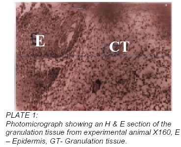
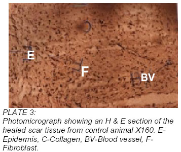
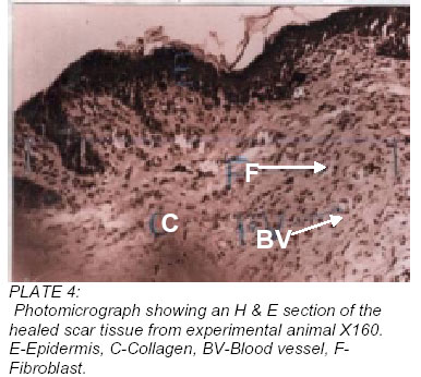
Histology of granulation tissue There was no significant difference in the histological appearance of the haematoxylene and eosin stained section of the granulation tissues randomly collected from the two animal groups. Both showed adequate inflammatory response with numerous inflammatory cells in the extracellular matrix. New blood vessel formation was also observed and adequate (Plates 1 and 2). Histology of the end scar tissue The only significant difference observed in the end scar tissue from the two groups is the relative abundance of fibroblasts in the control group of animals compared with the experimental group in haematoxylene and eosin stained sections of the end scar tissues which were randomly collected from the two animal groups. The mean fibroblast count was 90.2 ± 17.4 / high power field in the control compared with 44.2 ± 5.8 / high power field in the experimental group. This was found to be statistically significant at P < 0.05. There was no significant difference in the mean blood vessel count between the control group 13.4 ± 2.4 and the experimental group 25.8 ± 5.7, P < 0.05. Table 2 shows the values obtained for the fibroblast and blood vessel counts in the two animal groups, plates 3 and 4. DISCUSSION Although many indigenous tribes around the world have long suspected that this ubiquitous, annual, herbaceous plant might have medicinal wound healing properties, It has not really gotten the attention of orthodox medical practitioners as a potential source of a healing agent which may prove to be useful in the treatment of intractable cutaneous ulcers. Durodola in 1977 carried out a bacteriological study of a crude extract of the plant. The extract, which was obtained with Petroleum ether, was found to exhibit antibacterial activity against staphylococcus aureus (a major wound pathogen) in-vitro (Durodola, 1977). This study was carried out to test the wound-healing claim of this herb in an in-vivo situation. It was also our intention to elucidate the mechanism by which it exerts its wound healing effect if any. The wound was allowed to heal by secondary intention and therefore, the extract could exert its effect either by fibroblast proliferation, angiogenesis or keratinocyte migration. We could not however determined to what extent keratinocyte migration contributed to the wound healing effect because we could not determine exactly when re-epithelization took place. The attempt to find out the contribution of fibroblast mitogenicity and angiogenic effects of the extract was the rational behind our assessment of the blood vessel count as well as the fibroblast count. As can be seen from the above results, there was a sixty-percentage increase in the rate of wound contraction at day 10. Unfortunately we could not determine the exact day of wound healing due to the frequency of the wound dressing. However the wound contraction in an animal has been found to be proportional to the rate of re-epithelization (Billingham, 1956). The exact significance of the reduction in fibroblast count is not certain. It could be that the scar tissue that was harvested from the experimental animal was at a more advanced stage in terms of scar remodeling than the control scar. It is well known that fibroblast population in scar tissue reduces gradually over time as part of the remodeling process (Cotran, 1994)). There was no significant difference in the blood vessel count between the two groups. The wound healing effect therefore cannot be ascribed to increased angiogenesis in the experimental group. The only rational explanation for the accelerated wound healing is the antibacterial effect of the extract, which is a proven fact. Contents of Ageratum conyzoides include alkaloids, cumarins, essential oils and tannins. More specifically, it contains Ageratochromone, 2,6-dimefloxyageratochromone, Eugenols (Oliver-Bever, 1983; Kasturi and Manithomar, 1967) flavonoids such as conyzoigun, dotriaconthene, 7-mefloxy-2-2dimethylchromene (Oliver-Bever, 1980, Adesogan and Okunade, 1979), phenol and phenolic esters which are known disinfectants, as well as other antimicrobials (Durodola, 1977). Many of these are pharmacologically active and could have been responsible for the antimicrobial effect of Ageratum conyzoides. Another important point to note is the homogeneity of the dimensions of the experimental wound compared to the controls. The fact that infectious organisms were effectively contained is most likely responsible for this observation. Limitations: The experiment was limited by the fact that the exact day when wound healing occurred could not be determined. As earlier stated, this was due to the five daily interval of the wound dressings carried out. We were not able to determine exactly which of the constituents of the dressing agent was responsible for the would healing effect noticed. This analysis can be carried out at a later date when facilities for such analysis become available. REFERENCES
© Ibadan Biomedical Communications Group The following images related to this document are available:Photo images[md03005p3.jpg] [md03005p1.jpg] [md03005p4.jpg] [md03005p2.jpg] | |||||||||||||||||||||||||||||||||
| |||||||||
