 |
| About Bioline | All Journals | Testimonials | Membership | News |
|
||||||
|
||||||
African Journal of Biomedical Research, Vol. 6, No. 3, Sept, 2003, pp. 151-153 Case Report LIVER CIRRHOSIS ASSOCIATED WITH A NON-RESPONSIVE ASCITES IN A 10 MONTH OLD ALSATIAN DOG NOTTIDGE H.O1, AJADI R.A2, CADMUS S.I.B3, SHONIBARE O4, OKEWOLE E.A1, TAIWO V.05; EMIKPE B5, ADEDOKUN R.A.M4, ODUYE O.O.1 1Departments of Veterinary Medicine, 2Veterinary Surgery/Reproduction, 3Veterinary Public Health/Preventive Medicine, 5 Veterinary Pathology and 4Veterinary Teaching Hospital, University of Ibadan, Ibadan, Nigeria Received: June 2002 Code Number: md03059 A ten month old Alsatian bitch presented with complaint of recurring ascites over a period of three months and had ‘been refractory to diuretic therapy. The condition was diagnosed as liver cirrhosis by serum chemistry, exploratory laparotorny and histopathology of the liver. Result of the serum chemistry showed a progressively decreasing serum albumin and liver, enzymes. Similarly the Albumin /Globulin (A:G) ratio was progressively decreasing. Haematological findings were that of anaemia of chornic disorder (mild normocytic, normochromic, non responsive). The PCV, HB and RBC also decreased progressively. Exploratory laparotomy findings were that of a slightly enlarged liver with diffuse miliary nodules on .both the parietal and visceral surfaces. Few larger nodules ‘were also present. ‘The liver was firmer in consistency and two separate masses of fibrinous tissue measuring about 5 cm in length and 2cm in thickness were seen floating in the abdominal transudate. The transudate which measured about 15 litres was colourless and slightly cloudy. The bitch was euthanised following laparotomy and on the owners request due to the non-responsiveness of the animal to diuretic and other supportive therapies. Keywords: Liver cirrhosis, Heart failure, ascites, frusemide, dog. INTRODUCTION The liver accounts for approximately 3-8% of the total body weight in the carnivores and and occupies a central role in diverse metabolic activities that help maintain the body’s normal. homeostatic mechanism (Center, 1989; Strombeck and Guildford, 1990). It has been shown that the liver has a high degree of heart differentiation and at the same time retains the capacity to regenerate (Roger, 1993). Hence, it is able to maintain normal activities over a long period of the dog's life span. The liver responds to injury either by hepatocellular regeneration, bile duct hyperplasia, hepatic fibrosis, cirrhosis or acquired portosystemic shunting (Fausto and same Meade, 1999; Rogers, 1993). Inability’ of the was liver to maintain an adequate hepatic function as a result of any of these responses often result in a syndrome termed liver failure (Rogers, 1993). This may be single and specific or in most cases multiple. It may be and characterized by cholestasis and icterus, photosensitization, hepatic encephalopathy, (Manderino and Devries, 1985). Chronic liver failure resulting from cirrhosis has been reported in adult dogs (Williams, 1991). Very often, the cause is usually undetermined. However, it may be associated with parasitism, congestive heart disease and lesions that impair the flow of blood through the liver (Herrtage, 1991). This report presents a 10 month old puppy with liver cirrhosis and a non-responsive ascites associated with congestive heart the failure. HISTORY A 10 month old Alsatian puppy was presented at the Veterinary Teaching Hospital of the University of Ibadan with a complaint of depressed appetite and distended abdomen. It had been presented and treated for the same complaint two months earlier. The puppy was the only one affected form a pack of three dogs in the same house. They were all fed with enough table scraps. They had all been vaccinated against rabies, canine distemper, infectious canine hepatitis, leptospirosis and parvovirus and dewormed quarterly against round worms and hook worms using mebendazole (VitamebR, Vital pharmaceuticals). Physical examination of the puppy revealed bilateral mucopurullent ocular discharge with congested ocular mucous membranes. The coat was rough, dry and lusterless and ‘the abdomen was severely distended. In addition, she was observed to be exercise intolerant. Urination was irregular, reduced in quantity and only when walked. There was tachycardia with low amplitude, the heart beat often becoming, regularly irregular after ‘little exercise. Respiratory rate was normal but usually panted after little exercise. The, temperature was constant between 38.6oC - 39.3°C throughout the five weeks of hospitalization. Based on these findings a tentative diagnosis of ascites of hepatic origin and/or congestive heart failure was made. MANAGEMENT
Blood samples were taken at two weekly intervals for blood profile from the cephalic vein using a 21 gauge needle. Samples of the voided urine were analyzed weekly using the dipstick method. Abdominal transudate obtained by paracentensis was also analysed by the dip stick method. The puppy was treated with furosemide by intravenous injection at the rate of 3mg/kg body weight twice daily. In addition about 200mls of abdominal transudate were removed daily through paracentensis. Oxytetracycycline was administered intramuscularly: at a dosage rate of 5mg/kg body weight. Livjivan (Animal care company Ltd), a herbal liver tonic was also administered orally at a dosage of 2 tables daily to improve liver function. Exploratory laparotomy was performed three weeks after presentation because of the poor resolution of the ascites despite the diuretic therapy. Liver biopsy was taken during the exploratory laparotomy for histopathology. RESULT
Abdominal, distension was observed not to decrease significantly - despite the therapy and the daily paracentensis. Instead, a moderately extended, subcutaneous oedema of the ventral abdomen and bilateral pedal oedema developed. These however resolved when the dosage of the furosemide administered was increased to 4mg/kg but with no significant effect on the ascites. The results of the serial blood samples are shown in table 1. The haematocrit (PCV), haemoglobin (Hb) and erythrocyte values (RBC) progressively, decreased during the period of management. A normocytic, normochromic non-regenerative anaemia was observed. Similarly, the values of plasma proteins (albumin) and the liver enzymes (SGOT, SGPT & ALP) decreased progressively. The albumin/globulin’ (A:G) ratio . was persistently below 0.55 throughout this period. The results of the urine and the abdominal transdusate analysis revealed no abnormalities. At laparotomy, about 15 litres, of. colourless but slightly cloudy exudate was obtained. Two fibrinous masses measuring about. 5cm in length cm in thickness each were found floating in the abdominal transudate. The liver was slightly enlarged, firmer than normal with diffuse milliary nodules on both the visceral and parietal surfaces. Few larger nodules measuring about 2-3mm in diameter were also present. Table 1 - Complete blood profile of serial blood samples of the Dog.
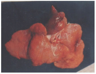
NECROPSY Following exploratory laparotomy, the ascites recurred within seven days. The non-responsiveness of the ascites to furosemide and findings during exploratory laparotomy made the owner of the dog request for euthanasia. Euthanasia was carried out using pentobarbitone sodium (6% Saggatal) at the rate of 60mg/kg body weight administered intravenously. Postmortem examination following euthanasia revealed a slightly flabby heart with no clot in the chambers. The liver was not different from that observed at exploratory laparotomy (Plate 1). Both kidneys were of normal size and consistency but with the capsule adhering to the cortex. The stomach showed few discrete areas of ulcers Histopathological finding on the liver revealed proliferation of fibrous connective tissue around the portal areas and the surrounding lobules. Cellular infiltration with lymphocytes and mononuclear leukocytes were also found. DISCUSSION AND CONCLUSION Liver cirrhosis has been reported to commonly affect older dogs of both sexes (Herrtage, 1991). The subject in this report was a 10 month old Alsatian puppy which appeared unusually too young to develop such condition. The cause of liver cirrhosis in dogs have remain largely undetermined (Herrtage,1991). However, it has been associated with toxic principles, parasitism, congestive heart failure and other lesions that impair blood flow to the liver (Popper, 1977). This case report could not be associated with toxins or parasitism because other dogs in the same environment were not affected. The fact that the puppy showed decreased tolerance to exercise, pedal oedema and irregular heart beat when exercised suggested that the liver cirrhosis was more likely to be a result of congestive heart failure. An interest finding in this case was the non responsiveness of the patient to diuretics. This was confirmed by the persistent oliguria and the non-significant reduction in the volume of the abdominal transudate. This might have been due to poor renal perfusion resulting from, reduced renal blood flow associated with congestive heart failure (Herrtage, .1991). The analyses of the serial blood samples were that of liver cirrhosis (centre, 1989). These included hypoalbuminemia, a non-regenerative normocytic nomochromic anaemia (anaemia of chronic disorder) and persistently decreasing liver enzymes. This was due to the replacement of active hepatocytes by fibrous connective tissue. Hence, the inability of the liver to secrete these substances. The progressively decreasing values of these parameters were an indication that the condition of the liver despite therapy progressively deteriorated. The differences in the result of the urine and abdominal transudate analyses indicated that the abdominal transudate is different from urine hence ruling out the possibility of bladder rupture. Gastric ulceration have been reported to develop secondary to chronic liver disease as a result of altered gastric acid production and decrease epithelial cell turnover associated with hypoalbuminemia (Stanton & Bright, 1989). A similar finding was noticed in the puppy reported here. Also the firm, slightly enlarged liver with multiple miliiary nodules due to fibrosis is pathognomonic with liver cirrhosis (Popper, 1977; Royers, 1992). REFERENCES
© Ibadan Biomedical Communications Group The following images related to this document are available:Photo images[md03059p1.jpg] |
| |||||||||
