 |
| About Bioline | All Journals | Testimonials | Membership | News |
|
||||||
|
||||||
African Journal of Biomedical Research, Vol. 7, No. 3, Sept, 2004, pp. 133 - 138 Full Length Research Article COMPLETE REGRESSION OF TRANSMISSIBLE VENEREAL TUMOUR (TVT) IN NIGERIAN MONGREL DOGS WITH VINCRISTINE SULPHATE CHEMOTHERAPY M.A. TELLA1, O.O. AJALA1 and V.O. TAIWO2[*] Departments of 1Veterinary
Surgery & Reproduction and 2Veterinary Pathology, University of
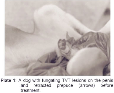
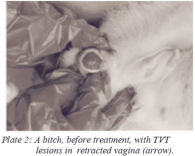
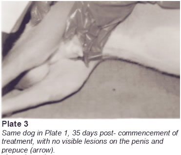
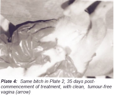
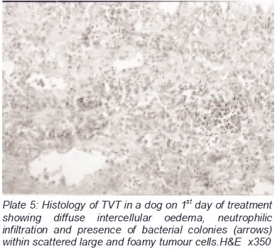
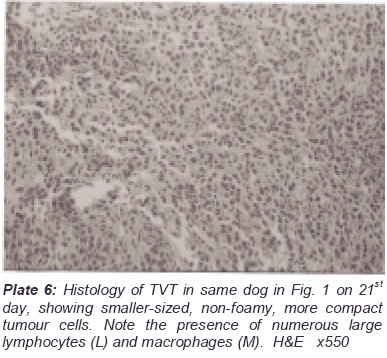
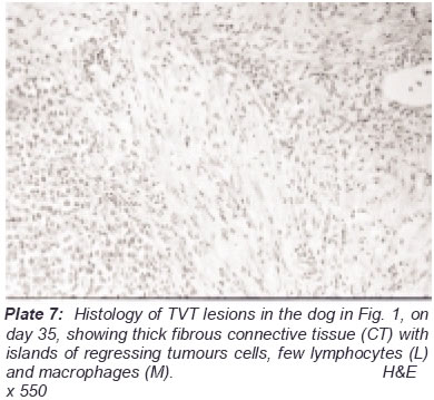
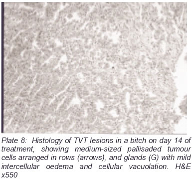
Ibadan, Ibadan. Nigeria. Received: February, 2004 Code Number: md04029 Intravenous administration of 0.025mg/kg body weight vincristine sulphate, in normal saline, in four weekly doses led to complete regression of lesions, within 35 days, in 4 mongrel dogs and 6 bitches with histologically diagnosed transmissible venereal tumour (TVT). Early side effects observed in the dogs, such as anorexia and dehydration disappeared after complete regression of the tumours. There was a progressive decrease in the number of mitotic figures, intercellular edema, bacterial contamination and inflammatory reaction within the tumour masses indicating that vincristine sulphate has direct effects on tumour cell division and bacterial multiplication. The tumour cells, initially very large (18-25µ; average 21.8±3.2µ diameter), with very abundant vacuolated cytoplasm and fine nuclear chromatin pattern, progressively became smaller cells (10-12µ; average 11.2±10.9µ) with heterochromatic nuclei and scant cytoplasm. Arrangement of tumour cells ranged from the initial glandular appearance to pallisading and compacted cells, which disappear as islands within thick fibrous connective tissue. Vascularization of the tumour masses also progressively waned. The presence of large numbers of lymphocytes, plasma cells and activated macrophages in the regressing tumours strongly suggests a role for immune-mediated control of TVT complimentary to that of vincristine sulphate. The slight and transient normocytic normochromic anaemia and leucopenia observed in the dogs may be related to the anti-mitotic effect of vincristine on bone marrow haemopoiesis. These untoward effects of vincristine may have been tolerated by the dogs because of the low dosage, short and well-spaced duration of therapy; an advantage over the adverse effects of some previously used combined anti-tumour drugs. Key words: Canine transmissible venereal tumour; progressive regression; vincristine sulphate, single chemotherapy. INTRODUCTION Transmissible venereal tumour (TVT) or Sticker’s sarcoma, a contagious venereal tumour of dogs, has continued to be a serious problem around the world (Moultan, 1961), especially in towns and villages all over Nigeria, occurring at same frequencies in both male and female dogs (Smith and Washbourn, 1998). It is most commonly observed in dogs that are in close contact with one another, or in stray and wild dogs that exhibit unrestrained sexual activity (Cangul, 2003). The lesions of TVT are confined to the mucous membranes of the external genitalia of dogs of both sexes and can be found in adjacent skin, brain, oral, nasal and conjunctiva mucosae and inguinal lymph nodes (Bostock and Owen 1975; Rogers et al., 1998; Ferreira et al., 2000). It is a multi-nodular, invasive, poorly circumscribed tumour usually closely attached to the penis and prepuce, while its site is dreadful and the accompanying hemorrhagic discharge produces offensive odor. The management of TVT has not been very easy in dogs, especially in developing countries, as most owners cannot afford the cost of surgical intervention and/or radiotherapy. This has made the control of this tumour almost impossible in this region. Combined use of anti-tumour drugs have been described as a more effective method of treatment (Brown et al., 1980; Idowu et al., 1984; Oni, 1994), but the long-duration therapy have not only led to adverse side effects, but is economically tasking for most dog owners. Vincristine sulphate is the salt of an alkaloid obtained from the common periwinkle (Vincarosa livia); it is also known as leurocristine. It has a wider application in human medicine in combined chemotherapy with other anti-tumour drugs (Takita et al., 1979; Nathan et al., 1993). The exact mechanism of its action is still of much debate, but it has been found to cause mitotic arrest and act on intercellular tubules (Whitehead et al., 1980). The need for cost-effective, affordable and minimal side effect course of treatment of TVT necessitated the trial of the efficacy of single low dosage intravenous administration of vincristine sulphate in this study. MATERIALS AND METHODS Animals Ten mongrel dogs, 4 males and 6 females, 2 to 4 years of age and mean body weight of 16.5±1.5kg with histologically proven progressive TVT lesions were used for this study. The male dogs (Plate 1) had the fungating lesions on the penis and the preputical mucosae, while the bitches (Plate 2) had the lesions extending 2-6cm into the vagina, heavily swollen and pendulous vulvae, with serosanguimous fluid exuding from the vulva orifice. The animals were kept in the separate kennels throughout the period of treatment and observation. Administration of Vincristine sulphate Lyophilised vincristine sulphate (Vinstin; Neon Antibiotics PVT Ltd., India) in 1mg pack was purchased from a veterinary pharmacy in Ibadan. It was reconstituted with the 10 millilitres (ml) normal saline provided by the manufacturer in a vial to make a final dilution of 0.1mg vincristine sulphate/ml. The dogs, irrespective of their sexes, were given appropriate volumes of the reconstituted drug at 0.025 mg/kg body weight intravenously (i/v) weekly for a period of 4 weeks. The dogs were monitored daily for 35 days and a further 6 months after complete remission of lesions. Five ml. of blood from each dog was collected into heparinized tubes by radial venipuncture before and thereafter weekly for 5 weeks post-commencement of treatment for routine haemogram (Jain, 1986). Histology of the tumour Tumour biopsy specimens (3-5mm2) aseptically excised before and weekly post-commencement of treatment from each dog, were fixed in 10% phosphate-buffered formalin and routinely processed for light microscopic evaluation of the lesions and photomicrography as described by Taiwo and Anosa (2000). Cellular morphomeric characteristics, counting and measurement of sizes were carried out using a microscope (Olympus, Japan) fitted with an eye-piece micrometer vernier scale as described by Taiwo et al., 1999). RESULTS Clinical evaluation of the dogs revealed mild but progressive anorexia, dehydration and polydypsia. These symptoms waned from 21 days post-commencement of treatment onwards with the animals showing progressively improving body conditions. The dogs developed transient mild to moderate normocytic normochromic anaemia and moderate leucopenia between on days 7 and 14 post-commencement of therapy (data not shown) which returned to normal from day 21 onwards. Progressive decreases and firmness of tumour masses and stoppage of fluid exudation became evident from 14 days onwards, and by the 35th day of the commencement of treatment, all the tumour masses had completely regressed with no visible scars or tumour mass found on the preputial sheaths and penis of the males (Plate 3), and the mucosae of the vulva and vagina in the bitches (Plate 4). Table 1 shows the cellular composition and morphometry of the tumour biopsy samples per high power view (x100 oil immersion; 0.78mm2) taken on the 1st and 28th day post-commencement of treatment for the male dogs and bitches. Histologically, in the male dog, the tumour on the 1st day of treatment was very oedematous with many focal areas of necrosis and neutrophilic cellular infiltration and presence of bacterial colonies (Plate 5). The tumour cells were very large (18-25µ; average 21.8±3.2µ diameter), with very abundant cytoplasm, some with multiple clear vacuoles, very few lymphocytes and macrophages (14.8±2.4µ) and few mitotic figures. On the 21st day, the tumour was very compact with considerably fewer, smaller (10-12µ; average 11.2±0.9µ) tumour cells with heterochromatic nuclei, large number of lymphocytes, plasma cells and enlarged and activated macrophages (17.2±1.8µ) (Plate 6). By the 35th day, thick bands of fibrous connective tissue were found traversing the poorly vascularized tumour (Plate 7) and no mitosis was observed. The cellular morphometry in the bitches were similar to those of the males, with the tumour cells initially large (12-16µ; average 19.8±1.2µ) with fine nuclear chromatin pattern and low nuclear:cytoplasmic ratio. Cytoplasmic vacuolation is however, not as marked as in the males. Pallisaded tumour cells, some with glandular arrangement (Plate 8) accompanied by moderate interstitial oedema and presence of considerable number of capillaries per unit area characterized the earlier stages of the tumour in the bitches. Mitotic figures, between 1-2 per high view, were also present. Table 1: Qualitative and quantitative analyses of the cellular composition of
transmissible venereal
*Data calculated as Mean
± Standard error One the 28th day of treatment however, the tumour was more compact, with small-sized cells (9.5-14µ; average 11.4±2.2µ) with high nuclear:cytolasmic ratio. There were fewer tumour cells, few capillaries, considerable fibrosis and no mitotic figures on the 35 th day. DISCUSSION This study has shown that four weekly regimen of intravenous administration of vincristine sulphate at 0.025mg/kg body weight alone was very effective in the complete remission of TVT in dogs. This translates to intravenous administration of a total of 1.6mg vincristine sulhate over 4 weeks for a dog with an average weight of 16kg. Our findings show a tremendous reduction in the dosage of similar therapeutic regimen by Amber et al., (1990) who used 0.5mg/m2 body surface area (BSA) weekly for 12 weeks. These workers (Amber et al., 1990) even recorded a recurrence in one of 20 dogs within 12 months; no remission was observed in the current study, even after one year. Our findings are however, similar but less tasking on the dogs given combined cyclophosphamide (0.1mg/kg BW) and vincristine sulphate (0.025mg/kg BW) daily for between 10 and 12 days by Idowu et al. (1984), and Oni (1994). Previous combination therapies using vincristine, cyclophosphamide and methotrexate (Brown et al., 1980) were not as successful, probably because of the high dosage and toxic side effects of these anti-mitotic drugs (Price et al., 1991) on the myocardium, intestines and bone marrow, all organs with very high cellular turnover rate. The early untoward effects observed in the dogs during this study, such as mild transient anaemia and leucopenia, anorexia and dehydration disappeared after complete regression of the tumours. There was a progressive decrease in the number of mitotic figures, intercellular oedema, bacterial contamination and inflammatory reaction within the tumour masses, indicating that vincristine sulphate has either direct effects on both tumour cell division and bacterial multiplication or enhanced the healing process within the tumour mass. The tumour cells, initially very large (18-25µ; average 21.8±3.2µ), with very abundant vacuolated cytoplasm and fine nuclear chromatin pattern, progressively became smaller (10-12µ; average 11.2±10.9µ) and had heterochromatic nuclei and scant cytoplasm. Tumour cells arrangement ranged from the initial glandular or loose appearance to pallisading and compacted cells, which disappear as islands within thick fibrous connective tissue. Vascularization of the tumour masses also progressively waned. The presence of large numbers of lymphocytes, plasma cells and activated macrophages in the regressing tumours strongly suggests a role for localized antibody-mediated control of TVT complimentary to that of vincristine sulphate. This agrees with the findings of Yang (1987) and Den Ottter et al. (1999), who reported the significance of interleukin-2 and antibodies in the immunotherapy of TVT. However, Ferreira et al. (2000) reported the absence CD3, IgG, IgM and lambda light chains in TVT cells, hence concluded that there was no local immune response in a five-year old cross-bred dog. Mizuno et al. (1994) reported that major histocompatibility complex (MHC) class II-associated cytotoxic T lymphocytes might play a significant role in TVT regression. We however, think that both antibody-mediated and cell-mediated responses are integral parts and mutually inclusive processes of TVT regression, as opposed to fibroblastic differentiation proposed by Kennedy et al. (1977). The slight and transient normocytic normochromic anaemia and leucopenia observed in the dogs may be related to the anti-mitotic effect of vincristine on bone marrow haemopoiesis. These untoward effects of vincrisitine may have been tolerated by the dogs because of the low dosage, well-spaced short duration of therapy. This is obviously an advantage over the adverse effects of some previously used combined anti-tumour drugs, which necessitated giving the dogs fluid and/or antibiotic supportive therapies (Brown et al., 1980; Amber et al., 1990; Oni, 1994). The rapid reduction in the mitotic figures was observed to have occurred at the second dosage regimen, while on the 4th and last administration (28th day) the tumour cells have become more compact with considerable fibroplasias, no bacterial contamination and no mitotic figures present. REFERENCES
© 2004 - Ibadan Biomedical Communications Group The following images related to this document are available:Photo images[md04029p3.jpg] [md04029p8.jpg] [md04029p1.jpg] [md04029p4.jpg] [md04029p5.jpg] [md04029p6.jpg] [md04029p2.jpg] [md04029p7.jpg] | |||||||||||||||||||||||||||||||||||||||||||||||||||
| |||||||||
