 |
| About Bioline | All Journals | Testimonials | Membership | News |
|
||||||
|
||||||
African Journal of Biomedical Research, Vol. 8, No. 1, 2005, pp.35-39 Full Length Research ArticleWeight Changes and Organ Pathology in Rats Given Edible Larvae of Cirina Forda (Westwood) O.O. AKINNAWO1, V.O. TAIWO2, A.O. KETIKU1 and J.O. OGUNBIYI3 Departments of 1Human Nutrition, 2Veterinary Pathology
and 3Pathology, University of Ibadan. Ibadan, Nigeria. Received: September, 2004
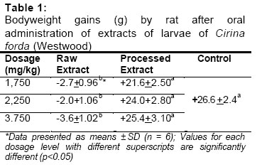
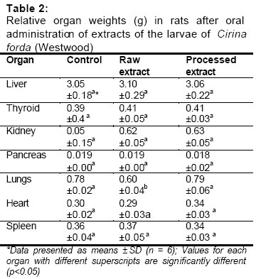
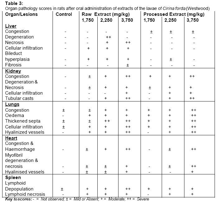
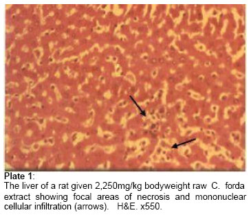
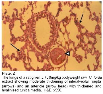
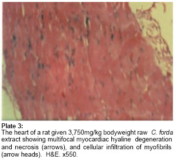
Code Number: md05006 Abstract The effects of oral administration of extracts of raw and processed larvae of Cirina forda (Westwood) on morphometry and histopathology were studied in albino rats. Weights of rats in the control group and in the group that were fed the processed larvae were significantly higher (p<0.05) than those of the group that received raw extract of the larvae. The relative weights of liver, heart, thyroid, pancreas and spleen were similar (p>0.05) in all the groups. The relative weight of lungs was significantly higher (p<0.05) in the control group of rats and the group that received the processed larvae than in the group given the raw larvae. Also, the relative weights of the kidneys in the rats that received the processed extract and the raw extract were similar but significantly higher (p<0.05) than the relative weight of kidney in the control group. Histopathological changes observed in the tissues of rats given the raw extract of the larvae include hepatocellular degeneration and necrosis, bile duct hyperplasia, congestion and tubular degeneration in the kidney and hyaline degeneration of myocardial fibres. The lung showed pulmonary congestion, thickened interalveolar septa, thick perivascular lymphocytic cuffs, and thickened and hyalinized tunica media of arterioles. The histopathological changes observed in the organs of the rats suggest that the raw larva of Cirina forda (Westwood) was toxic and produced a vascular circulatory disturbance resulting in organ damage in the animals. Processing the larvae by boiling and sun-drying reduced the toxicity on the liver and heart but not in the kidney. More research is needed on the toxicological aspects of the consumption of Cirina forda larva. Keywords: Insect larvae, processing, entomophagy, histopathology, rats INTRODUCTION The use of insects as human food (entomophagy) is widely practiced especially in the developing countries. The insects normally consumed include locusts, grasshoppers, termites, flying insects and caterpillars (Ene, 1963; Ashiru, 1988; DeFoliart, 1989). Studies have shown that these insects are good sources of high quality proteins, fats, minerals and vitamins (Oliveira et al., 1976; Kodouki et al., 1987; Ramos-Elorduy et al., 1997). However, some insects contain powerful pharmacodynamically active substances, which are known vertebrate toxins. Examples are toluene and O-cresol present in longhorn beetles (Moore and Brown, 1971), anabolic steroids in some species in the family Dysticidae (Schildknecht, 1970), cyanogenetic glycosides in the larva of moth, Zygena trifolli (Jones et al., 1962), and alkaloids in fire ants (Moore and Brown, 1971). A seasonal ataxic syndrome associated with the consumption of the edible larva of Anaphe venata Butler has been reported in southwest Nigeria (Adamolekun, 1993). This syndrome is characterized by sudden onset of severe muscular tremors and gait ataxia. The larva of Cirina forda Westwood is another edible insect larva that is widely consumed as an essential ingredient of vegetable soup (Fasoranti and Ajiboye, 1993). The wide acceptability and consumption of this larva warrants an in-depth study of the safety aspect of its consumption. This paper presents the morphological effects of the consumption of the larva of Cirina forda Westwood on some organs of rats. MATERIALS AND METHODSSample Collection and Preparation The larvae of Cirina forda Westwood were handpicked from the crowns of sheabutter tree, Butyrospernum paradoxum in Batati Village, Lavum Local Government Area of Niger State, Nigeria. The identity of the larvae was confirmed at the Department of Crop Protection and Environmental Biology, University of Ibadan, Ibadan, Nigeria. Two hundred and fifty grammes of the live insect larvae were boiled in 500ml of distilled water on a low heat for 2 hours. The boiled larvae were sun-dried for 72 hours, their hairs removed, and the hairless body milled into powder. Another set of live larvae, weighing 250g, was killed by freezing as described by Finke et al. (1989), thereafter thawed and dried in a dry oven at 40oC for 48 hours. The dried larvae were milled into powder after the removal of body hairs. One hundred and fifty grammes of each larvae powder were dissolved separately in 100ml of distilled water at room temperature and filtered to remove undissolved particles using cheesecloth. Animals and Sub-lethal Dose Administration Forty-two (42) adult albino rats of both sexes weighing between 150-180g were used for the study. The rats were weighed individually and then randomly divided into seven groups of six rats each after equalization of weights. They were allowed access to feed and water ad libitum. Noting that in a previous study (Akinnawo et al., 2002), an LD50 of 7,000mg/kg body weight was recorded for rats, sub-lethal doses of 1,750, 2,250 and 3,750mg/kg body weight of the Cirina forda larval solution, in calculated volumes, were used. Each rat in groups B, C and D received 1,750, 2,250 and 3,750mg/kg body weight raw larval solution orally, respectively, while those in groups E, F and G received 1,750, 2,250 and 3,750mg/kg body weight processed larval solution, also orally. Group A rats, which received corresponding volumes of distilled water by the same route, served as the control group of rats. Administration was carried out daily for 14 consecutive days. Organ Collection and Pathological Studies At the end of the experiment, the rats were weighed and anaesthetized in a chloroform jar. The organs were removed after dissecting each rat through a central incision on the abdomen. The liver, kidney, lungs, heart, spleen, pancreas and thyroid gland were removed and weighed immediately. Each tissue sample was fixed in 10% phosphate-buffered formalin for 24hrs. Thereafter, the tissues were dehydrated in graded concentrations of xylene, embedded in molten paraffin wax and sectioned at 5μ. The sectioned tissues were fixed on grease-free glass slides and stained with haematoxylin and eosin (H&E) for light microscopic examination. Organ pathological changes were scored in the rats according to Fadina et al. (1999). Photomicrographs of some of the lesions were taken using an OrthoLux Microscope fitted with a Leitz camera unit, and processed routinely in a colour photo laboratory. Statistical Analysis Means and standard deviations of body and organ weights in each group of rats were calculated and differences between group means determined using the 2-way analysis of variance (ANOVA) (SAS, 1987) and Duncan’s multiple range test (Duncan, 1959) at 95% confidence interval (that is, p<0.05). RESULTS AND DISCUSSIONTable 1 shows the body weight changes in rats administered with the raw and processed larval extracts. The rats that received the raw extract consistently lost weight during the period of study, while the control group and the groups that received the processed larval extracts gained weight. Processing of the larva by boiling and sun-drying reduced the effect of this factor as evidenced in the weight gains obtained in rats given the processed larval extract. However, the weight gains in the control group were significantly higher (p<0.05) than in the groups receiving the processed larval extract indicating that the toxic factor is heat-resistant. The relative organ weights of the rats after oral administration of the larva extracts are presented in Table 2. The relative weights of liver, heart, thyroid, pancreas and spleen were similar (p>0.05) in all the groups. However, the relative weight of the lungs was significantly higher (p<0.05) in the group that received the processed larval extract than in the group given the raw larva extract. Also, the relative weights of kidneys were similar in the groups fed with either raw or processed larva extracts but these values were significantly higher (p<0.05) than those of rats in the control group. The organ pathology scores of the rats after oral administration of the raw and processed larva extracts are presented in Table 3. The liver, kidney, heart and lungs of the rats given either raw or processed C. forda larvae showed variable histological changes, while the rats in the control group showed only mild to moderate pulmonary congestion and oedema. Notable lesions in the rats that received raw larval extracts include bile ductular hyperplasia, multifocal hepatocellular degeneration and necrosis (Plate.1) and moderate periportal fibrosis. Others include vascular congestion in both the renal cortex and medulla, multifocal areas of glomerular degeneration and necrosis, and presence of pinkish proteinaceous casts in the tubules of the renal medulla. There were widespread pulmonary congestion, thickened perivascular lymphocytic cuffs, and a few vasa vasori and most pulmonary arterioles had swollen endothelial cells, and thickened and hyalinized tunica media (Plate 2). The interalveolar septa were thickened with cellular infiltrates consisting of lymphocytes and few macrophages. There was mild to moderate congestion of the vasa vasori, and multifocal areas of hyaline degeneration of cardiac myofibrils and mononuclear cell infiltration in the heart (Plate 3). The spleen showed moderate lymphoid depopulation in the white pulp, presence of considerable numbers of macrophages, erythrophagocytosis and haemosiderosis. No lesions were observed in the pancreas and thyroid glands of all the rats used in this study. The rats that received the processed larva showed milder and less widespread histopathological changes than the rats that received the raw extract. The observable lesions became more severe with increasing dosage of the larval extract, whether raw or processed. The severe histopathological changes observed in the liver, kidney and heart of rats given the raw C. forda extract showed that it is extremely toxic. The liver, being the first organ that encounters all absorbed materials from the gastrointestinal tract, has been shown to respond to toxicological insults in a number of ways including cellular degeneration and necrosis, bile duct hyperplasia and fibrosis (Jubb et al., 1995). The kidney is an excretory organ that removes metabolised and non-metabolised toxic materials from the body (Robbins et al., 1985); hence this organ would be exposed to high concentrations of the noxious materials that could have caused the lesions. The control rats exhibited mild to moderate pneumonia, which was more severe in rats given varying doses of the larval extract. Though rats are highly susceptible to cold weather conditions, especially snuffles (pasteurellosis) during the rainy season, when this study was carried out, the condition may have been exacerbated by the larval toxicosis. It is noteworthy that arteriolar vascular damage, characterized by endothelial cell swelling with thickened and hyalinised tunica media is one of the hallmarks of the toxicosis of the larval extracts of C. forda. This was found notably in the lungs and to a lesser extent in the heart. Vascular damage, especially at the arterial side, will lead to hypoxia and anoxia through ischaemia, resulting in tissue degenerative and necrotic lesions observed in most organs. The constituents of the raw larvae may themselves be directly toxic to body organs. However, processing of the larva by boiling in water for 2 hours and sun-drying may have reduced the toxic effects, considerably. The processing method employed would have removed most of the heat labile and/or water-soluble toxins from the larvae. In this study, the raw larvae used still had their viscera intact. In some other traditional modes of processing of edible larvae, the gut contents are removed or eviscerated by applying pressure on each caterpillar from the head between the collector’s fingers (Mbatta and Chidumayo, 1999). If such a procedure is incorporated as an addition to the boiling and sun-drying of Cirina forda caterpillars, the toxicity of the larva might be considerably reduced. CONCLUSIONSThe raw edible larva of Cirina forda (Westwood) is toxic to rats and the liver, kidney and less so the heart, appear to be the target organs. The pathological changes observed in these organs suggested that the larval extract caused some degree of destruction to blood vessels and reduction of basic circulation consistent with shock syndrome in the rats. This could result in loss of or diminished effective circulation of blood leading to ischaemia and tissue necrosis. Processing of the larva by boiling in water for 2 hours and sun-drying considerably reduced the toxicity in the liver and the heart. However, the processing had little effect on the toxic effects of the extract on the kidney, suggesting that the some heat-resistant substances in the boiled larvae are nephrotoxic. More research work is still needed on the toxicological principles and aspects of the entomophagy of this edible insect larva. REFERENCES
The following images related to this document are available:Photo images[md05006p1.jpg] [md05006t3.jpg] [md05006p2.jpg] [md05006t1.jpg] [md05006p3.jpg] [md05006t2.jpg] |
| |||||||||
