 |
| About Bioline | All Journals | Testimonials | Membership | News |
|
||||||
|
||||||
African Journal of Biomedical Research, Vol. 8, No. 3, 2005, pp. 169-178 Full length Research Article Consumption of Aqueous Extract of Raw Aloe Vera Leaves: Histopathological and Biochemical Studies in Rat and Tilapia Taiwo, V.O1*, Olukunle, O.A. 2, Ozor, I.C.2 and Oyejobi, A.T.1 1Department
of Veterinary Pathology, University of Ibadan, Ibadan, Nigeria Received: November, 2004 Code Number: md05031
Abstract Forty-five juvenile tilapia and 30 weanling albino rats exposed to water containing 50, 100 and 150ppm of aqueous extract of Aloe vera leaves for 96 hours and 28 days, respectively were used for this study. Fifteen tilapia and 10 rats exposed to clean water (0 ppm A. vera) served as controls. Clinical signs, mortality, gross and histologic organ pathology in the tilapia; weekly haematology, plasma biochemical parameters and organ pathology were monitored in the rats. Fish cultured in water containing A. vera exhibited erratic swimming patterns, rapid opercular movements, skin depigmentation and died within 24-96 hours. Gross and histologic tissue lesions in the test fish include skin depigmentation, pale and shriveled gills, dull, opaque and sunken eyes, stunting and clubbing of gill filaments, vacuolar degeneration and necrosis of gill epithelial cells, hyaline degeneration and necrosis of myofibrils, calcification of vasa vasori, hepatocellular vacuolar degeneration and necrosis. Haematologic and plasma biochemical changes in test rats include moderate to severe normocytic normochromic anaemia, hypoproteinaemia, increased AST levels, and decreased cholesterol and triglyceride levels. Gross and histologic tissue lesions include mild to moderate pulmonary congestion, flabbiness of the heart, hepatomegaly, mottling of kidneys, vacuolar degeneration and necrosis of hepatocytes, Kupffer cell hyperplasia, periportal fibrosis, glomerular and tubular degeneration and necrosis, matting and clubbing of small intestinal villi, catarrhal enteritis and goblet cell hyperplasia. The severity of these changes increased with increasing concentrations of A. vera. No mortality, gross or histologic changes were observed in both control fish and rats. Results from this study show that consumption of water containing extracts of raw A. vera is very toxic to fish and rats. The serious health implication for human consumption of raw A. vera is discussed. Key words: Aloe vera, tilapia, rats, alternate medicine, human health implications INTRODUCTION Aloe vera (L), a member of the family Lilieceae, is a popular perennial succulent plant that is cactus-like in its characteristics (Tyler, 1993). The plant has a long history as a multipurpose folk remedy (Reynolds and Dweck, 1999), and has been associated with myth, magic and medicine since pre-biblical times (Balter, 1992). Historical evidence indicates that A. vera originated in the warm, dry climate of southern and eastern Africa, and was subsequently introduced into northern Africa, the Arabian Peninsula, China, Gibraltar, the Mediterranean countries, and the West Indies (Haller, 1990). Hedendal (2000) described A. vera as one of the most talked about, yet most misunderstood plants in history. Modern clinical use of A. vera began in the 1920s and claims now abound, in numerous research and commercial literature in journals and on the Internet, regarding its numerous therapeutic potentials when used both topically and parenterally. It is acclaimed to cure ailments ranging from mild fever, wounds and burns, gastrointestinal disorders, diabetes, sexual vitality and fertility problems to cancer, immune modulation and AIDS (Woo et al., 1981; Sebastian and Bhandari, 1984; Agarwal, 1985; Koo, 1994; Kim and Lee, 1997; Kemper and Chiou, 1999; Kim et al., 1999; Reynold and Dweck, 1999; Davis, 2001; Ritter, 2003). In Nigeria, there is a very strong cultural belief in herbal medicare, most often due to the latter’s economic advantage and easier reach compared to the high cost of orthodox medicine. This is more compounded by low literacy levels and often epileptic and grossly inefficient orthodox healthcare delivery system. Since the sudden introduction and widely acclaimed megatherapeutic potentials of A. vera and its products (the cure-all craze) in the mid 1990s, and the highly expensive “processed” A. vera products, it is common site to see homestead A. vera “plantations” (Fig. 1) at every corner in most towns and villages. This has led to unrecommended and uncontrolled consumption of raw A. vera leaves by the lowly and mighty in the society. Conflicting reports on the therapeutic potentials of A. vera (Schmidt and Greenspoon, 1993), its toxicity, especially when used parenterally (Brusick and Menge, 1997; Balter, 1992), and the fact that most advertised A. vera products have no specific approval for use by the Food and Drug Administration of the United States of America (FDA) (FLP, 2003; Changes International, 2004) necessitated this current research on the effects of aqueous extracts of raw A. vera leaves on rats and fish, as animal models. The implication of unguided human consumption of A. vera leaves in our healthcare system is discussed. Materials and Methods Preparation of Aqueous Extract of Aloe vera Aloe vera plant was obtained from the Department of Agronomy, University of Ibadan, Ibadan, Nigeria. The leaves were washed with clean water, cut into pieces and weighed. 250gm of the cut leaves was pulverized in an electric blender (Philip Electrical, UK), soaked in 2 litres of water for 3 hours and later filtered through a 1mm mesh sieve. The filtrate was made up to 10 litres with water, making a working dilution of 25,000ppm of the water extract of A. vera. Fish Experiment Sixty juvenile tilapia, Oreochromis niloticus (mean length 9.5±1.5cm; body weight 30.0±5.0g) were purchased from Tropical Aquaculture Products Limited, a commercial fish multiplication center in Ibadan, Nigeria. The fish were divided into four groups of 15 juveniles each in aerated four 40.5 litre plastic aquaria. Each of the aquaria contained 20 litres of water to which appropriate volumes of the working stock of the aqueous extract of A. vera were added to make 0ppm (no A. vera), 50ppm, 100ppm and 150ppm. Fish in the 0ppm tank served as the controls. Water temperature, dissolved oxygen and ammonia levels were maintained at acceptable levels (Boyd, 1981; Moses, 1983). The pH value of the water in each tank was determined using a pH probe (Gallenkamp, GmBh, Germany) and recorded. The fish were fed at 5% of their body weight, daily in two equal rations, in the morning (7.00hr) and evening (18.00hr). The feed was compounded to contain 28% crude protein and 3,400 calories. Immediately after the fish were placed in the tanks, observations of clinical signs (swimming patterns and response to stimuli), skin colouration and mortality were monitored every 6 hours in a 96-hour biostatic assay. Rat Experiment Forty white albino rats, aged 8 weeks, purchased from the Experimental Animal Laboratory of the Faculty of Veterinary Medicine, University of Ibadan, Nigeria were used for this study. The rats were stabilized for 1 week during which they were fed commercial rat pellets (Ladokun Feeds, Ibadan, Nigeria) and clean drinking water ad libitum. At the commencement of the experiment, the rats were divided into four groups of 10 rats each and given drinking water containing 0ppm, 50ppm, 100ppm and 150ppm aqueous extract of A. vera, as described for the fish experiment. Clinical signs were monitored every 6 hours. Two rats per group were randomly selected on days 0, 7, 14, 21 and 28 and mildly sedated in a diethyl ether fume chamber for 1-2 minutes. They were thereafter put on dorsal recumbency and the skin on the ribs and sternum was held taut using the left thumb and index fingers. Using a 22G needle attached to a 5ml syringe, 3ml of blood was aspirated from the heart into labeled tubes containing the sodium salt of ethylenediamine tetraacetic acid (Na-EDTA) anticoagulant, and gently mixed to prevent clotting. Packed cell volume (PCV), haemoglobin (Hb) concentration, erythrocyte (RBC), total leucocyte (WBC) and differential leucocyte counts, and corpuscular volume (MCV) and mean corpuscular haemoglobin concentration (MCHC) were determined by standard techniques (Jain, 1986). After the centrifugation of the unclotted blood at 1,200g for 10 minutes, the plasma was carefully removed and used for the estimation of the following biochemical parameters: total protein, albumin, globulin, urea and creatinine as described by Ogunsanmi et al. (1994). Others include activities of plasma aspartate aminotransferase (AST) and alanine aminotransferase (ALT), measured by the procedures of Reitman and Frankel (1957), alkaline phosphatase (ALP) by a modified method of Frajola et al. (1965) and γ-glutamyl transferase (GGT) by the method of Szas (1969). The plasma total cholesterol and triglyceride were determined as described by Toro and Ackermann (1975). Thereafter, the rats were dissected and gross changes in the organs observed and recorded. Sections of the intestines, liver, lungs, kidney, heart and brain of each rat were harvested into labelled sample bottles containing 10% phosphate-buffered formalin fixative for 24 hours. They were thereafter, trimmed and dehydrated in graded concentrations of xylene, embedded in wax and sectioned at 5µ and fixed on to clean, grease-free glass slides. The thin sections were stained with haematoxylin and eosin (H&E) for histologic examination under the light microscope. Photomicrographs of organ/tissue lesions were taken with Authotek Camera (Leitz GmBh, Germany), attached to the light microscope and processed routinely. Statistical Analyses Data obtained were subjected to statistical analysis using 2-way analysis of variance (SAS, 1987), and means were compared for significant differences (if any) using the Duncan’s multiple range test (Duncan, 1959). Results Fish Experiment No untoward clinical changes were observed in the control tank (0ppm A. vera) throughout the 96 hours of the experiment. Varying degrees of changes in swimming patterns, consisting of erratic swimming and reversal movements, attempts at jumping out of water, rapid opercular movements, gasping, staying for long periods of time under water, and blanching (depigmentation) of skin were observed in fish in the test tanks. These changes were progressively more severe with increase in concentration of A. vera extract in tank water, that is, 50ppm, 100ppm and 150ppm, with the latter being most severe. The pH of the stock aqueous extract of A. vera was 5.8, while those of the water in the aquaria are Tank 1 (0ppm A. vera) – pH 7.2, Tank 2 (50ppm A. vera) – pH 6.9, Tank 3 (100ppm A. vera) – pH 6.7 and Tank 4 (150ppm A. vera) – pH 6.6. The mortality patterns of the test fish are as shown on Table 1. None of the control fish died throughout the period of study. Similarly, no deaths were recorded in all the fish tanks within 24 hours of the experiment. The first sets of mortalities (40%) were recorded after 24 hours in the Tank 4 (150ppm A. vera). 100% mortality was recorded in Tank 3 (100ppm) after 48 hours, while 80% of the fish in Tank 2 (50ppm) died after 72 hours. Table 1: Mortality patterns in the control and test tilapia exposed to varying concentrations of aqueous extract of A. vera
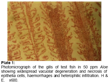
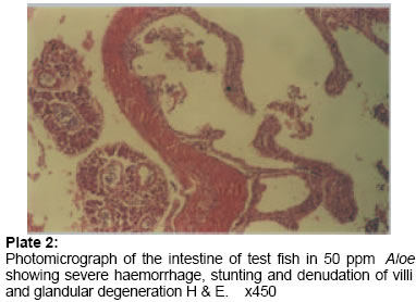
No significant gross and histopathological changes were observed in the tissues and organs of control fish (0ppm A. vera) throughout the period of experiment. Table 2 shows the scores of gross and histologic changes in tissues/organs of the fish in the various experimental tanks. Generally, gross lesions include depigmentation of skin and pale and shriveled gills, and dull, opaque and sunken eyes. Histologic changes include varying severities and spread of stunting and clubbing of gill filaments vacuolar degeneration and necrosis of gill epithelial cells and heterophilic infiltration of the gill submucosae (Plate 1). Others include hyaline degeneration and necrosis of myofibrils and calcification of vasa vasori, vacuolar degeneration and necrosis of hepatocytes, and to a lesser degree, pancreatic cells and infiltration by melanomacrophages. There was severe villous atrophy, characterized by denudation and stunting of villi, congestion, glandular degeneration, necrosis and lymphocytic infiltration in the submucosae of the intestines (Plate 2) and focal areas of neuronal degeneration, neuronophagia and gliosis in the cerebrum. The most severe histologic lesions were observed in fish in the tank containing 50ppm A.vera. Table 2: Pathology scores of control and test tilapia exposed to varying concentrations of aqueous extract of A. vera
Key to scores:- - = No lesions observed; ± = Mild, focal lesions; + = Mild, multifocal lesions; ++ = Moderately severe diffuse lesions; +++ = Very severe diffuse lesions Table 3: Haematology of rats given oral administration of the water extract of A. vera
* Rats in Groups 1, 2, 3 and 4 received 0, 50, 100 and 150ppm aqueous extract of A. vera, respectively Table 4: Plasma proteins and enzyme activities of rats given oral administration of the water extract of A. vera
* Rats in Groups 1, 2, 3 and 4 received 0, 50, 100 and 150ppm aqueous extract of A. vera, respectively Rat
Experiment Table 6 shows the scores of gross and histologic changes in tissues/organs of the experimental rats. Apart from slightly congested lungs in one of rats on day 14 of the experiment, no observable gross lesions were observed in all the organs of the control rats (0ppm A. vera) throughout the 28-day period of the study. Table 5: Plasma lipids and metabolites (mg/dl) of rats given oral administration of aqueous extract of A. vera
* Rats in Groups 1, 2, 3 and 4 received 0, 50, 100 and 150ppm aqueous extract of A. vera, respectively Table 6: Pathology scores of rats given oral administration of varying concentrations of the aqueous extract of A. vera
Key to scores:- - = No lesions observed; ± = Mild,
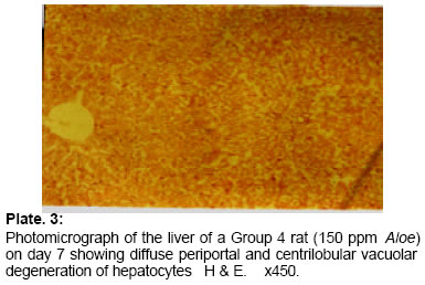
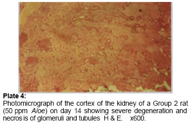
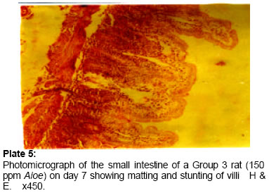
focal lesions Similarly, no gross lesions were observed on the lungs, hearts, brain, intestines, spleen and kidneys of Groups 2 and 3 rats up till 14 days of the study. However, there was a progressive increase in the paleness and sizes of the livers of the rats in Groups 2, 3 and 4 from day 7 of the study onwards. Hepatomegaly was most severe in the rats in Groups 3 on day 14. There was a moderate flabbiness of the heart and increasing mottling of the kidneys of rats in Groups 3 and 4. These were most severe in rats of Group 4 on the 28th day. No gross lesions were observed on the brains and intestines, except that the intestinal contents became progressively pasty and later watery in of all the rats of Groups 2, 3 and 4 from day 7 till the end of the study. Histopathological changes include very mild to moderate pulmonary congestion, mild peribronchiolar and perivascular lymphocytic cuffings and thickened interalveolar septa in the lungs, varying severities of vacuolar degeneration and necrosis of hepatocytes. (Plate 3), Kupffer cell hyperplasia and mild periportal fibrosis, especially on day 28 in rats of Group 4. There was moderate to severe glomerular and tubular degeneration and necrosis (Plate 4). Intestinal lesions include matting and clubbing of villi (Plate 5), catarrhal enteritis and goblet cell hyperplasia. There were focal areas of hyaline degeneration and necrosis of myofibrils and mononuclear cellular infiltration in rats of Groups 3 and 4 on day 28. Brain lesions include meningeal congestion and haemorrhages, perivascular oedema, neuronal degeneration and moderate gliosis. DISCUSSION AND CONCLUSIONS Essentially two products can be extracted from A. vera leaves; the clear gel that forms naturally in the leaf's hollow interior is used to treat skin irritation, and it is an active ingredient in hundreds of skin lotions, toilet soaps, sun blocks and cosmetics (Grindlay and Reynolds, 1986; Kemper and Chiou, 1999; Foster, 2004; Moody et al., 2004). The resin canal cells found in thick leaf epidermis produce a yellow juice (latex) that is used as a laxative and disinfectant (Tyler, 1993; Ghazanfar, 1994; Foster, 2004), and in experimental and folklore medicine for liver complaints, piles, emetic, anti-pyretic, enlarged spleen, cooling agent, skin diseases, tuberculosis, fungal diseases, peptic ulcers, reduction of blood glucose levels, asthma, AIDS, anti-cancer, and as an immunomodulator (Shah et al., 1989; Kemper and Chiou, 1999; Reynolds and Dweck, 1999; Hedendal, 2000). In fact the list is endless. The terms “gel” and “juice” are not clearly defined by manufacturers and often are confused by consumers. The mechanical separation process is not always complete, so A. vera latex can be found in some A. vera gels; hence it is desirable to make the gels as pure as possible, because A. vera latex contains the anthraquinone glycosides - aloin A and B, which are potent laxatives (Tyler, 1994). The results obtained in this study have shown the water extract of A. vera as a very potent toxic substance to fish and rats when consumed. It is especially lethal to fish at as low as 50ppm in water causing 100% mortality within 96 hours. There was severe depigmentation, destruction of the gills, intestines, liver and less so the kidneys and brain, with the worst organ damage occurring in fish with longer exposure of the A. vera extract (50ppm) over 96 hours. The organ and tissue damage in the experimental fish may be due to the direct toxicity of the A. vera extract on organs such as the gills, intestines, liver and heart. Damage, especially to the gills, will affect oxygen exchange and tissue respiration, culminating in organ and tissue hypoxia, degeneration and necrosis. The pH of the various concentrations of the water with A. vera extract in the aquaria are slightly in the acidic range (pH 6.6 – 6.9). A. vera gel is 99% water with a pH of 4.5 (Kemper and Chiou, 1999). Even though, no mortality was recorded in the experimental rats in this study, diarrhoea, catarrhal enteritis, villous atrophy, liver, kidney and heart damage were the hallmarks of the intoxication of rats with A. vera extract, and these were more severe as the concentration of the extract increased. The normocytic normochromic anaemia and significantly increased plasma activity of AST in the experimental are the direct consequencies of several organ damage, especially of the liver, heart and kidneys (Jubb et al., 1995). The hypoalbuminaemia may be related to the profuse diarrhoea and protein-losing gastroenteropathy, as well as hepatic damage (Jubb et al., 1995). One interesting finding however is the lowering of plasma lipids in rats that consumed the aqueous extract of A. vera. This could be interpreted to mean that A. vera may be useful for treating artherosclerotic problems and reduction of plasma lipids. This claim however, needs to be further examined. The major objective of this research was to investigate the effect of unguided and often unrecommended oral administration of A. vera leaves in human, using the tilapia and rats as animal models. This is very important because of the current craze, in Nigeria, by middle and low income, literate, and especially illiterate, old and young men and women who consume often large amounts of raw A. vera leaves due to spurious advertisements and belief in the plant as a “cure all” and “magic” plant, all in the name of “naturopathy” and “alternate” health methods. It is to be noted that most advertisers and users of this plant (and probably so for numerous other herbs) are oblivious of the fact that the parenteral administration of raw A. vera plant is not approved by the FDA (Smith and Struck, 1997; FLP, 2003; Changes International, 2004). Death has been reported of four cancer patients who were treated with A. vera intravenously by a physician whose license was subsequently revoked after investigation and trial by the Maryland Police (Smith, 1997a; Smith and Bloom, 1997; Smith and Lipton, 1997; Smith and Struck, 1997). Brusick and Menge (1997) expressed concern over the safety of A. vera stating that genotoxicity studies showed that A. vera-containing laxatives pose cancer risks to human when used as directed. Acute toxicity with A. vera gel caused severe cramping, diarrhoea and nausea, while long-term ingestion has been reported to lead to potassium deficiency, muscle weakness and cardiac arrhythmias (Kemper and Chiou, 1999). Some of the known contraindications of products containing A. vera include intestinal obstruction or stenosis, atony, severe dehydration with electrolyte depletion, chronic constipation inflammatory intestinal diseases, cardiovascular diseases, pregnancy or lactation, in patients with cramps, colic, haemorrhoids, nephritis, or any undiagnosed abdominal symptoms such as pain, nausea, or vomiting (Hedendal, 2000). While folk medicine has its roots in the very annals of human history, caution must be exercised in the use of some of these herbs, and that claims such as have been attributed to A. vera must be backed by unequivocal scientific evidence. There are many areas of contradictions in most of the advertised therapeutic potentials of A. vera, especially when used parenterally. A few of the notable contradictions include: its use as an emmenagogue and abortifacient (Saha et al., 1961; Singh et al., 1979; Nath et al., 1992) vis-à-vis its use for the prevention of miscarriage (Bhattharai, 1992) and its anti-oxytotic activity (Andrade et al., 1996); the use of A. vera gel in the treatment of peptic ulcers (Blitz et al., 1963) vis-à-vis the ineffectiveness of both the exudate and gel components of A. vera in the treatment of gastric and duodenal ulcers experimentally induced in rats (Parmar et al., 1986); and its acclaimed use in lowering blood glucose level in diabetics (Ghannam et al., 1986; Ajabnoor, 1990; Yongchaiyudha et al., 1996), contradicted by Koo (1994) who reported not only the ineffectiveness of the A. vera gel in lowering blood glucose levels of alloxan-treated rats, but that it actually seemed to have caused an increase. Most of the Nigerians who consume raw A. vera leaves have very strong belief in its alleged megatherapeutic efficacy (personal communication), but are unable to purchase the numerous commercially available “processed” products because of their prohibitive costs, as they belong to the low income group. They then resort to the unguided and continuous consumption of the raw leaves, oblivious of its implications on their health and productivity, not only in the short, but long term. There is no unified recommended dosage information for most A. vera products, whether raw or “processed”, and provision of dosage information by most marketers and advertisers of aloe products does not constitute a recommendation or endorsement (Kemper and Chiou, 1999). Avila et al. (1997) confirmed that aloin and a low molecular weight fraction of the A. vera leaf (Aloe-emodin) are cytotoxic, and should be removed from products processed for commercial use. A high molecular weight fraction of A. vera was shown to deplete complement, while the low molecular weight fraction interfered with processes in activated polymorphonuclear leukocytes, leading to the production of oxygen free radicals and subsequent cytotoxicity (t’Hart et al., 1988, 1990). Very many undiagnosed illnesses, inexplicable deaths and conditions in humans, especially in the under-developed and developing economies of the world, may be related to the consumption of unapproved herbal preparations or products such as from A. vera. While discussing the need for alternative medical practitioners to give evidence for their acclaimed “alternative” health methods, Smith (1997b) stated that the scientific approach to drug or medicant testing is designed to weed out ideas that users wish were true, but most often are not, or in most cases unproved. Herbalists, naturopathics and even drug companies want their products used and sold, but they have to prove that these drugs work and are safe enough (Smith, 1997b). Skeptics may not be willing to accept the plausibility of most paranormal claims, especially in the case being discussed here, unless the evidence is extremely strong, hence the saying that "extraordinary claims demand extraordinary proof" (Gracely, 1998) is justifiable. In conclusion, the oral consumption of raw A. vera leaves, especially when unrecommended, should be discouraged (or at best handled with utmost caution) because of its potential deleterious effects on human and animal health and productivity. It is therefore recommended that the appropriate Food and Drug Regulatory bodies in the country should closely monitor and regulate the administration of A. vera, in any form. The dumping into our land and water bodies of A. vera “wastes” by those who cultivate and “process” the plant should be discouraged in order to prevent the bioaccumulation and magnification along food chains of A. vera’s toxic constituents, and hence cause mass mortality and morbidities in man and animals. Mass education and the provision of adequate and accessible basic medicare to the people are also very imperative. It is to be noted that the areas (or situations) where alternative therapies seem to have most appeal is in the very ones where conventional therapies are either not available, or are not able to satisfy the expectations of the consumer (Alcock, 2001). Even when available, it may be too expensive and unaffordable by the less privileged. REFERENCES
© 2005 Ibadan Biomedical Communications Group
The following images related to this document are available:Photo images[md05031p4.jpg] [md05031p3.jpg] [md05031p2.jpg] [md05031p1.jpg] [md05031p5.jpg] | ||||||||||||||||||||||||||||||||||||||||||||||||||||||||||||||||||||||||||||||||||||||||||||||||||||||||||||||||||||||||||||||||||||||||||||||||||||||||||||||||||||||||||||||||||||||||||||||||||||||||||||||||||||
| |||||||||
