 |
| About Bioline | All Journals | Testimonials | Membership | News |
|
||||||
|
||||||
African Journal of Biomedical Research, Vol. 9, Vol. 1, 2006, pp. 23-29 Full Length Research Article Starch Based Hydrogel with Potential Biomedical Application as Artificial Skin Kunal Pal#, #@Banthia A.K. and *Majumdar D.K. # Materials Science Centre, Indian Institute Of Technology,
Kharagpur-721302, India. Received:
August 2005 Code Number: md06004
ABSTRACT The wound is a biosynthetic environment in which numerous cellular processes are interlinked in the process of repair. Modern dressings are designed to facilitate wound healing rather than just to cover it. Hydrogel dressing can protect injured skin and keep it appropriately moist to speed the healing process by absorbing exudates while maintaining the products of tissue repair, including growth factor and lysosomes, in contact with the wound. The design and development of novel membrane of hydrogel prepared by crosslinking of polyvinyl alcohol with starch suspension using glutaraldehyde as a crosslinking agent was attempted. The membrane was characterized by FTIR spectroscopy. The mechanical property of the hydrogel membrane was characterized by tensile tests. The diffusion coefficient of salicylic acid through the membrane was also evaluated. FTIR spectra of the membrane indicated the absence of free aldehydic groups of glutaraldehyde. The membranes had sufficient strength. The diffusion coefficient of the analgesic drug salicylic acid, used as a model drug, in the prepared starch hydrogels has been measured using a diaphragm cell technique. At 30 °C, the measured value of the diffusion coefficient was approximately 5.03×10⁻¹⁴ m2/s. Keywords: Wound Healing; Hydrogels; Crosslinking; Polyvinyl Alcohol; Starch. INTRODUCTION Hydrogels are hydrophilic natured three-dimensional networks, held together by chemical or physical bonds. Water absorbed by hydrogel is not be released under ordinary pressure. Hydrophilic groups such as hydroxyl (OH) and carboxyl (COOH) in principal and their side chains absorb and store water. If enough interstitial space exists within the network, water molecules can become trapped and immobilized, filling the available free volume [LaPorte, 1997; Mark et al, 1966]. This is the quality that brings about the specific benefit of hydrogels in wound treatment. They are immediately functioning as moist wound dressings and do not need further wound secretions to attain a gelatinous consistency. At the same time they are capable of absorbing the surplus contaminated exudates and safely retaining them within the gel structure. The absorption of secretions causes an expansion of the cross-links in the polymer chains, making room for the inclusion of foreign bodies such as bacteria, detritus, and odor molecules that are irreversibly taken up along with the liquid. The basic physical features of hydrogel dressings can be specifically modified, according to the properties of the polymers used and the additional special equipments of the products. Hydrogels save the wound from fluid loss, are capable of providing the lesion with additional moisture, and securely protect it against external noxae [Krasner, 2001; Falanga, 2000; Mulder and Vande Berg, 2002]. Under the dressing a microclimate is developed, that stimulates and regulates all cellular activities and nutritional processes during the individual phases of wound healing. Additional advantages such as transparency, cushioning effect, cooling effects etc. considerably increase the utility value of hydrogels, in particular concerning patient comfort and ease of application. The high moisture content and the soft-elastic, cushioning properties of the hydrogel almost act like a “second skin”. The dressing adapts perfectly to the wound and has a light cooling effect, which is agreeable to the patient and helps to ease pain. This feature is of special significance in the treatment of superficial epithelial wounds such as donor sites for split skin grafts, which can often be extremely painful, due to the concomitant exposure of free nerve endings lying underneath the epidermis [Hartmann International, 2005]. Even the dressing removal itself is almost painless because hydrogel does not adhere to the wound. Hydrogel stays permanently moist and can even after prolonged application be removed without pain and risk of wound irritation [7-9]. Moreover, malodor is reduced with hydrogel, because the odor molecules are retained in the gel structure along with the absorption of secretions. For the wounds concerned, treatment with hydrogels may bring about great relief for both the patient and the nursing staff. The acceptance of a wound therapy with hydrogel is thus, in general, very high on the part of the patient. Hydrogels of natural polymers, especially polysaccharides, also have been used recently because of their unique advantages. Polysaccharides are, in general, non-toxic, biocompatible, biodegradable, and abundant (Cascone et al, 2001). However, as polysaccharides dissolve easily in water, cannot form stable hydrogel, an effective method is to make them into a synthesized polymer gel networks to form natural and synthesized polymer blend hydrogels, which is becoming a subject of academic as well as of industrial interest. Hydrogels can be applied as an interface between bone and an implant (Netti et al, 1993), as artificial skin (Young and Wu, 1998), as contact lenses (Brinkman et al, 1991), as blood contact materials (Taguchi et al, 1998) and in-controlled release applications for delivery of enzymes, hormones, contraceptives, anticoagulant, etc. (Abusafieh et al, 1997). Biodegradable polymers such as for instance poly(lactic acid) (PLA), poly(glycolic acid) (PGA) and their respective copolymers are already applied in several drug delivery systems (Zhu et al, 1990; Youxin et al, 1993). However, only a few attempts (Heller et al, 1990; Pereira et al, 1998) have been reported in trying to use starch-based polymers in these type of applications; despite being well known that they are biodegradable materials (Bastioli, 1995), they have been proposed in several works to be used as biomaterials [Reis et al, 1996; 1997). Starch is one of the most abundant and cheap polysaccharides. Usually starch includes about 30% amylose (a linear α-(1,4) glucan) and 70% amylopectin (dendritically branched version). Chemically modified starches with improved properties are becoming more and more important in industry application not only because they are low in cost, but mainly because the polysaccharide portion of the product is biodegradable. Chemical modification of starch via graft copolymerization of vinyl monomers onto it has been studied widely in recent years [Athawale and Vidyagauri, 1998; Kiatkamjornwong et al, 2000). But only a few studies on starch polymer blend hydrogels have been reported [Hashim et al, 2000]. In this work PVA/starch blend hydrogels will be prepared by chemical crosslinking technique, and in the meantime the efforts will be maid to characterize the hydrogel. MATERIALS AND METHODS Materials Corn starch (CS), salicylic acid (SA), ethanol and glutaraldehyde (GA) were obtained from Loba-Chemie Indoaustranal Co., Mumbai, India. Polyvinyl alcohol (PVA), mol. wt. 125000, was obtained from s.d. fine- chem. limited, Mumbai, India. Hydrochloric acid 35% pure was obtained from Merck Limited, Mumbai, India. Double distilled water was used throughout the study. GA reagent was prepared by mixing 0.5ml of GA in a solution mixture of 10ml ethanol and 0.05ml Hydrochloric acid. Preparation of hydrogels Fifty ml of 10% PVA solution was taken in a beaker. To the PVA solution 50ml of 5% starch suspension in water was added with constant stirring to get a homogeneous mixture. To this mixture GA reagent (10.55ml) was added with constant stirring. Care was taken to eliminate entrapment of air bubbles during mixing and the mixture was used to obtain a membrane by the conventional solution casting method. The membrane was dried at room temperature. The membrane so obtained was named as CSPVA(rt). Both the membranes were washed thoroughly with distilled water to wash off the hydrochloric acid and GA, if any. Then the membranes were dried at room temperature. Characterization Corn starch (CS) and PVA were subjected to FTIR spectroscopy in the range of 4000-400cm-1 as KBr pellets and the membrane was subjected to Attenuated Total Reflectance FTIR (ATR) spectroscopy in the range of 4000-400cm-1. FTIR spectrophotometer (NEXUS –870, Thermo Nicolet Corporation) was used for the study. The raw materials and the patches were subjected to X-ray diffraction (XRD-PW 1700, Philips, USA) using CuKα radiation generated at 40KV and 40 mA; the range of diffraction angle was 10.00-70.00o 2θ. The tensile strengths of the membranes were determined in the Hounsfield H10KS tensile testing machine. The following test conditions were maintained: a) Cross-Head
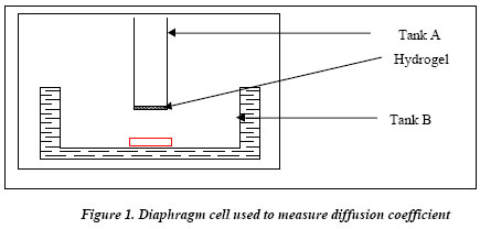
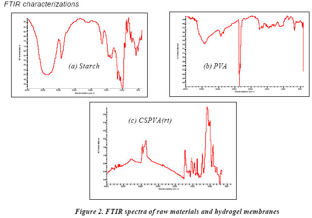
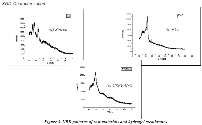
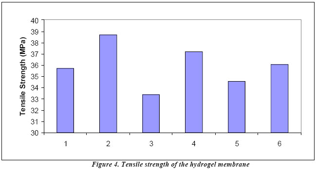
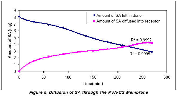
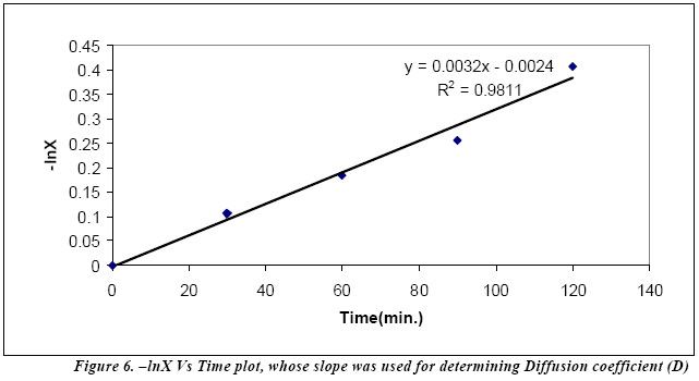
Speed: 50mm/min Measurement of diffusion coefficient: A diaphragm cell shown in Figure 1 was used to measure the diffusion coefficient. The cell consisted of two chambers separated by a film (~ 0.2-mm thick) of the hydrogel. The first chamber contained a known concentration of SA in water (Tank A). The other chamber consisted of filtered deionized water (Tank B). The system was placed in a constant-temperature water bath. A pipette was used to draw 0.1ml sample from donor and 1.0ml sample from the receptor compartment periodically. The samples were analyzed by acidic ferric chloride solution to determine the concentration of SA in each chamber as a function of time. The diffusion coefficient, D, was calculated from these results. The experimentation was conducted at room-temperature (30oC). Distinct peaks of hydroxyl groups can be observed at around 3300 cm-1 from the FTIR spectra of CS and PVA (Figure 2a and 2b). FTIR spectra of CSPVA(rt) (Figure 2c) indicates the absence of free hydroxyl group in the hydrogel membrane. Also there are no free carbonyl group peaks at 1740-1720 cm-1 in the membranes indicating that all the carbonyl groups of GA have been used up for crosslinking. XRD patterns of raw materials and membranes are shown in Figure 3. It can be observed that CS (Figure 3a) had peaks at 15o, 17o, 18o, 23o and 26.5o 2θ where the peak at 18o 2θ was most intense while PVA (Figure 3b) had peaks at 19.5o, 22.5o, and 28.6o 2θ and the peak at 22.5o 2θ was most intense. In the case of PVACS(rt) (Figure 3c) there was only one peak at 19.25o 2θ indicating that the crystallinity of the membrane is mainly due to PVA. (Figure 4) In order to evaluate the mechanical property of the hydrogel membrane, the tensile strength of the membrane was measured. The tensile strength of the hydrogel membrane was found to be 35.92±1.87 MPa that is comparable to the failure strength of skin (34 MPa) (Bhat, 2002). So, the membrane developed can be used as artificial skin that can give a cushioning effect to the wound. Measurement of the diffusion coefficient The desired hydrogels can be produced consistently with the technique outlined above. Typical variations of the concentration of SA in the two tanks during a single experiment are shown in Figure 5. As can be expected, the concentration of the drug in Tank A decreases over time, while there is a corresponding increase in the concentration of SA in Tank B. At any time, t, the concentration values in the two tanks can be used to calculate the diffusion coefficient, D, of the drug in the hydrogel from the following equation: D = 1/(βt) x ln[{C1(t)-C2(t)}/ {C1(0)-C2(0)}] (1) where β = (AH/WH) [(1/V1) + (1/V2)] (2) where: C1(0) =initial concentration of SA in Tank 1; C2(0)=initial concentration of SA in Tank 2; C1(t)=concentration of SA in Tank 1 after time t; C2(t)=concentration of SA in Tank 2 after time t; AH=effective cross-sectional area of diffusion in the hydrogel sample; WH=width of the hydrogel sample; V1=Volume of Tank 1; and V2=Volume of Tank 2. A plot of - ln[{C1(t)-C2(t)}/{C1(0)-C2(0)}] (denoted by –lnX) with time yielded a straight line as shown in Figure 6. The slope of this line was used to calculate the diffusion coefficient, D as indicated in Eq. 1. The diffusion coefficient, D, of the SA through the crosslinked heat-treated CS and PVA membrane was found to be 5.03x10⁻¹⁴ m2/s. The membrane can be used as an artificial skin and various nutrients/healing factors and medicaments can be delivered directly by the process of diffusion to the site of action (wound surface) by putting a swab/hydrophilic matrix containing the nutrients/healing factors and medicaments over the artificial skin. RESULTS AND DISCUSSIONS FTIR characterizations XRD Characterization Conclusion Membrane obtained by crosslinking of corn starch and PVA with GA showed sufficient strength. FTIR spectra of the membrane indicated the absence of free aldehydic groups of GA. The XRD studies suggest that the crystallinity imparted in the crosslinked product of CS and PVA was mainly due to crystallinity of PVA. The diffusion coefficient, D, of the SA through the crosslinked CS and PVA membrane was found to be 5.31x10⁻¹⁴ m2/s. The prepared hydrogel membrane can be used as artificial skin and at the same time various nutrients/healing factors and medicaments can be delivered to the site of action. Acknowledgement The authors are thankful to Indian Institute of Technology, Kharagpur, India for funding the research. The first author is also grateful to his lab-technician and lab-mates (Mr. N. K. Mallick, Mr. A. H. Bhat, Mr. Arfat Anis, Mr. Dibakar Behara, Ms. H. Satpathee, Mrs. S. Mondal and Ms. R. Bana) for their constant encouragement and support during the completion of the work. REFERENCES
The following images related to this document are available:Photo images[md06004f6.jpg] [md06004f3.jpg] [md06004f5.jpg] [md06004f4.jpg] [md06004f2.jpg] [md06004f1.jpg] |
| |||||||||
