 |
| About Bioline | All Journals | Testimonials | Membership | News |
|
||||||
|
||||||
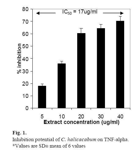
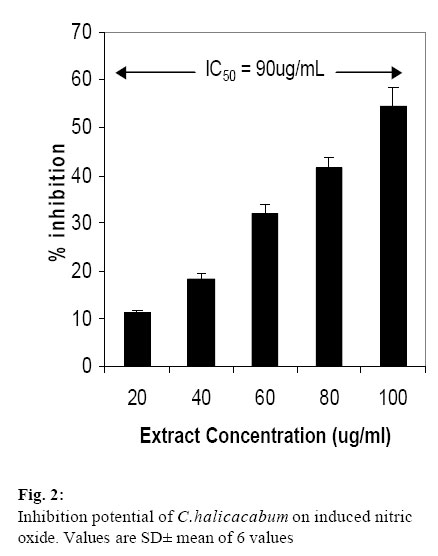
African Journal of Biomedical Research, Vol. 9, Vol. 2, May, 2006, pp. 95-99 Full length Research Article Cardiospermum halicacabum suppresses the production of TNF-alpha and Nitric oxide by Human Peripheral Blood Mononuclear cells Venkatesh Babu K.C. and Krishnakumari S Kongunadu Arts and Science College, Bharathiar University, G.N.Mills (PO), Coimbatore-641 029, Tamilnadu, India *Address for Correspondence: e-mail: drskrishnakumari123@rediffmail.com Phone (office): 91-422-2642095; Fax (office):91-422-2644452 Received: January, 2006 Code Number: md06017 ABSTRACT Cardiospermum halicacabum is an Asian and African herb known to possess anti-inflammatory activities. The inhibitory effects of the ethanolic fraction of Cardiospermum halicacabum leaves extract,(EFC) on the production of pro-inflammatory mediators, nitric oxide(NO) and tumor necrosis factor-alpha (TNF α) were studied in a lipopolysacchride (LPS) activated human peripheral blood mononuclear cells. TNF α production was measured based on the selective sensitivity of TNF α for L929 tumorigenic murine cells. Similarly LPS stimulated macrophages cell line RAW 264.7 was used for NO inhibition studies. EFC potently inhibited TNF α (5-40 μg/mL) with IC50=17 μg/mL, and NO (20-100 μg/mL) with IC50 value being 90 μg/mL.The viability of cells at all the concentration was unaffected as determined by the MTS cytotoxicity assay.C.halicacabum thus exhibits the anti-inflammatory properties, justifying its use in rheumatoid arthritis treatment. Keywords: Cardiospermum halicacabum, nitric oxide, TNF α. INTRODUCTION C.halicacabum is a plant of Sapindaceae family wide spread in tropical and sub-tropical Asia and Africa.In India, C.halicacabum leaves are commonly consumed leafy vegetable. Indian system of medicine recommends C.halicacabum leaves for rheumatism, chronic bronchitis, stiffness of limbs and snakebite (Chopra et al,1980). According to World Health Organisation (W.H.O) still about 80% of the world’s populations rely mostly on plant based drugs. Low cost and easy availability, these factors has generated a renewed interest in plant medicine in the last decade.The traditional practitioners in India prescribe the leaves to the patients without regard to any possible adverse effects in the view of its many uses. In the line of the pharmacological validation of this plant, the toxicological evaluation of C.halicacabum revealed that the drug is safe and is not toxic up to 40g/Kg in rats (Santhakumari et al,1981). It is known to contain saponin, quebrachitol, apigenin, proanthocyanidin and stigmosterol (Dass,1966 and Satyavathi et al,1976). Several experimental arthritis models have been used to mimic human RA, ranging from immunization with cartilage components to infection with joint trophic organisms (Griffiths,1995 and Wooley,1991). These have contributed to the basic understanding of joint disease and to the development of effective anti-arthritic agents. Recent research has demonstrated the importance of the increased production of TNF α and NO in the perpetuation of the inflammatory process of this disease. TNF α is a multifunctional cytokine with important role in immune response and inflammation.TNF α regulates expression of factors including IL-1, IL-6, as well as a group of eicosanoids (Huang et al, 2002) in addition TNF α induces NO in a variety of cells (Tominaga et al, 1996).Blocking TNF α in the experimental models has routinely shown benefit (Williams et al,1992). NO,a free radical derived from the oxidation of the terminal guanidine nitrogen atom of L-arginine by nitric oxide synthase (iNOS).Activated macrophages transcriptionally express the inducible NOS, which is responsible for the prolonged and profound production of NO (Salvemini et al,1996).This uncharacteristic release of NO can lead to amplification of inflammation, as well as tissue injury .Therefore, inhibition of NO production is a very important therapeutic target in the development of anti-inflammatory agents. Due to scanty information regarding this plant’s mechanism of action in the inflammatory conditions, this study was under taken considering the occurrence of TNF α and NO at inflammatory sites and their ability to induce many of the hallmarks in the inflammatory response. MATERIALS AND METHOD Plant material C. halicacabumwas collected in March 2005 from the Kongunadu Arts and Science College campus (Bharathiar University, India) in March 2005 and plant was identified by Dr.V.Balasubramaniam, Department of Botany. Voucher specimen deposited, accession No: 958 .Fresh mature leaves of the plant were dried at 40ºC for 24 hours and powdered. The powder was soaked in 95% alcohol for 72 hours. The supernatant was separated and dried under vacuum. Standard sample solution used in the assay was prepared in DMSO; in the controls DMSO alone was used. Cell Cultures Human peripheral blood mononuclear cells (HPBMC) was obtained by laying diluted blood on Histopaque 1077(Sigma) and centrifuged at 1400 rpm for 30 minutes at room temperature. Murine L929 cells were cultured in Minimum Essential Medium (MEM) containing 2 mM L-glutamine, 1.0mM sodium pyruvate, 0.1mM non-essential amino acids,100 units/ml penicillin, 100 µg/mL streptomycin, 1.5 g/L sodium bicarbonate and 10% horse serum. The cells were harvested using 0.25% (w/v) trypsin and 0.03% (w/v) EDTA. Effect of extract on TNF α production: TNF-alpha production was assessed as reported by Ali (1986) and Taverne et al (1986). HPBMC count was adjusted to 1x10⁶ cells/ml using RPMI 1640 medium (containing 10% foetal calf serum). 100µL of this suspension was added to each well of 96 well plates. The extract was added to this cell suspension at log doses. After incubation for 30 minutes, the cells were stimulated with LPS (1µg/mL) for 4 hours at 37oC in an atmosphere of 5% carbondioxide (CO2). Each concentration was assayed in triplicates. Triplicates of an unstimulated cell control were also maintained. After 4 hours, the plates were spun at 2000 rpm for 5 minutes, the supernatant collected and stored at –80o C until further use. A duplicate plate is also maintained to assess the cytotoxicity of the extract by 3-[4,5-dimethylthiazole-2-yl]-2,5-diphenyltetrazolium bromide (MTS) assay. Murine L929 cell population were suspended to a concentration of 8 x 10⁵ cells / mL. 50 μL of this cell suspension was dispensed into each well of 96 well tissue culture plates. The plates were incubated overnight (20 hours) at 37 °C in a humidified, 5% CO2 atmosphere. 40 µL of the cell supernatant (obtained after centrifugation of plates containing HPBMC) was added to corresponding wells of the plate with L929 cells. Actinomycin D (0.1µg/well) was added to the L929 cells and the volume made up to 100µL/well. The plates were incubated overnight (20 hours) at 37°C in a humidified, 5% CO2 atmosphere. Cell viability assay Cell viability was determined by MTS assay. After the incubation period, the cells were washed twice and incubated with 110 µL of 0.5mg/mL 3-[4,5-dimethylthiazole-2-yl]-2,5-diphenyltetrazolium bro-mide for 2 h at 37°C. The medium was discarded and 100 µL DMSO were then added. After 30 min incubation, the absorbance at 490 nm was measured. Percent cytotoxicity of the compound is calculated by the following equation: % Cytotoxicity = (A-B)/A x100 Where, A is the absorbance of cultures treated with DMSO alone and B is the absorbance of cultures treated with the plant extract. Data are presented as mean of two blood donor. Each assay point is carried out in triplicate. The percent inhibition of TNF-α release was calculated relative to cultures treated with DMSO alone, by the following equation: % Inhibition = (A-B)/A x100, Where, A= percent cytotoxicity of LPS-stimulated cultures treated with DMSO and B = percent cytotoxicity of LPS-stimulated cultures treated with plant extracts. Data are presented as mean of two blood donor. Each assay point is carried out in triplicate. Effect of extract on Nitric oxide (NO) production. The method described byGreen (1982) was essentially followed. The murine macrophage cell line, RAW 264.7 cells were seeded at 8x10⁵/mL in 24 well plates and were activated by incubation in medium containing LPS (1µg/mL) and test samples (extract) at 20 µg/mL to 100µg/mL.NO released from the culture was assessed by determination of NO-2 concentration in the culture supernatant.100µL of culture media were incubated with 150µL of Griess reagent (1% sulfanilamide,0.1% naphthylethylene diamine in 2.5%phosphoric acid solution) at room temperature for 10 minutes in 96-well micro plates. Absorbance at 540nm was read using ELISA plate reader. Standard calibrations were prepared using sodium nitrite. Statistical analysis The results of the experiment were expressed as mean ± S.E.M. The statistical significance of differences was estimated by Student’s t-test for unpaired comparison. When P< 0.05, the difference was considered to be significant. RESULTS TNF α was measured with a cytotoxicity assay using specifically L929 tumourigenic murine cell lines. EFC inhibited TNF α release from LPS stimulated human peripheral blood mononuclear cells significantly, with IC50 value being 17µg/mL. EFC inhibited in a concentration-dependent way, the production of TNF α (Fig1).The results are the SD± mean value of two triplicates each of two blood donor. Similarly the EFC exhibited 54% inhibition of LPS-induced NO production by RAW 264.7 murine cell line at the concentration of 100 μg/mL in cell culture media (Fig 2).The concentration required to inhibit the production of NO by 50 %( IC50 value) was found to be 90 μg/mL. DISCUSSIONS Sera and synovial fluid of RA patients with increased levels of TNF α are found, suggesting that TNF α may play a role in the pathology of the disease (Feldmann and Maini,2001). Apart from TNF α ,numerous factors that play an important role in such disease include, complement, eicosanoids and nitric oxide (Deborah A Ribardo et al,2002 and Anggard,1994).Influence of complement and eicosanoids on inflammation has been well established, however the relevance of NO as mediator of inflammation is more recent (Di Rosa,1996).TNF α , which has been reported to induce the activation of iNOS gene transcriptional factor (Schutze et al,1995)and this resultant induced production of NO has been implicated for immunological and inflammatory diseases including septic shock, rheumatoid arthritis ,graft rejection, and diabetes(Anggard,1994). Blocking of TNF α interrupts the inflammatory process, by inactivation of T cells, macrophages, and fibroblasts (Yazdani-Biuki B et al, 2005). Anti-TNF α therapy induces a rapid improvement in multiple clinical assessment of disease activity including morning stiffness, pain score and swollen joint count (Camussi et al,1998). Thus the cytokine blockers made its presence felt in the treatment of RA and are routinely used to treat patients not adequately responding to traditional disease modifying drugs. However the cytokine blockers havelimitations in that they have many undesired effects, and thereis therefore a need for improved treatments. As a result, new anti-inflammatory and analgesic drugs are being searched all over the world as an alternative to existing drugs. During this process, the investigation of the efficacy of plant based drugs has been paid great attention because they have little side effects. Results of this in vitro inhibition studies on induced expression of TNF α and NO support the pharmacological basis of C.halicacabum used as a medicinal herb for the treatment of rheumatoid arthritis and related inflammatory disorder. REFERENCES
Copyright 2006 - Ibadan Biomedical Communications Group The following images related to this document are available:Photo images[md06017f1.jpg] [md06017f2.jpg] |
| |||||||||
