 |
| About Bioline | All Journals | Testimonials | Membership | News |
|
||||||
|
||||||
African Journal of Biomedical Research, Vol. 10, No. 2, 2007, pp. 165-173 Pharmacological evidences for antiamnesic potentials of Phyllanthus amarus in mice *Hanumanthachar Joshi and Milind Parle Pharmacology Division, Department of Pharmaceutical Sciences,
Guru Jambheshwar University (State Technical University)
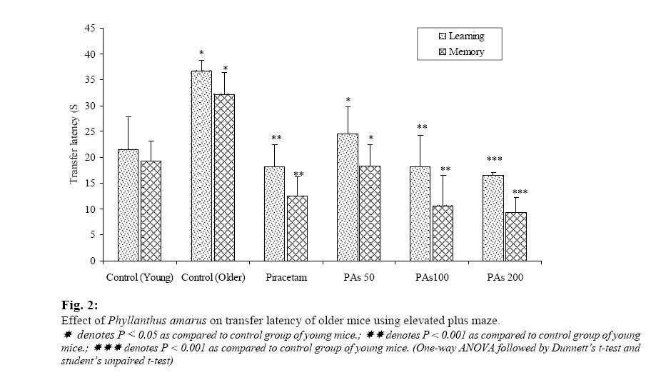
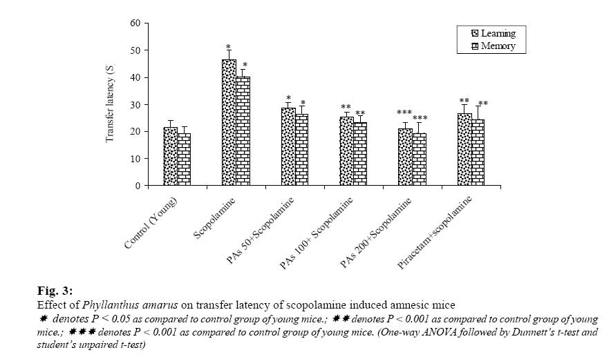
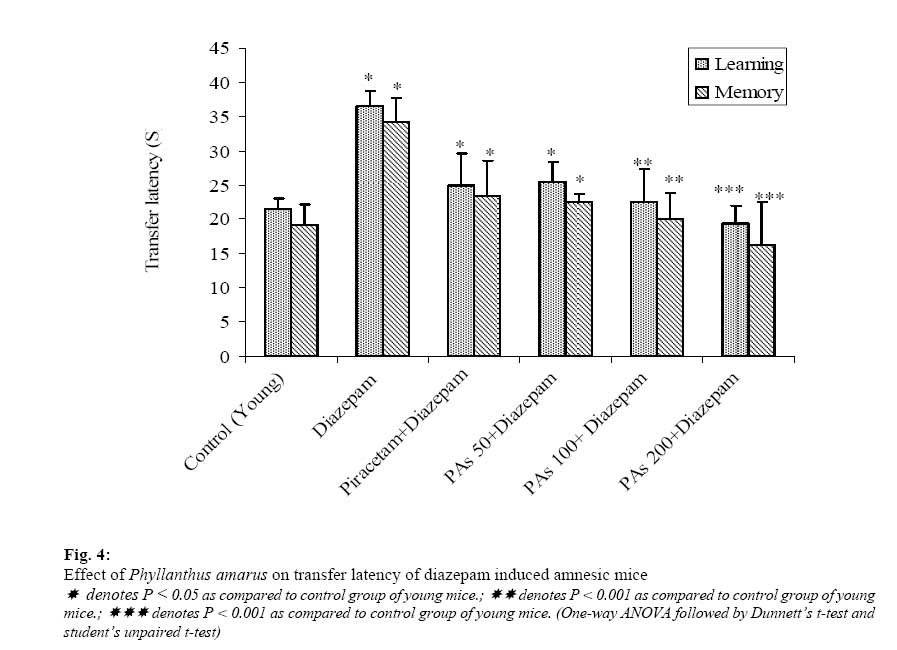
Post Box – 38, Hisar, (Haryana) -125 001, India. Received: September 2006 Code Number: md07022 ABSTRACT Phyllanhus amarus (PAs) is commonly known as Bhumi amla and is traditionally used since centuries in ayurveda. The present study was undertaken to investigate the effects of PAs on cognitive functions and brain cholinesterase activity in mice. PAs (50, 100 and 200 mg/kg) produced a dose- dependent improvement in memory scores of young and older mice. PAs also reversed successfully the amnesia induced by scopolamine (0.4 mg/kg, i.p.) and diazepam (1 mg/kg, i.p.). Interestingly, brain cholinesterase activity was also reduced. The underlying mechanism of action for the observed nootropic effect may be attributed to pro-cholinergic activity exhibited by PAs in the present study. Therefore, it would be worthwhile to explore the therapeutic potential of PAs in the management of patients with cognitive disorders. Keywords: Phylantus amarus, mice, antiamnesic, memory INTRODUCTION Phyllanthus amarus Linn. is commonly known as bhumi amla and is traditionally used to treat flu, dropsy, diabetes, and jaundice (Foo, 1993). It is also used to treat hepatic and urolitic diseases and have diuretic activity. P. amarus is reported to possess antiviral (Lee et al., 1996), anticancer (Joy et al., 1998; Rajesh et al., 2000), hepatoprotective (Prakas et al., 1995), antioxidant (Joy et al., 1995), antiinflammatory (Kassuya et al., 2005) activity. P. amarus mainly contains phyllnathin and hypophyllanthin as active ingredients (Sharma et al., 1993; Somanabandhu et al., 1993). The aqueous extract of P. amarus had been employed for treatment of nervous debility, epilepsy, as medhya (intellect promoting) and in vata disorders. In the present study, the potential of antiamnesic effects of P. amarus was investigated. Enhancement in the life-span of human beings in developed and developing countries has resulted in proportionate increase in the number of patients suffering from senile dementia. Alzheimer’s disease (AD) is said to be the leading cause of dementia in elderly individuals. AD individuals exhibit deterioration in mental functions rendering them incapacitated to perform normal daily activities. However, evidence shows that AD can also afflict young individuals as early as 40 years of age (Sugimoto et al., 2002; Joshi et al., 2005a). AD patients exhibit marked decline in cognitive functions and severe behavioral abnormalities such as irritability, aphasia, apraxia, agnosia and restlessness (Parle et al., 2004a; Khachaturian, 1985). Neuritic plaques (consisting of a core of β-amyloid aggregates covered by dead neurons, microglia and apolipoprotein E) and neurofibrillary tangles are the major pathological lesions of an Alzheimer brain (Selkoe, 2001). Since the allopathic system of medicine is yet to provide a radical cure for Alzheimer’s disease, it is worthwhile to explore the utility of traditional medicines. In the light of above, the present study was undertaken to investigate the influence of P. amarus on memory of mice. The effects of P. amarus on brain acetylcholinesterase activity were also studied. MATERIALS AND METHODS Collection of the plant material: Leaves and stems of P. amarus Linn. were collected from Dehradun, Uttaranchal and were dried at 50 ◦C. A voucher specimen of the plant was identified by Taxonomists at Botanical survey of India, Dehradun. The voucher specimen (HKJ/PA-23) has been kept at Department of pharmaceutical sciences, Guru Jambheshwar University, Hisar, Haryana, India Preparation of aqueous extract: Powdered P. amarus (500 g) was extracted twice overnight with 2000 ml of distilled water at room temperature. The supernatant was collected and evaporated to dryness at 50 ◦C under reduced pressure. The yield of the extract was 10.4% w/w. Animals All the experiments were carried out using male, Swiss Albino mice procured from the disease-free small animal house of CCS Haryana Agricultural University, Hisar (Haryana), India. Young (3-4 months old) mice weighing around 20 g and older (12-15 months old) mice weighing around 35 g were used in the present study. The animals had free access to food and water, and they were housed in a natural (12h each) light-dark cycle. Food given to mice consisted of wheat flour kneaded with water and mixed with a small amount of refined vegetable oil. The animals were acclimatized for at least 5 days to the laboratory conditions before behavioral experiments. Experiments were carried out between 0900 h and 1800 h. The experimental protocol was approved by the Institutional Animal Ethics Committee (IAEC) and the care of laboratory animals was taken as per the guidelines of CPCSEA, Ministry of Forests and Environment, Government of India (registration number 0436). Chemicals The drugs used in this study were obtained from following drug houses. Scopolamine hydrobromide (Sigma-Aldrich, USA), Diazepam injection (Calmpose®, Ranbaxy, India), 5,5-dithiobis-2nitrobenzoic acid (DTNB), acetylcholine iodide, eserine salicylate, sodium dihydrogen phosphate, disodium hydrogen phosphate (Hi-Media, India). Scopolamine hydrobromide and Diazepam injection were dissolved separately in normal saline and injected i.p., volume of i.p. injection was 1 ml/100 g of mouse. Experimental Protocol In the present investigation, the mice were divided in to 52 different groups (n=6) for investigations using various interoceptive as well as exteroceptive memory models and for biochemical estimations. PAs (50, 100 and 200 mg/kg, p.o.) was administered to young and older mice of different groups. These mice were exposed to the training session using elevated plus maze or passive avoidance apparatus on 8th day after 90 min of the last dose. Retention (memory) of the learned task was recorded after 24 hours i.e. on 9th day. Amnesia was induced in separate groups (interoceptive model) of young mice by scopolamine (0.4 mg/kg, i.p.) or diazepam (1 mg/kg, i.p.) on 8th day after 90 minutes of the last dose. Piracetam (400 mg/kg, i.p.), an established nootropic agent was injected for eight days to positive control group of animals. The whole brains were collected after 9 days of administration of PAs. Exteroceptive Behavior Models Elevated Plus-Maze Elevated plus-maze served as the exteroceptive behavioral model to evaluate memory in mice. The procedure, technique and end point for testing memory was followed as per the parameters described by the investigators working in the area of psychopharmacology (Itoh et al, 1990; Joshi et al., 2005b). The elevated plus maze for mice consisted of two open arms (16 cm × 5 cm) and two covered arms (16 cm × 5 cm × 12 cm) extended from a central platform (5 cm × 5 cm), and the maze was elevated to a height of 25 cm from the floor (Joshi et al., 2005c). On the first day (i.e. 8th day of drug treatment), each mouse was placed at the end of an open arm, facing away from the central platform. Transfer latency (TL) was defined as the time (in seconds) taken by the animal to move from the open arm into one of the covered arms with all its four legs. TL was recorded on the first day (training session) for each animal. The mouse was allowed to explore the maze for another 2 min and then returned to its home cage. Retention of this learnedtask (memory) was examined 24 h after the first day trial (i.e. 9th day, 24h after last dose). Significant reduction in TL\ value of retention indicated improvement in memory. Passive avoidance paradigm Passive Avoidance Behavior based on negative reinforcement was used to examine the long-term memory (Sharma and Kulkarni, 1990; Joshi et al., 2006a). The apparatus consisted of a box (27 cm × 27 cm × 27 cm) having three walls of wood and one wall of Plexiglass, featuring a grid floor (made up of 3 mm stainless steel rods set 8 mm apart), with a wooden platform (10 cm × 7 cm × 1.7 cm) in the center of the grid floor. The box was illuminated with a 15 W bulb during the experimental period. Electric shock (20 V, A.C.) was delivered to the grid floor (Parle et al., 2004b). Training (i.e. 30th day of drug treatment) was carried out in two similar sessions. Each mouse was gently placed on the wooden platform set in the center of the grid floor. When the mouse stepped-down placing all its paws on the grid floor, shocks were delivered for 15 seconds and the step-down latency (SDL) was recorded. SDL was defined as the time (in seconds) taken by the mouse to step down from the wooden platform to grid floor with all its paws on the grid floor. Animals showing SDL in the range of 2-15 seconds during the first test were used for the second session and the retention test. The second-session was carried out 90 min after the first test. During second session, if the animals stepped down before 60 seconds, electric shocks were delivered once again for 15 seconds. During the second test, animals were removed from shock free zone, if they did not step down for a period of 60 seconds and were subjected to retention test. Retention (memory) was tested after 24 h (i.e. 9th day, 24h after last dose) in a similar manner, except that the electric shocks were not applied to the grid floor observing an upper cut-off time of 300 seconds (Joshi et al., 2006b, 2006c). Significant increase in SDL value indicated improvement in memory. Biochemical estimations Collection of brain samples: The animals were sacrificed by cervical decapitation under light anesthesia on the 8th day, 90 mins after administration of the last dose of PAs. Immediately after decapitation whole brain was carefully removed from the skull. For preparation of brain homogenate, the fresh whole brain was weighed and transferred to a glass homogenizer and homogenized in an ice bath after adding 10 volumes of 0.9% w/v sodium chloride solution. The homogenate was centrifuged at 3000 rpm for 10 min and the resultant cloudy supernatant liquid was used for estimation of brain acetylcholinesterase activity. Estimation of brain cholinesterase: Brain cholinesterase activity was measured by the method of Ellman et al with a slight modification (Ellman et al., 1961; Voss and Sachsse, 1970). 0.5 ml of the cloudy supernatant liquid was pipetted out into 25 ml volumetric flask and dilution was made with a freshly prepared DTNB (5,5-dithiobis-2nitrobenzoic acid) solution (10 mg DTNB in 100 ml of Sorenson phosphate buffer, pH 8.0). From the volumetric flask, two 4ml portions were pipetted out into two test tubes. Into one of the test tubes, 2 drops of eserine solution was added. 1 ml of substrate solution (75 mg of acetylcholine iodide per 50 ml of distilled water) was pipetted out into both the tubes and incubated for 10 min at 30 ° C. The solution in the tube containing eserine was used for zeroing the colorimeter. The resulting yellow color is due to reduction of DTNB by certain substances in the brain homogenate and due to non-enzymatic hydrolysis of substrate. After having calibrated the instrument, change in absorbance per min of the sample was read at 420 nm (Joshi et al., 2006d). Statistical analysis All the results were expressed as Mean ± Standard Error (SEM). Data was analyzed using one-way ANOVA followed by Dunnett’s t-test and student’s unpaired t-test. P-values < 0.05 were considered as statistically significant. RESULTS Effect on transfer latency using elevated plusmaze Transfer Latency (TL) of second day (day 9th of drug treatment) reflected retention of learned task or memory. The young animals treated with PAs (50, 100 and 200 mg/kg, p.o.) showed dose- dependent reduction in TL of 9th day, indicating significant improvement in memory, when compared with control group (Fig.2). These concentrations of PAs (50, 100 and 200 mg/kg, p.o.) also produced significant improvement in memory (P < 0.001) of older mice (Fig.2). Scopolamine (0.4 mg/kg, i.p.) and diazepam (1 ml/kg, i.p.) injected before training significantly increased (P < 0.05) the TL of 9th day indicating impairment in memory (amnesia) (Fig.3). The mice treated with PAs (50, 100 and 200 mg/kg, p.o.) for 9 successive days) reversed successfully the amnesia induced by both scopolamine and diazepam (Fig.4). Piracetam (used as the positive control) at the dose of 400 mg/kg, i.p. improved memory (P < 0.05) of both young and older mice and reversed the amnesia induced by scopolamine and diazepam as expected. Effect on step-down latency using passive avoidance paradigm Step Down Latency (SDL) of second day (9th day of drug treatment) reflected the long-term memory of animals. Various concentrations of PAs (50, 100 and 200 mg/kg, p.o.) administered to young and older mice for 9 days, showed dose-dependent increase in SDL values as compared to respective control groups (Fig.5 and 6). PAs (50, 100 and 200 mg/kg, p.o.) administered for 9 days reversed memory deficits due to ageing induced amnesia. The groups of mice, which were treated with piracetam (400 mg/kg, i.p.) for seven successive days showed improvement in memory of young as well as older mice. Effect on brain cholinesterase activity PAs (50, 100 and 200 mg/kg, p.o.) showed a remarkable reduction in brain cholinesterase activity in young and older mice, as compared to respective control groups by using Ellman’s kinetic colorimetric method (Fig.7). PAs (50, 100 and 200 mg/kg, p.o.) reduced cholinesterase activity in young and older mice as expected. DISCUSSION There has been a steady rise in the number of patients suffering from Alzheimer’s disease (AD) all over the world. Alzheimer’s disease is a genetically heterogenous neurodegenerative disorder, which is slow in onset but relentless in progress (Palmer, 2002; Parle et al., 2004b). There are around 35 million patients suffering from Alzheimer’s disease all over the world, out of which United States of America alone has around 4.5 million patients (Hebert et al., 2003). Despite the severity and high prevalence of this disease, Allopathic system of medicine is yet to provide a satisfactory antidote. Therefore, neurobiologists all over the world are looking for new directions and alternative strategies for managing this disease of senior citizens. In India AD patents are estimated to be less than 3.5 million (Shaji, 2005). These prevalence figures however, point out that the number of patients suffering from AD are considerably small in India when compared to USA. Therefore, we were motivated to explore the potential of certain nutrients from Indian dishes responsible for this protection against AD. In the present study, P. amarus administered for 8 days improved the memory of mice as reflected by diminished TL and enhanced SDL values as compared to control animals. Furthermore, pretreatment with PAs for 8 days protected the animals from memory deficits produced by scopolamine and diazepam. These findings suggest the possible neuroprotective role for P. amarus. Acetylcholine is considered as the most important neurotransmitter involved in the regulation of cognitive functions. There is an extensive evidence linking the central cholinergic system to memory (Ghelardini et al., 1998; Peng et al., 1997; Olney, 1990; Parle et al., 2004a). Cognitive dysfunction has been shown to be associated with reduced cholinergic transmission and the facilitation of central cholinergic transmission with improved memory (Bhattacharya et al., 1993). Selective loss of cholinergic neurons and decrease in cholinacetyltransferase activity was reported to be a characteristic feature of senile dementia of the Alzheimer’s type (Agnolli et al., 1983). Our research findings using Glycyrrhiza glabra, Myristica fragrance and Zingiber officinale have displayed a link between memory improving effect and cholinesterase inhibition (Joshi et al., 2006e). In the present study, the PAs when administered for 9 days by young and older mice showed significant reduction of brain acetylcholinesterase activity thereby probably facilitating cholinergic transmission and improving memory of animals. Oxygen free-radicals are implicated in the process of age-related decline in cognitive performance and may be responsible for the development of Alzheimer’s disease in elderly persons (Sinclair et al., 1998; Berr, 2002; Butterfield and Lauderback, 2002; Floyd and Hensley, 2002; Perry et al., 2002). Oxygen-free radicals and other byproducts of oxidative metabolism have been shown to be neurotoxic (Sayre et al., 1997; Rogers et al., 2003) and antioxidant rich diets improved cerebellar physiology and motor learning in olderrats (Bickford et al., 2000). Anti-oxidant constituents of P. amarus may be favorably contributing to the memory enhancing effect seen in the present study. Thus, the protective effect of PAs may be attributed to its antioxidant property by virtue of which susceptible brain cells get exposed to less oxidative stress resulting in reduced brain damage and improved neuronal function. In the present study, we observed that PAs inhibited brain acetylcholinesterase enzyme, thereby elevating acetylcholine concentration in brain homogenate and ultimately improved memory of both young and older mice. Immunohistochemical studies suggested the existence of chronic inflammation in certain regions of the brain in Alzheimer’s disease patients. Since inflammation can be damaging to host tissue, it was hypothesized that anti-inflammatory drugs might be inhibiting both the onset and the progression of Alzheimer’s disease. This hypothesis is supported by the observation that indomethacin (NSAID) halted the progressive memory loss seen in Alzheimer’s disease patients (Rao et al., 2002). Moreover, it has also been observed that elderly patients suffering from Alzheimer’s disease showed reduction in symptoms of Alzheimer’s disease upon chronic use of anti-inflammatory drugs. Indomethacin, a nonsteroidal anti-inflammatory drug exhibited a memory protective effect against electro convulsive shock induced retrograde amnesia and also against amyloid deposits in the brain (Stephen et al., 2003). Anti-inflammatory action of P. amarus might also be contributing to the observed memory-enhancing activity (Kassuya et al., 2005). Thus, a combination of anti-inflammatory, neuroprotective, anticholinesterase and antioxidant effects exhibited by PAs may all be eventually responsible for the memory improving effect observed in the present study. Conclusion In the present study, we observed that PAs lowered serum cholesterol levels of mice, inhibited acetylcholinesterase enzyme, thereby elevating acetylcholine concentration in brain homogenate and ultimately improved memory of both young and older mice. Therefore, it would be worthwhile to explore the therapeutic potential of P. amarus in the management of Alzheimer patients. REFERENCES
© Ibadan Biomedical Communications Group The following images related to this document are available:Photo images[md07022f2.jpg] [md07022f1.jpg] [md07022f3.jpg] [md07022f4.jpg] |
| |||||||||
