 |
| About Bioline | All Journals | Testimonials | Membership | News |
|
||||||
|
||||||
African Journal of Biomedical Research, Vol. 10, No. 3, 2007, pp. 249 - 255 Full Length Research Article Pharmacological Evaluation of a Nigerian Polyherbal Health Tonic Tea in Rat *, aAdeneye A A and bBenebo AS aDepartments of Pharmacology and bPathology
/ Forensic Medicine, Lagos State University
College of Medicine, Ikeja, Lagos, Nigeria. Received: December
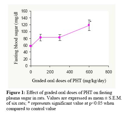
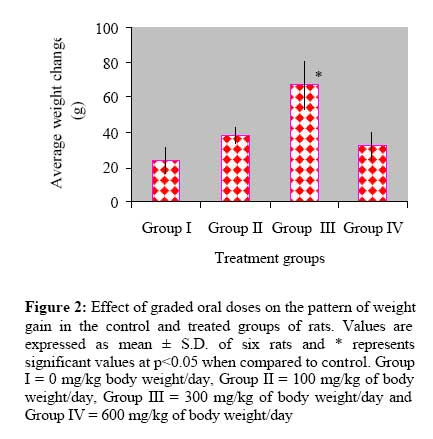
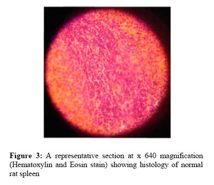
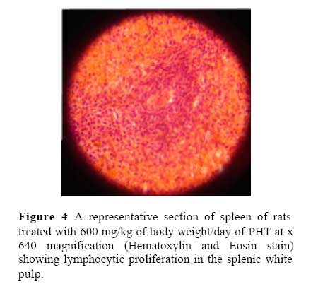
2006 Code Number: md07035 ABSTRACT The present study is a fourteen day study, designed to investigate the hematological and biochemical effects of single, daily oral doses of 100 – 600 mg/kg of a Nigerian Polyherbal Tonic Tea (PHT) in four groups of adult Wistar rats. Acute oral toxicity test of PHT at the limit dose of 5000 mg/kg was also conducted using Up-and-Down Procedure on statistical software program (AOT425StatPgm, Version 1.0.). Results showed PHT to induce significant (p<0.05) dose-related elevation in the packed cell volume (PCV), platelet, total leukocyte counts and lymphocyte differentials, while causing significant (p<0.05) suppression of granulocyte differentials in dose-related fashion. PHT, also, induced a significant (p<0.05) dose-dependent rise in the fasting blood sugar which was at variance with its folkloric use as an oral hypoglycemic agent. PHT did not induce mortality at the tested limit oral dose, indicating its relative oral safety up to 5000 mg/kg on acute exposure. Key words: Nigerian Polyherbal Tonic Tea; Hematological indices; Fasting blood sugar; Acute Oral Toxicity; rats INTRODUCTION In the last two to three decades, there has been a renewed interest into research and utilization of medicinal plants, particularly, flora of the tropical rainforest (Soejarto, 1996). The revived interest in plant-derived drugs is attributed to the current widespread belief that “green medicine” is cheap, safe, more dependable and accessible than the costly synthetic drugs many of which are associated with intolerable effects (Parek and Chanda, 2006; Venkatesh and Krishnakumari, 2006). Despite achievements recorded in drug discovery and development from plant sources, Phytomedicine continues to be highly valuable for developing synthetic pharmaceuticals employed in the treatment of both human and animal diseases. According to the World Health Organization (WHO), about 80% of the world’s population depends wholly or partly on plant-derived pharmaceuticals (WHO, 1996) Correspondingly, in most developing countries, including Nigeria, there is a heavy dependence on herbal preparations for the treatment of human and animal diseases despite the availability of conventional pharmaceuticals (Nwabuisi, 2002). This is because the exorbitant cost of most conventional pharmaceuticals prevents most people from being able to acquire them. Thus, the need for pharmacognostic and pharmacological evaluation of these medicinal plants may lead to discovery of novel effective compounds cannot be underscored (Janovska et al., 2003). Such research could lead to new drug discovery and or advance utilization of indigenous herbal medicine for orthodox treatment (Parek and Chanda, 2006). Although medicinal potentials and attributes of many medicinal plants have been investigated and validated, drug development from these plants has been slow, which can be related to factors such as non-availability of appropriate co-ordination in the chemical, pharmacological and clinical aspects of the various scientific investigations (Ajaiyeoba et al., 2004). Polyherbal Health Tonic Tea (PHT) is one of the several polyherbal remedies in Nigeria, used in folkloric medicine in South-West Nigeria for the treatment of an array of diseases affecting humans. It is composed of pulverized, dried leaves of Persea Africana (Lauraeae), Morinda lucida (Rubiaceae), Magnifera indica (Anacardiaceae), Carica papaya (Caricaceae), Vernonia amygdalina (Compositae)and Cassia occidentalis (Caesalpiniaceae), all combined in equal weight ratio.PHT is used for the treatment of pain, blood deficiencies, hypertension, diabetes mellitus, malaria, fever, inflammations, as immune booster and in the improvement of blood circulation. However, despite the extensive use of PHT in the treatment of blood deficiencies, scientific data on the validation of its folkloric use in the treatment of this clinical condition is lacking. Thus, the present study aims at evaluating the hematological and biochemical effects of the PHT in young adult Wistar rats. MATERIALS AND METHODS Plant materials and aqueous extract preparation Three 60g packets of the polyherbal formula were purchased from a retail outlet for PHT in Maryland, Lagos State, Nigeria in September, 2006. 25 g of PHT was extracted with 250 mL of distilled water by hot extraction for 1 hr at 100 °C. The decoction was allowed to cool, after which it was filtered with a sterile, white cotton cloth. The filtrate was then completely dried to deep-brown, fine powder using an aerated oven (Genlab Laboratories, U.K.) preset at 50 ºC for 48 hours. The extraction was repeated 3 times. The average % yield was 12.0 ± 1.0 %. The crude extract obtained was stored in tight-fitted capped containers and stored at 4 ºC for 2 days before the commencement of the experiment. From this stock, fresh concentration of 100 mg/ml of the extract was prepared daily and corresponding oral doses (100, 300 and 600 mg/kg body weight) for each rat was calculated based on individual rat weight. Experimental Animals and their care Experimental procedures involving the experimental animals and their care were conducted in compliance with the Guidelines for Care and Use of Laboratory Animals in Biomedical Research as promulgated by the Canadian Council of Animal Care (1984) and United States National Institutes of Health (1985).A total of forty, young adult Wistar rats, weighting 105 - 120 g were obtained from the Nigerian Institute of Medical Research, Yaba, Lagos, Nigeria, after ethical approval had been obtained from the ad hoc Animal Ethical Committee of the Lagos State University College of Medicine, Ikeja, Lagos State, Nigeria. The rats were fed a standard rat chow (Livestock Feeds, Ikeja, Nigeria) and water ad libitum and were maintained at standard laboratory conditions (12/12 hr dark/light cycle, 23 ± 1 ºC temperature, and 55 ± 3 % humidity). Acute oral toxicity studies of PHT in rats using limit dose test of Up-and-Down Procedure Acute oral toxicity study was conducted using the limit dose test of Up-and-Down Procedure according to Organization for Economic Co-operation and Development (OECD) Test Guidelines on Acute Oral Toxicity under a computer-guided Statistical Program (AOT425statPgm, version: 1.0.), at a limit dose of 5000 mg/kg body weight per oral route and default of Sigma at 0.5., as adopted by Adeneye et al. (2006a). Behavioral manifestations of acute oral toxicity such as tremors, convulsions, sleep, altered feeding, salivation, altered somatomotor activities, diarrheal etc., were also noted. All observations were systematically recorded and individual records being maintained for each rat. Oral administration of PHT Before the experiment began, rats were fasted overnight for 14 – 16 hours. Group I, which was the control group, received 10 mL/kg distilled water, orally, throughout the study period while Groups II - IV were orally administered single, daily doses of 100, 300 and 600 mg/kg of body weight of the extract dissolved in 10 mL/kg distilled water, respectively, for 14 days. Body weight Measurement The body weights of rats were measured on days 0 and 15, respectively, with a mettler weighing balance (Mettler Toledo Type BD6000, Mettler-Toledo GmbH, Greifensee, Switzerland) and the difference in weight in reference to the initial weight per group was calculated. Blood sample collection and bioassays Prior to termination of the experiment on day 15, the rats were fasted overnight but distilled water was made available ad libitum. Blood samples were collected through cardiac puncture under halothane anesthesia, using 21 gauge (21G) needles mounted on a 5 ml syringe (Hindustan Syringes and Medical Devices Ltd., Faridabad, India) into Ethylene Diamine Tetra-acetic Acid (EDTA)-coated sample bottles for full blood count (FBC). After the blood collection, the rats were sacrificed. Blood samples for FBC which included packed cell volume (PCV), platelet count, total and differential leukocyte counts, were assayed using standard procedure as described by Schlam et al. (1975). PCV was determined using micro-hematocrit method after spinning the blood-filled, heparinized Vitrex NRIS micro-hematocrit tube (Modulohm A/S, Herlev, Denmark) in Hawksley micro-hematocrit centrifuge (Hawksley and Sons Ltd., Sussex, England) at 12,000 g for 15 min. The total leukocyte counts were determined using improved Neubaeur hemocytometer and FBS was determined using One Touch Basic Blood Glucose Monitoring System (LifeScan Inc., Milpitas, California, U.S.A.), respectively. Histopathological studies of rat spleen After the animals were sacrificed, postmortem examination was performed and the spleens were identified and carefully dissected out en bloc for histopathological examination. After rinsing the dissected spleen in normal saline, sections were taken from each harvested spleen. The tissue was fixed in 10% formo-saline, dehydrated with 100% ethanol solution and embedded in paraffin. It was then processed into 4-5 µm thick sections stained with hematoxylin-eosin and observed under a photomicroscope (Model N - 400ME, CEL-TECH Diagnostics, Hamburg, Germany). Statistical Analysis Results were presented as mean ± standard deviation (S.D.) for body weights while data for hematological and biochemical indices were expressed as mean ± standard error of mean (S.E.M.) of six observations. Data were statistically analyzed using two-way analysis of variance, then subject to Student’s t-test, to determine their level of statistical significance on statistical computer software program, SYSTAT 10.2. Statistical significance was considered at p<0.05. RESULTS Results and sequence of acute oral toxicity of PHT in rats using limit dose test of Up-and-Down Procedure Table 1 shows sequence and results of PHT in rats sequentially treated with oral 5000 mg/kg of body weight of the extract. The extract was not associated with mortality in any of the 3 sequentially treated rats. However, the dose was associated with behavioral abnormalities such as decreased appetite, decreased exploratory behavior, lethargy, tremor, diarrhea and initial hyperthermia followed by sustained hypothermia which lasted for 4-24 hour post-oral administration (rectal temperature of 38.5 ± 2.5 °C and 35.5 ± 1.2 °C, respectively). Effect of 14-days of oral administration of PHT on packed cell volume, platelet count, total and differential leukocytes count in normal rats Table 2 depicts effect of 14-days of oral PHT treatment on the investigated hematological indices in treated rats. As shown, PHTinduced significant (p<0.05) elevations in the PCV, platelet count, total leukocyte counts and the lymphocyte differentials dose dependently. Conversely, PHT caused dose-dependent suppression of granulocyte differential, the most significant (p<0.05) of which was seen at the highest oral dose. Table 1:
Table 2:
Values are expressed as mean ± S.E.M. of six rats; *, a represents significant increase while *, b represents significant decrease at p<0.05 when compared to control value Effect of 14 days oral administration of PHT on fasting blood sugar (FBS) in rats Figure 1 depicts effect of 14-days of oral treatment of PHT on the fasting blood sugar in rats. PHT was noted to have induced significant (p<0.05) elevation in the FBS in dose related pattern. Effect of 14 days oral administration of PHT on average body weight in rats Figure 2 depicts effect of oral graded doses administration of PHT on the mean body weight gain of treated rats over 2 weeks of treatment. PHT significantly (p<0.05) elevated the pattern of weight gain in a dose-unrelated fashion. Histopathological results of rat spleen treated with oral PHT for 14 days Figures 3 and 4 are photomicrographs (at x640 magnification, Hematoxylin and Eosin stain) depicting effects of 2 weeks of oral treatment with PHT on the spleens of control and rats treated with 600 mg/kg of body weight/day of PHT. The extract induced diffuse lymphocyte proliferation around the splenic white pulp (Figure 4). DISCUSSION Blood as an index of physiological and pathological status in humans and animals is well documented (Schlam et al., 1975; Effraim et al., 1999; Ogwumike, 2002) and the most frequently investigated hematological parameters include hemoglobin, packed cell volume, white blood cell count, and platelets count (Schlam et al., 1975). In accord with this principle, packed cell volume, platelet count, total and differential leukocyte counts were measured in the present study. The critical role of stem cells or progenitor cells in hematopoiesis is well documented (Weissman, 2000). The hematopoietic stem cells are the progenitor cells for erythrocytes, platelets and the various subsets of leucocytes, which develop in the bone marrow throughout life (Baker et al., 1979). In the rat marrow population, erythroid cells constitute 39%, myelopoietic 34%, lymphopoietic 24% and reticulum cells 3% (Hulse, 1964). Production of monocytes and neutrophilic granulocytes from the myelomonocytic stem cell develops through an antigenically heterogenous (Ferrero et al., 1983)progenitor population known as the colony forming unit granulocyte macrophage (CFU-GM) [17] (Bradley and Metcalf, 1966). In Wistar rats, the mean normal value range of PCV and total leukocytes count are documented to be 40.5 – 53.1% (Shaw and Maclean, 1971) and 7,063 – 8,760 cell/mm3, respectively (Iranloye, 2002). However, literature has shown that oral ingestion of medicinal compounds or drugs can alter the normal range of these measured hematological parameters (Abatan and Arowolo, 1989; Ajagbonna et al., 1999). In the present study, 14-days of oral administration of 100-600 mg/kg of body weight/day of PHT elevated PCV from 34.7 ± 1.4 % to 41.3 ± 1.5 % and total leukocyte count from 4,533 ± 363 to 6,550 ± 700 cells/mm3, in a dose dependent pattern. Similar pattern was recorded for platelet count and lymphocyte differentials from 254,000 ± 37,000 to 408,000 ± 21,000 cells/mm3 and 73.3 ± 2.11 to 91.3 ± 2.4 %, respectively. It is possible that PHT possesses active constituent(s) containing erythropoietin-like substance(s) or contain active biological principle(s) stimulating erythropoietin synthesis or release either by inducing hypoxia or directly acting on the kidneys to cause erythropoietin secretion. Also, the dose-related increase in the platelet and total leukocyte counts could be due to release of the glycoproteins, thrombopoietin and leukopoietin, in a similar way as impaired oxygenation of the kidney induces erythropoietin release (Erslev and Gabuzda, 1979). However, these hypotheses still require validation. On the contrary, graded oral doses of PHT lowered the granulocyte differentials of the leukocytes in a dose-related fashion (Table 2). This suggests that PHT could have either selective toxicity for the granulocyte lineage or inducing vascular margination of the granulocytes while promoting proliferation of lymphocytes. The lymphocyte proliferative effect of PHT was corroborated by the histopathological findings which showed some degree of lymphocyte proliferation around the splenic white pulp (Fig. 4). The dose-related elevation in fasting blood sugar is also significant. Oral administration of PHT was shown to elevate fasting blood sugar in dose-related pattern; the most significant (p<0.05) effect was observed at 600 mg/kg of body weight/day (Fig. 1). This result is at variance with its folkloric use as an oral hypoglycemic agent. The exact mechanism(s) of inducing hyperglycemia was not elucidated in the present study. Further studies are required to validate the diabetogenic hypothesis. Results of the current study also showed that PHT induced a dose-unrelated weight gain in the PHT-treated group of rats with a significant (p<0.05) value seen at 300 mg/kg day when compared to the control group (Fig. 2). Result of the acute oral toxicity for PHT also indicated that 5000 mg/kg of body weight of PHT was not associated with death. According to Bruce (1985, 1987) and American Society for Testing and Materials (1987), any chemical substance with LD50 estimate greater than 2000 – 5000 mg/kg of body weight/oral route could be considered of low toxicity and safe in humans. Thus, PHT can be considered relatively safe on acute exposure even if taken in doses up to or more than 2000 – 5000 mg/kg of body weight. In conclusion, results of our study lend supports to the folkloric use of PHT in the treatment of blood deficiencies. Acknowledgements The authors are highly indebted to Mr. M.O. Arogundade of Haematology and Blood Transfusion Department, Lagos State University College of Medicine, Ikeja, Nigeria, for his technical assistance. REFERENCES
The following images related to this document are available:Photo images[md07035f4.jpg] [md07035f3.jpg] [md07035f1.jpg] [md07035f2.jpg] | ||||||||||||||||||||||||||||||||||||||||||||||||||
| |||||||||
