 |
| About Bioline | All Journals | Testimonials | Membership | News |
|
||||||
|
||||||
African Journal of Biomedical Research, Vol. 11, No. 1, Jan, 2008, pp. 115-118 Short communication Histopathology of Tilapia tissues harbouring Clinostomum tilapiae parasites Adeyemo A.O1* and Agbede, S.A2 1Department of
Fisheries Technology, Nigeria
Delta University, Wilberforce
Island, Bayelsa State. Received: December
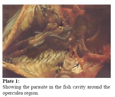
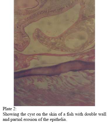
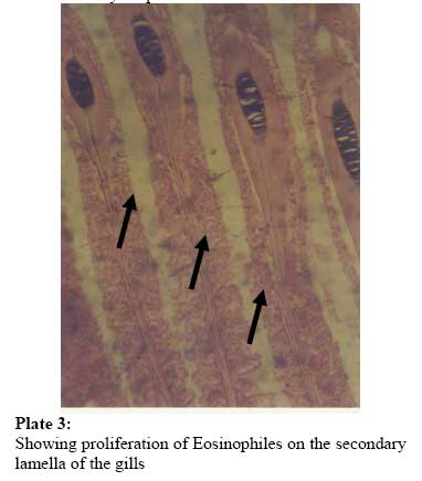
2006 Code Number: md08016 ABSTRACT Tissues obtained from infected Oreochromis niloticus were processed sectioned and stained with haemotoxylin and eosin. Good sections were selected, studied and photographed. The histopathology revealed a proliferation of eosinophiles at the secondary lamellar of the gills. The site of attachment on the fish skin showed the cyst to be double walled, the metacercariea is suspected to produce this cyct as a form of defensive mechanism to wall off and prevent dislodgement. Other tissues did not show observable lesions. Key words: - Oreochromis niloticus, infection, Clinostonum tilapiae, histopathology INTRODUCTION The impact exerted by parasites on host could be mechanical, chemical or physical. Effects of parasitic infection on fish are of notable importance, for instance respiratory function of the skin and gills of fish are disturbed by gyrodactylus, dactylogrus and argulosis infections, causing the fish to become dull, feeble, frequently swimming to water surface with erratic movement and may die of exhaustion (Moore, et al 1984) Metacercariea of the trematode Clinostomum marginatum were known to cause considerable damage to the viscera and musculature of many fish species both wild and cultivated from North America (Hoffman & Meyer 1974), Although the pathogencity of Clinostomum species has not been given prominency in the past, but insignificant conditions have been reported on fish being second intermediate host of the parasite. This study presented a histopathological aspect of the Clinostomum tilapiae infection on Oreochromis niloticus tissues, harbouring the parasites namely the skin and the gills. MATERIALS AND METHODS Tissues of Oreochromis niloticus that were infected by Clinostomum tilapiae were obtained from the Dept. of Wildlife and fisheries Management fish farm, University of Ibadan. The tissues were fixed in Bouin’s fluid for 24hrs. the fixed materials were transferred and processed through ascending graqdes of alcohol, dried in a wax miscible agent and impregnated in wax. Sectioning was carried out on a rotary microtome at 5mm. Sections were floated on warm water at 48oC and mounted on chemically clean slides coated with egg albumin. The mounted, unstained sections were dewaxed in three stages of xylene at 1 minute each and actual staining was carried out using the haemoxylin and eosin staining was carried out using the haemoxylin and eosin staining technique. (Bullock, 1978). Stained mounted sections were examined under light microscope for good ones that were selected for photomicrography. Photographs were taken at x40 magnification of microscope eye piece using the camera at 50mm focal length. RESULT AND DISCUSSION Skin, body cavity and phaygeal region of Clinostomum tilapiae was found as cysts, on the niloticus, shown in plate I and II Helminthes generally harm their host through mechanical damage produced by devouring host tissues. The metacercariea cyst of clinostomo tilapiae are produced by the epithelia of the fish in reaction to infection, this may also cause inrritation on the skin and excess mucus secretions leading to an inflammatory response at the site of attachment. The cyst is double walled as shown in PLATE II. Containing the metacercane. The skin produced this cyst as a defensive mechanism to prevent further penetration. Amlacher (1966) observed that adult trematode may not invade organs by embedding in the tissues but only attaches to convenient site where they may be able to obtain all its nutrient requirements. In this study the cysts were obtained on the skin and pharyngeal region and Ractiliffe, (1968) found that C tilapia actually has the physiological adaptation of upward turning towards the mouth rather than going into the intestine because of the oxygen requirement. Eosinophilic dermatitis associated with C complanatum infection on the skin of Tilapia species was found by Garcia et al, (1993). Eosinophiles proliferation on secondary iamella of las shown the gills in PLATE III can be explained by the findings of Barnett et al (1996), when eosihophilic granulocytes. Occurred abundantly on the skin, gut and haemopoietic tissues of many fishes as a result of handling stress and helminthic infections. This also showed that the defensive mechanism of the host was functioning adequately. Coulibay et al (1995) found no serious pathology on the fish infected by clinostomids except that the fish were unsightly causing its rejection. However, clinostomids infection may retard growth, cause weight loss and pronounced exophthalmus. The presence of the cysts on the skin and pharyngeal region predispose fish to stimulate wide range of responses varying from very mild inflammatory infiltrate to an extremely severe acute narcotizing lesion which can be fatal or just an acute inflammatory episode followed by encystment, fibrosis and ultimately death of the parasite if its life cycle does not continue by predation on the fish host by birds as the final host (Roberts 1978). Clinostomum tilapia was found as cysts on the skin, in the body Cvity and at the pharyngeal region of oreochromis niloticus on examination as shown in PLATE I AND II. The cyst is double walled as shown in PLATE III when attached to the fish skin and these cysts were produced by the epithelia of the fish in reaction to infection and also as a defense mechanism to prevent further penetration. The production of the cyst may also cause irritation on the skin and excess mucus secretions leading to an inflammatory response at the site of attachment. No gross lesions was observed in any of the internal organs having the cyst of C tilapia the metacercaue the was suspected to secrete substances at the pharynx, that causes the proliferation of oesinophiles at the gills and capillary congestion with the presence of melonomacro phages centees. REFERENCES
Copyright 2008 - Ibadan Biomedical Communications Group The following images related to this document are available:Photo images[md08016f3.jpg] [md08016f2.jpg] [md08016f1.jpg] |
| |||||||||
