 |
| About Bioline | All Journals | Testimonials | Membership | News |
|
||||||
|
||||||
Middle East Fertility Society Journal, Vol. 9, No. 1, 2004, pp. 58-65 Laparoscopic management of ovarian dermoid cysts Osama Shawki, M.D., Ihab Soliman, M.D., Alaa Ebrashy, M.D., Mustafa El Sadek, M.D., Abeer Bahnassy, M.D. Department of Gynecology, Cairo University and department of pathology, national cancer institute, Egypt Correspondence: Osama Shawki, Al Ebtesama Hopsital, 10 Aboul Magd Amer Street, Heliopolis, Cairo, Egypt, Tel: 202 4170111, Fax: 202 2912775, E mail: romeo@menanet.net Received
on July 2, 2003; Code Number: mf04009 ABSTRACT Objective: To evaluate the safety and potential
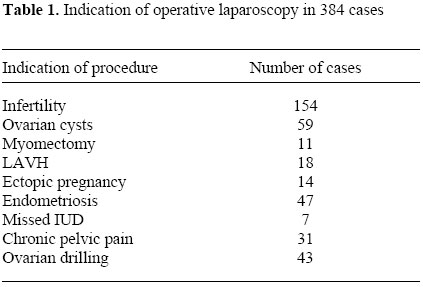
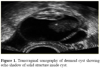
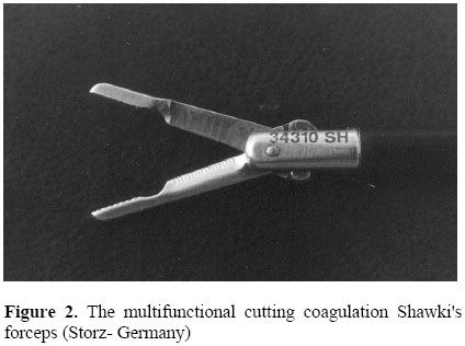
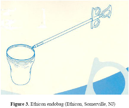
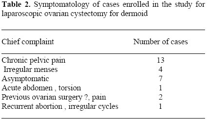
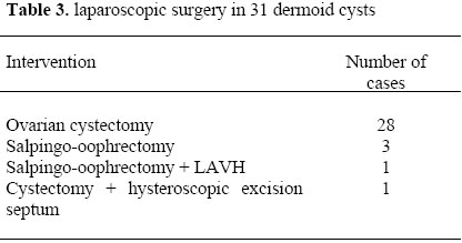
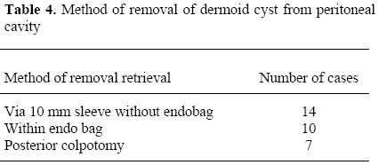
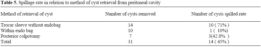
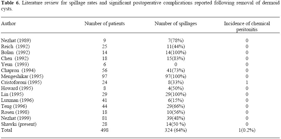
advantages of laparoscopic approach for management of ovarian dermoid cysts. Keywords: Laparoscopy, dermoid cyst, ovarian cystectomy, spillage. Benign cystic teratoma, or as commonly addressed dermoid cysts, are basically germ cell tumors of the ovary. Pathologically, they are enrolled under group of benign mature teratomatas. They account for about 20 - 25% of all ovarian neoplasm and occur bilaterally in 10 - 15% of cases (1). The prevalence of malignant transformation in dermoid cysts has been reported as 1- 3 % (2, 3). Most of dermoid cysts occur without significant clinical symptoms and they are often discovered incidentally during pelvic examination or routine ultrasound. The potential for complications such as torsion, spontaneous rupture, risk of chemical peritonitis, and malignancy usually makes surgical treatment quite necessary upon diagnosis (4). Most operative textbooks describe classical treatment for dermoid cysts have been ovarian cystectomy or oophrectomy through laparotomy with utmost care to avoid spillage of cyst contents. Although laparoscopic surgery has replaced many standard laparotomy techniques, however many skeptical opinions were hesitant about role of laparoscopic surgery for dermoid cyst removal. The potential fear of spillage of cyst material and possible development of chemical peritonitis imposed fears of adopting laparoscopic approach (4, 9). The phobia of spillage complications existed for long time until recently challenged by many laparoscopists (4, 9). Accordingly, laparoscopic approach has become increasingly accepted and more commonly adopted since 1989 (4). Because most cases with benign cystic teratoma are of reproductive age and wish to preserve fertility, a conservative approach is ideal to minimize post operative adhesions and thus decrease chances to compromise fertility (4). Trained endoscopic surgeons became more confident to approach dermoid cysts via endoscopic rout and reported satisfactory results and no complications (4, 16). In our study, we evaluate the safety and efficacy of laparoscopic management of benign cystic teratoma and present some guidelines and tips to improve results of surgery and avoid possible complications that may result from cyst spillage. We also demonstrated that the use of endobag creates satisfactory and easy removal of cysts however; removal without impermeable bag gives the same safety and results if certain guidelines were followed. MATERIALS AND METHODS Our study included Twenty-eight patients with diagnosis of unilateral or bilateral dermoid cysts. Cases were recruited among 384 operative laparosocpies conducted during the period from May 1999 to February 2002 (Table 1). All patients did preoperative evaluation including transvaginal sopnography and Doppler studies to confirm nature of cysts (figure 1). Conclusive confirmation for the cysts was confirmed by the pathological examination for the specimens removed in all cases. Patient charts were revised for demographic data, chief complaint, obstetric history, preoperative investigations, details of operative technique, method of cyst removal, cyst incidence of spillage, blood loss, operative time, postoperative complications, duration of hospital stay, postoperative complications and pathology reports. All patients were counseled for the procedure and informed consent was obtained to do laparoscopic management. All operations were conducted under general anesthesia with endotracheal intubation. We utilized Karl - Storz operative endoscope and camera (Tuttlingen, Germany). We used an infraumbilical 10 mm trocar for the telescope and two 5 mm trocars for secondary punctures and operative instrumentation. Pneumoperitoneum was achieved using Storz Laparflator and then bowels were retracted to upper abdomen using fan retractor. Then diagnostic laparoscopic inspection was conducted thoroughly to evaluate the pelvis and upper abdomen. We strongly recommend against steep Trenedelenberg's position to avoid any chance for migration of spilled material to upper abdomen during surgery. This might be little awkward to us during surgery but we compensate for this by proper bowel retraction and positioning of bowels to upper abdomen using the fan retractor. After we obtain clear view for pelvis, we do lysis of any existing adhesions to allow free mobilization and dissection of cysts. In cases designed for ovarian cystectomy, we applied the rules recommended by Nezhat et al (4). We added some modifications from our side to facilitate surgery and add safety in case any spillage happen. A grasper forceps was used to apply traction on ovarian ligament and steady the ovary. Combined uterine manipulation (Zummi uterine manipulator) plus grasping ovarian ligament allows keeping the ovarian cyst steady during steps of cystectomy. This was facilitated by squeezing the ovary between body of uterus and lateral pelvic wall to maintain steady and easily accessible dissection of the cyst. First, cleavage plane was created by diathermy spatula or Maryland's forceps. A plane is widened between cyst capsule and stroma and hydro dissection continue the enucleation steadily. Combined hydro dissection with blunt pealing of capsule will complete the job easily. We used the Shawki's cutting coagulation forceps (Karl-Storz, Tuttlingen, Germany) for dissection and haemostasis (Figure 2). It has a great value for multifunction as its narrow tip allows precise dissection of cyst capsule, also grasping and traction with its serrated edge was quite secure. The blade of the instrument allows monopolar coagulation of any bleeding points added to its sharp scissors on its proximal end of the blade that was used for precise incision of tissues. Actually, it saves time that may be consumed for exchanging instruments as well as preserves the pneumoperitoneum stable throughout surgery. Grasping the edges of the cyst and slaw traction apart will undress the cyst capsule and deliver the dermoid out of its bed. Eventually, the cysts were enucleated easily and haemostasis was performed for any bleeding spots encountered during dissection. Due to thick nature of dermoid cyst capsule, blunt dissection and pealing was quite easy and risk of cyst puncture was seldom occurring if keeping in proper tissue planes. In case spillage occurs, we immediately resort to vigorous jet wash suction irrigation using warm ringer's solution. Jet irrigation dislodge and clear any sticky debris from surface of peritoneum and push them towards cul de sac. We use two wide bore suction irrigation canulae simultaneously from both secondary puncture sites. This will ensure rapid and immediate clean up of spilled material and avoid any spread to upper abdomen and contact with viscera. A copious amount of fluid was utilized not leas than 8 - 12 liters (meaning up to 24 bottles of half liter solution). Additionally, avoidance of Trendelenberg's position will help to keep any spillage material to be confined and collected in Cul de sac with no spread to upper abdomen. Indeed, suction irrigation consumed most of the time of surgery, but we find it of paramount importance to achieve goals of clean surgery and avoid chemical peritonitis. The evacuated cyst, together with its contents were shelled out from normal ovarian tissues and removed via trocar sleeve. We had to replace the 5 mm trocar to ten mm one after dilatation of port entry to facilitate retrieval of tissues. In the endo bag group, we used Ethicon endobag (Ethicon, Somerville, NJ) to contain the cysts prior to its aspiration or puncture (Figure 3). In this method, the cyst was placed in the impermeable bag and only then punctured and aspirated while contained inside the protective bag. Any spillage material will be securely contained inside the bag and avoid any risk of spilled material. Then the deflated cyst within the endo bag was extracted via the 10 mm trocar sleeve. In the last group, a posterior colpotomy was performed giving attention not to loose much of pneumoperitoneum. The cyst or adenexa was delivered via the vagina easily. After closure of posterior colpotomy, pneumoperitoneum was resumed to allow complete and careful inspection of site of surgery. Homeostasis was performed using bipolar forceps, Shawki's multifunction forceps or the roller ball electrode designed by author also (Karl Storz, Germany). We did not place any sutures and no attempt was made to approximate ovarian edges. A golden rule that we adopt was continuous jet wash irrigation with prompt suction throughout surgery, taking care to avoid any spread of fluid to upper abdomen. This kept the pelvis crystal clear and no microscopic residue from cyst material or contents were left in the pelvis. Additionally, copious fluid dilutes the irritant effect of dermoid cyst contents. No conversion to Laparotomy occurred in any of the operations. RESULTS Twenty -eight patients with diagnosis of benign cystic teratoma of the ovary (Dermoid cysts) underwent operative laparoscopic removal of the dermoid cysts. The mean patients age was 31.7 (range 18 -47 years) and mean parity was 2 (range 0-5). The chief complaint was pelvic pain in 13 patients, irregular menstrual cycles and pain in 4 cases,10 cases were asymptomatic and discovered incidentally during routine ultrasound examination and one case presented with acute abdomen and torsion. (Table 2) One of the cases had history of recurrent abortion and complete uterine septum on hysterography. Two cases had previous surgery for ovarian cysts but no documents were available to confirm pathological nature of these cysts removed. These two cases were subjected for salpingo-oophrectomy as there were no enough residual ovarian tissue to preserve. Accordingly, we decided to remove the whole adenexa for convenience. Twenty -five cases had unilateral cysts while 3 had bilateral cysts. Different procedures were performed to manage the cysts according to patient age, fertility and clinical presentation. 28 cysts (90%) were removed by ovarian cystectomy and enucleation, 3 cases (9.7%) were subjected to salpingo-oophrectomy (one of them adenexa was removed as step in LAVH). In one case, concomitant hysteroscopic excision of uterine septum was carried for treatment of recurrent abortion (Table 3). In the cases we removed the adenexa, the indication was inadequate ovarian tissue to preserve after removal of cyst and patient was near menopause. LAVH was performed for associated fibroid uterus and irregular perimenpousal bleeding in one case. Of all cysts removed via either cystectomy or salpingo-oophrectomy, 14 (45%) removed via trocar sleeve without use of endobag, and 10 (32%) were removed within the impermeable pouch of the endobag (Table 4). Inside the endobag pouch, puncture and suction of cyst contents and or morcellation of cyst tissues was performed safely to facilitate retrieval of material via trocar sleeve. Eight cases were removed via posterior colpotomy, including the 2 cases of salpingo-oophrectomy and 5 cases of considerably larger cysts. In the LAVH case, the cyst was delivered along with the uterus via vaginal wound of posterior colpotomy. The spillage rate per patient was 50% (14/ 28), but the total spillage rate was 45% (14/31) for all cysts removed. Spillage rate varied with removal method; (10/14) 71.4% for enucleation and removal via trocar sleeve without endobag pouch, (1/10) 10% for removal of cysts within the endobag and (3/7) 42.8% in colpotomy group. In LAVH case, no spillage happened (Table 5). Astonishingly, spillage was not correlated with cyst size, mean SD cyst diameter was 5.4 -+ 2.1 cm and 5.7 -+ 2.3 cm for spilled and unspilled cysts respectively (P > 0.05). Concomitant surgical procedure consisted of hysteroscopic excision of complete uterine septum (1), adhysiolysis (3), Myolyisis (4) and LAVH (2). Mean cyst diameter, blood loss and operative time were 7.5 cm (range 3 - 11 cm), 120®70 mL, and 140®55 min respectively. In fact, straight forward cystectomy without additional operative intervention lasted 90®35 min. Most of time consumption during surgery was dedicated for the irrigation step to ensure complete pelvic clean out. There were no intraoperative significant complications apart from inferior epigastric injury during conversion of 5 mm trocar to 10 mm one. Pathological confirmation of definite mature cystic teratoma without atypia were quite clear in all cases. Overall hospital stays was 0.9 days. Only the case with torsion stayed 3 nights because of postoperative ileus and delay of bowel motion. Three cases suffered periumbilical infection at trocar entry site, treated efficiently by antibiotics. Five cases reported spontaneous pregnancy during the first year follow up. DISCUSSION Dermoid cysts are the most common benign ovarian neoplasm occurring in the childbearing age. Conservative surgical approach with the least chance of adhesions is demanding to preserve future fertility. There is a common sense among gynecologic surgeons, that spillage of cyst contents potentially leading to complications such as chemical peritonitis or spread of infection, is the most important risk in laparoscopic management of dermoid cysts. Spillage rates in laparoscopy are (15 - 100%) (4-16), compared to only (4 - 13%) via laparotomy (11-13). Obviously, spillage rates with laparoscopic approaches are higher than with laparotomy. However, the puzzling question is: Does spillage during laparoscopy affects the prognosis? Apart from sporadic case reports, review of the literature reveals a total of 14 studies documenting 470 laparoscopic dermoid cystectomies. Spillage occurred in 310 cases (66%). The significant postoperative complications were seen in only one case (11), with chronic granulomatous peritonitis occurring nine months postoperatively. However, in some of these studies there was no mention about precaution techniques to avoid spillage or measures to clean up pelvis after surgery. We presume that, sticking to the rule of jet wash irrigation with copious amounts of fluid removing up to microscopic particles of cyst contents is the gold standard to keep away from complications. Even more, there was no significant difference in complication rates among spillage and non spillage groups in our study. Additionally, cyst size has no impact on spillage rate. Astonishingly, we encountered easier dissection enucleation for larger cysts than small ones. Considering our results in addition to literature, we can confidently conclude that the rate of clinical peritonitis following spillage from laparoscopic ovarian cystectomy in dermoid is 0.2%. In our series, spillage rate was 50% per patient with no single case of evident clinical chemical peritonitis after the procedure. However, spillage during laparotomy has been recently reported (11, 13). We do believe that laparoscopic approach allows better rinsing cleaning and absolute pelvic clean out than laparotomy. The facilities of pneumoperitoneum and bowel retraction allow better exposure of Douglas pouch and collection of spilled material in confined space of cul de sac. Additionally, irrigation canulae with jet wash - lavage is an effective cleaning method that may not be available during laparotomy. We do believe that lavage during laparotomy will definitely carry the fluid to upper abdomen and probably to subphrenic recesses. It is quite difficult at laparotomy to aspirate back again all irrigation fluid. During laparoscopy, it is possible to aspirate the cyst after placing it intact within the endo bag. Although it seems satisfactory to contain all spilled material in the endo bag, however, even in case of pelvic spill happen, there is no risk of chemical peritonitis as long as we follow the rule for jet irrigation - aspiration policy. We found that spillage during enucleation of the cyst or during retrieval doesn't t affect prognosis. In fact, in cases who reported postoperative spontaneous pregnancy (3/5) had spillage of cyst contents during the surgery. A survey by American Society of Gynecologic Oncologists (18), revealed 42 cases of laparoscopic ovarian tumors subsequently found to be malignant. In our series follow up, we did not encounter any case of subsequent malignancy as confirmed by careful histopathologic examination. It was reported by Dembo et al (19, 20), that malignant ovarian cyst rupture may not affect the prognosis from spread of ovarian cancer. However, spread of malignancy is still a potential problem for laparoscopic management, probably due to effect of pneumoperitoneum. We recommend strict rules to avoid spillage in dermoid as much as possible during laparoscopic surgery. However, in case it happens, then should be no fear or anxiety of increased morbidity. We strongly advise against laparoscopic approach in case of any doubt of malignancy in ovarian cyst. A real dilemma about the possibility of existence of malignancy within a dermoid cyst. In our series, most patients were in the child bearing age and satisfactory transvaginal sopnography of no ascitis or risk factor for malignancy. We recommend doing frozen section in unanticipated suspected cases to avoid missing malignancy and incomplete surgery. Our operative time in laparoscopic dermoid ovarian cystectomy is comparable to laparotomy reports. Christoforoni (11) reported operative time for laparotomy 92±11 minutes. In our cases mean operative time excluding doing additional procedure was 90±35 minutes. A value approaching laparotomy time, bearing in mind that we spent most of the time in irrigation step. As has previously established, hospital stay, blood loss, patient morbidity, cosmetic and patient satisfaction results are better in laparoscopic surgery compared to laparotomy. There were few studies reporting laparoscopic dermoid cystectomy in pregnant women (22,23) and spillage occurred in some of them without compromise of pregnancy neither complications. We did not perform any case during pregnancy, but the patient with acute abdomen and torsion was recently aborted before surgery. CONCLUSION Review of 14 studies in the literature added to our study, we revealed only 0.2% incidence of chemical peritonitis following laparoscopic removal of dermoid cysts (Table 6). The unnecessary fear of dermoid content spillage was championed by the fact that, spillage never affects patient morbidity or prognosis in benign cystic teratoma as proved by literature. Laparoscopic approach allows proper exposure of Cul de sac and allows forceful jet lavage aspiration ensuring pelvic clean out from any microscopic material of the dermoid cyst. A situation may not necessary be available during open laparotomy. Thus we conclude that laparoscopic management of dermoid cysts is a safe and valuable method in selected cases. The phobia of complications due to cyst rupture during laparoscopy has no scientific base. Needless to mention that experience in operative laparoscopy is essential to perform the procedure and get the advantages of minimum access approach. REFERENCES
Copyright © Middle East Fertility Society The following images related to this document are available:Photo images[mf04009t2.jpg] [mf04009f2.jpg] [mf04009t1.jpg] [mf04009t6.jpg] [mf04009t3.jpg] [mf04009f3.jpg] [mf04009f1.jpg] [mf04009t5.jpg] [mf04009t4.jpg] |
| |||||||||
