 |
| About Bioline | All Journals | Testimonials | Membership | News |
|
||||||
|
||||||
Middle East Fertility Society Journal, Vol. 10, No. 3, 2005, pp. 219-222 Three dimensional sonohysterography versus standard hysterosalpingography in infertile women Abdel Meguid Ali, M.D., Mohamed Aboul Kassem, M.D., Shamel Moustapha Hefny, M.D., Badr Kair-Allah Amin, M.D. Department
of Obstetrics & Gynecology, CairoUniversity, Cairo, Egypt Code Number: mf05038 ABSTRACT Objective: to compare three-dimensional
hystero-salpingo contrast sonography (3D-Sonohysterography) versus X-ray
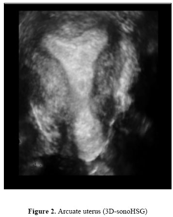
sonohysterography (XHSG) in the initial assessment of infertile women Keywords: sonohysterography, Hysterosalpingography, infertility Three dimensional ultrasound represents a new technique of imaging owing to its ability to register all three imaging planes simultaneously; to reconstruct new planes which are otherwise not visible as well as to visualize surfaces three-dimensionally (1). By instillation of contrast media (sterile saline) into the uterine cavity, the contour of the uterine cavity and the flow signals in Fallopian tubes can be visualized (2). Conventional two-dimensional Sonohysterography (2D-Sonohysterography) has limitations in that signals from the total length of the tube and full contour of the uterine cavity has rarely been depicted in a single scanning plane because of tubal tortuosity and limited projection angles of the ultrasound beam (3). Therefore, the outcome is mainly dependant on the skill of the examiner to see spillage at the fimbrial end and/or to mentally reconstruct an image of the tubes from partial visualization, which consequently requires repeated manipulations of the transducer and frequent injections of contrast medium. These time-consuming and occasionally painful procedures with 2D-Sonohysterography sometimes require sedation or anesthesia, which can be a burden to the patients (4). The aim of the study is to compare three-dimensional hystero-salpingo contrast sonography (3D-Sonohysterography) versus X-ray sonohysterography (XHSG) in the initial assessment of infertile women. MATERIAL AND METHODS The present study comprised fifty infertile women aged 22-40 years that were recruited after obtaining informed consent, attending fertility clinic; Kasr El-Aini hospital CairoUniversity, seeking advice for conception, from May 2003 to February 2004. The indication for investigations was more than one year of infertility that included primary infertility in thirty-five cases and secondary infertility in fifteen cases. All were subjected to full history taking, clinical examination, transvaginal ultrasonography was performed to identify any pelvic pathology followed by 3D hysterosonography. X-ray hysterosalpingography was performed later (within one week) except for those who had already a recent one (within the last 6 month). TVS was performed to identify any pelvic pathology. With the patients in lithotomy position, a speculum is inserted into the vagina and positioned such that the entire cervix is visualized and the os is easily accessible. The cervix and the vagina are swabbed with Betadine solution. A tenaculum is placed on the anterior lip of the cervix followed by application of a small and very thin uterine catheter (SILKOTAEX Size CH 08, Rush, 34600 Kamunting, Malaysia) fitted with a balloon for stabilization and occlusion of the internal cervical os (inflated with 2.0 ml of saline solution). After removal of the tenaculum, the three-dimensional trans-vaginal probe (MEDISON Kretz, Zipf, Austria, Voluson 530) is gently introduced into the posterior fornix of the vagina. The sterile saline is then injected slowly, under vision by ultrasound picture. Usually, no more than 5-10 ml of saline is instilled into the uterine cavity. At this stage one can observe the morphology of the uterus, and on detection of tubal flow signal by color Doppler, volume mode was initiated and the scanning plane was based on a coronal (horizontal) section of the uterus with targeted side of the ovary viewed. Two criteria have been proposed for the determination of tubal patency on conventional Sonohysterography: detection of spill at the fimbrial end and detection of steady flow signals in a segment of tube for at least 5 s (5). The tubal flow signals could be allocated to three categories. If continuous or intermittent flow signals from a uterine cornu and subsequent fimbrial spill could be depicted, the tube was diagnosed as patent. If the signals from the cornu were depicted but the fimbrial spill was obscure, the tube was still diagnosed to be patent. If no tubal signals arose from the cornu, the tubes were considered to be occluded (2). Statistical Analysis Results were evaluated for each group and data were compared using t-student test or chi-Square test where appropriate according to the type of the data to be compared. RESULTS Thirty-three cases with bilateral tubal patency and seventeen cases with unilateral occlusion were confirmed by 3D-Sonohysterography while on XHSG bilateral patency of tubes in thirty-six cases and unilateral occlusion in fourteen cases were identified (difference of three cases) All of the three cases were patent by XHSG ( Table 1). Nine cases with submucous fibroids diagnosed by transvaginal three dimensional sonography, all cases could be identified on 3D-Sonohysterography while only five cases could be diagnosed with XHSG, and four cases could not be confirmed on XHSG because of the unsatisfactory angle of x-ray projection (two cases were with submucous fibroid polyp and two posterior wall submucous fibroid) (Figure 1). Table 1. Representative pattern of tubal flow signals on 3D-Sonohysterography in 100 tubes
Analysis of the uterus by 3D-Sonohysterography could consistently (100%) provide an appropriate outline image of the uterine cavity where both uterine cornua and the cervical internal os were visualized simultaneously and this made precise measurements of the uterine cavity possible. Ten cases of arcuate uterus were diagnosed on 3D-Sonohysterography while only six were detected on XHSG (Figure 2). In addition, other uterine cavity abnormalities were diagnosed, however, no added advantage of 3D Sonohysterography on 3D transvaginal sonography. DISCUSSION Within the last years several new ultrasound techniques have appeared. Three-dimensional ultrasound scanning (3DUS), in which there has been great interest, is one of them. Especially within gynaecology. Recently, several authors have reported the use of ultrasound contrast media in the assessment of infertile women during trans-vaginal ultrasound, and have emphasized the place of three-dimensional hystero-salpingo contrast sonography (3D-Sonohysterography) in the imaging of infertile women (6). Analysis of tubal patency by 3D-Sonohysterography thirty-three cases with bilateral tubal patency and seventeen cases with unilateral occlusion were confirmed (Eighty-three tubes were patent and seventeen tubes were blocked) while on XHSG bilateral patency of tubes in thirty-six cases and unilateral patency in fourteen cases were identified (Eighty-six tubes were patent and thirteen tubes were blocked) . In the present study, in relation to the tubal patency, especially in selected cases having a low risk of pelvic pathology, the performance of 3D-Sonohysterography using saline should be recommended as an initial check-up for screening infertile women. However, in cases with high risk of pelvic pathology, chromolaparoscopy should be recommended to provide a precise evaluation. Analysis of the uterus by 3D-Sonohysterography could consistently (100%) provide an appropriate outline image of the uterine cavity where both uterine cornua and the cervical internal os were visualized simultaneously and this made precise measurements of the uterine cavity possible. Ten cases of arcuate uterus were diagnosed on 3D-Sonohysterography while only six were detected on XHSG. In cases of submucous uterine fibroids, it was shown that not only the distorted and enlarged uterine cavity could be visualized from many directions on 3D, but the information about the size, position, and the extent of submucous fibroids was also available. However, on XHSG, a fewer chances to obtain such images compared with 3D-Sonohysterography due to the limited angles in the X-ray projection and the view of X-Ray by which is taken. Nine cases with submucous fibroids diagnosed by transvaginal sonography, all of them could be identified on 3D-Sonohysterography, while only five cases could be diagnosed adequately on XHSG, and four cases could not be confirmed on XHSG. Thus, the potential of 3D-Sonohysterography for evaluating the uterine cavity was confirmed in the present study. Fibroids are not seen on X-ray HSG unless they are calcified or the uterine cavity is distorted and the cause will not usually be apparent) (7). REFERENCES
© Copyright 2005 - Middle East Fertility Society The following images related to this document are available:Photo images[mf05038f1.jpg] [mf05038f2.jpg] |
| |||||||||
