 |
| About Bioline | All Journals | Testimonials | Membership | News |
|
||||||
|
||||||
Malaysian Journal of Medical Sciences, Vol. 9, No. 1, January 2002, pp. 52-54 Ewing's 'Sarcoma Mimicking Tuberculosis- A Case Report
B. Shalini, S. Wahinuddin, M. Monniaty & S. Rosemi Department of Medicine, Hospital Kota Bharu,
15586 Kota Bharu, Kelantan, Malaysia
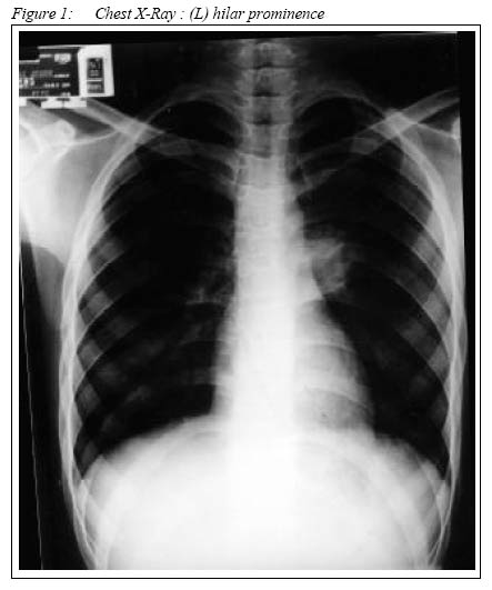
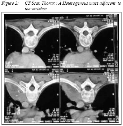
Submitted-20.2.2001 Code Number: mj02010 A 13-year old Malay school girl who had been apparently normal previously, presented with a three-month history of fever, malaise and loss of weight. She had anemia and raised values for ESR, lactic dehydrogenase, C-reactive protein, ferritin and a positive mantoux test. Her routine chest x-ray showed hilar prominence suggestive of hilar lymph node enlargement. C.T. Scan of the thorax revealed a posterior mediastinal mass, the histopathology of which was suggestive of Ewing's sarcoma. The rarity of the location of the tumour and its unusual mode of presentation prompted us to report this case. Key words : tuberculosis, posterior mediastinal mass, extraosseous Ewing's sarcoma. INTRODUCTION Ewing's Sarcoma (ES) is a malignant neoplasm of the bone and sometimes of soft tissues with characteristic radiological, morphological immuno-histochemical and cytological features. ES has now been shown to belong to a family of tumors with overlapping histopathological features and comprising of Ewing's sarcoma ( osseous and extra-osseous), peripheral primitive neuroectodermal tumor ( PPNET ) and Askin tumor (1). We report a teen-ager with extra-osseous ES who presented with prolonged fever, positive Mantoux test and a posterior mediastinal mass. Case Report : A 13 year-old Malay school girl was admitted with a three-month history of fever, loss of weight and generalized myalgia. She did not give a history of cough, chest pain, shortness of breath or night sweats. There was no history of joint pain, hair loss, rash or photosensitivity. On examination, she was thin, pale, febrile and tachycardic. There were no vasculitic lesions, alopecia or malar rash. The respiratory, cardio- vascular and abdominal examinations were normal and no lymph node was palpable. Laboratory evaluations revealed a hemoglobin value of 9.3 gm /dl, white blood cell count of 9.2 x 10 9/ dl and platelet count of 564 x 109/l. The erythrocyte sedimentation rate (ESR) was high (138 mm/1st hour) and the Mantoux reaction was positive (18 mm). The acute phase proteins like lactic dehydrogenase (916u/l), C-reactive protein (19.16 mg/dl) and serum ferritin (1000 ng / dl) were all elevated. The liver and renal functions were normal. Repeated blood cultures for pathogenic bacteria were negative. Sputum examintion for acid-fast bacilli was negative. Tests for connective tissue disease were negative. The chest X-ray revealed a left hilar prominence but the lungs were normal (figure 1). The ultrasound examination of abdomen and echocardiogram were normal. Since the clinical features and laboratory reports highly suggest of tuberculosis, the patient was started on empirical anti-tuberculous therapy (isoniazid 300 mg, rifampicin 300 mg and pyrazinamide 1000 mg). However there was no improvement after two weeks of anti-tuberculous therapy. She was then subjected to bronchoscopy which showed that the left upper lobe bronchus was slit-like, suggesting the presence of external compression. Bronchial brushings and washings were negative for acid- fast bacilli (AFB) and malignant cells. C.T. scan of thorax showed a heterogenous, enhancing lesion in the left hemithorax at the level of the carina, adjacent to the vertebral bodies. No calcification was seen within the mass. There was no evidence of parenchymal lesion in the lung or of pleural effusion (figure 2); and the adjoining vertebrae and ribs were intact indicating thereby that the tumor had not arise from bone and extended into the mediastinum. Meanwhile the patient continued to remain febrile and her hemoglobin level dropped to 8.4 gm/dl. The blood picture was in favour of microcytic hypochromic anaemia. Biopsy of the posterior mediastinal mass was done under C.T. guidance. The histopathology showed sheets and lobules of primitive small ovoid round cells which displayed high nuclear cytoplasmic ratio, fine nuclear chromatin and punched-out clear cytoplasmic vacuoles. The cells also showed periodic-acid-Schiff stain ( PAS) positive inclusions. These features were in favour of an Ewing's sarcoma. The patient was the referred to the oncologist for futher management. DISCUSSION The first large series (39 cases) of extraosseous Ewing's sarcoma (EOES) was published by Angervall and Enzinger (2) in 1975, of which 12 patients had the tumor in the paravertebral region mainly at the lumbar and sacral level. Subsequently it had been shown to occur at various sites in the human body including the soft tissues of the orbit, vagina, kidney and the posterior mediastinum (3,4,5,6,7). In many of the instances, the diagnosis has been made postperatively after excision of the mass or by biopsy procedures. The posterior mediastinal presentation of EOES is also uncommon. To our knowledge no similar case of posterior mediastinal EOES has so far been reported from Malaysia. In view of the posterior mediastinal and paravertebral location of the tumor, it is quite often mistaken for a neurofibroma (3). In our case, the mediastinal mass associated with a raised ESR, positive Mantoux test and other constitutional symptoms in a teen-ager led us to the diagonosis of tuberculosis. The histopathological report of the biopsy specimen of the mass was compatible with that of ES but for which the diagnosis would have eluded us. Elevated ESR, positive Mantoux reaction and raised acute phase proteins have not been reported in the EOES cases described in the current literature. The explanation remain obscure, though it does raises a possibility of an associated tuberculous illness in our case. Therefore in the differential diagnosis of posterior mediastinal tumors, EOES should also be borne in mind especially in young patients. Appropriate and early histological diagnosis is essential to plan appropriate management. REFERENCES
Copyright 2002 - Malaysian Journal of Medical Sciences The following images related to this document are available:Photo images[mj02010f1.jpg] [mj02010f2.jpg] |
| |||||||||
