 |
| About Bioline | All Journals | Testimonials | Membership | News |
|
||||||
|
||||||
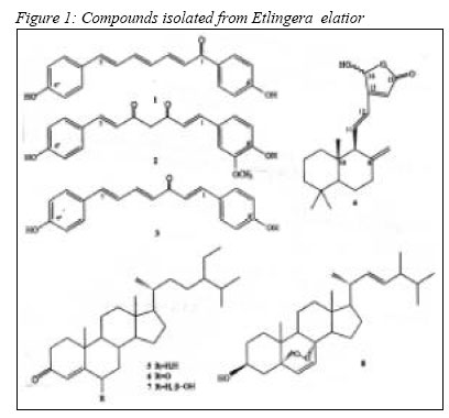
REVIEW ARTICLE ANTITUMOUR-PROMOTING AND CYTOTOXIC CONSTITUENTS OF ETLINGERA ELATIOR Habsah M*, Ali AM**, Lajis NH***, Sukari MA****, Yap YH4, Kikuzaki H*****, Nakatani N***** *1Department of Chemistry, Faculty of Science and Technology, University College of Science and Technology Malaysia (KUSTEM), Mengabang Telipot, 21030 Kuala Terengganu, Terengganu, Malaysia. Code Number: mj05003 Phytochemical studies on rhizome of Etlingera elatior have resulted in the isolation of 1,7-bis(4-hydroxyphenyl)-2,4,6-heptatrienone (1), demethoxycurcumin (2), 1,7bis(4-hydroxyphenyl)-1,4,6-heptatrien-3-one (3), 16-hydroxylabda-8(17),11,13trien-16,15-olide (4), stigmast-4-en-3-one (5), stigmast-4-ene-3,6-dione (6), stigmast4-en-6b-ol-3-one (7), 5α,8α-epidioxyergosta-6,22-dien-3β-ol (8). 1 and 4 were new compounds. Compounds 5 and 7 displayed high antitumour-promoting activity. Ethyl acetate extract showed a very significant cytotoxic activity against CEM-SS and MCF-7 cell lines (4 µg/ml and 6.25 µg/ml respectively). The antitumourpromoting activity was determined by EBV-EA assay and cytotoxic activity was determined by MTT assay. Key words : Etlingera elatior, antitumour-promoting, EBV-EA, cytotoxic, CEM-SS, MCF-7 Introduction Etlingera elatior belongs to the Zingiberacea family, and is classified under the genera of Etlingera. This plant is * Locally known as kantan. The mature fruits of Etlingera elatior are edible but sour, and are reputed for their antihypertensive activity. In many parts of Southeast Asia, the young inflorescence is used as “ulam” or as ingredients in laksa, curry, and mixed vegetables. A decoction of the fruits may be dropped into the ear to treat earache, and a decoction of leaves may be used to clean wounds (1). The decoction of young shoots is used to reduce body odour after giving birth. Mackeen et al. (1997) reported that the aqueous ethanol extract of the flower shoots of E. elatior possessed antimicrobial activity and was cytotoxic to HeLa cell line (2). Habsah et al. in 2003, reported the antioxidant activity and antitumour promoting activity of the crude dichloromethane and methanol extracts of E. elatior (3). The diarylheptanoids 1-3 from the ethyl acetate extract was reported to have high antioxidant activity (4). The previous screening of its flower shoot extract showed promising antitumour promoting activity (5). No phytochemical study has been done on this species except their essential oil of it young flower shoots (6), thus the objective of this study was to isolate cytotoxic and antitumour promoting compounds from the rhizome of E. elatior . Materials and Methods Plant material. One hundred kg of the fresh E. elatior rhizomes were collected in Klang and Banting, Selangor in October 1999. The rhizomes were cleaned, chopped into smaller pieces (3-5 mm thickness) and dried under the shade. A voucher specimen (No. SK 80/01) was deposited at the Herbarium of Laboratory of Phytomedicines (LF), University Putra Malaysia. Extraction and isolation. Sixteen kg of the
dried powdered rhizomes (16% w/w of fresh
rhizomes) were extracted three times each, first with
CHCl3, then with acetone, and finally with MeOH,
to give 120 g, 50 g and 8 g of extracts, respectively.
The CHCl3 extract was triturated with hexane and
filtered to give hexane (60 g) and CHCl3 soluble
extracts (60 g). The acetone extract was triturated
with ethyl acetate to give 8 g of ethyl acetate soluble
extract. Column chromatography (CC) of CHCl3
extract (40 g) on silica gel (5 x 40 cm) eluted with
hexane/diethyl ether, diethyl eter/ethyl acetate, ethyl
acetate/MeOH, gave combine fractions A-J
respectively. Repeated CC of fraction C (3 g) on
silics gel using diethyl ether in hexane (1:9) gave 7
(20 mg) and 8 (8 mg). Repeated CC of hexane
extract (20 g) on silica gel (5 x 40 cm), eluted with
hexane/diethyl ether, afforded eight fractions (A-H).
Repeated column of fraction F (3 g) afforded four
fractions (F1-F4), from which 5 (65 mg) was isolated
from fraction F2 (138 mg) after recrystalisation with
MeOH. Compound 6 (50 mg) was isolated from
fraction F4 (77.3 mg) after preparative TLC (20%
diethyl ether in hexane). Compound 4 (11.9 mg).
was isolated from fraction H (80 mg) after repeated
column chromatography on silica gel eluted with
10% ethyl acetate in CHCl3. Repeated CC of the
ethyl acetate soluble extract (8 g) on sephadex LH
20 (2.5 x 40 cm), eluted with MeOH, afforded 14
fractions (fractions EA-EN) Repeated CC of fraction
EK (160 mg) on silica gel, with 10% ethyl acetate 1,7-Bis(4-hydroxyphenyl)-2,4,6-heptatrienone (1): Yellow powder; UV (CH3OH) lmax (log e) 395 (4.51); IR (KBR) nmax 3300, 1653, 1578, 1511 cm-1; 1H NMR (CD3COCD3, 500 MHz) d 7.96 (2H, d, J = 8.8 Hz, H-2’,6’), 6.95 (2H, d, J = 8.8 Hz, H-3’, 5’), 7.41 (2H, d, J = 8.5 Hz, H-2”,6”), 6.85 (2H, d, J = 8.5 Hz, H-3”,5”), 7.20 (1H, d, J = 15.0 Hz, H-2), 7.46 (1H, dd, J = 15.0 Hz, J = 11.0 Hz, H-3), 6.64 (1H, dd, J = 15.0 Hz, J = 11.0 Hz, H-4), 6.94 (1H, dd, J =15.0 Hz , J = 11.0 Hz, H-5), 6.92 (1H, dd, J = 15.0 Hz, J = 11.0 Hz, H-6), 6.80 (1H, d, J = 15.0 Hz, H-7); 13C NMR (CD3COCD3, 125 MHz) d 131.3 (C-1’), 131.5 (C-2’, 6’), 116.1 (C-3’,5’), 162.5 (C- 4’), 129.5 (C-1”), 129.3 (C-2”,6”), 116.5 (C-3”,5”), 158.9 (C-4”), 187.9 (C-1), 124.9 (C-2), 144.1 (C- 3), 130.8 (C-4), 143.1 (C-5), 126.5 (C-6), 137.6 (C- 7); EIMS m/z 292 [M]+ (94), 171 (38), 121 (100); HREIMS m/z 292.1113 (calcd for C19H16O3, 292.1099) 16-Hydroxylabda-8(17),11,13-trien-15,16-olide (4): Gummy solid; UV (CH3OH) lmax (log e) 260 (4.51); IR nmax (KBr) cm-1: 1750 (a,b-unsaturated d -lactone), 3090, 892 cm-1 (exo-methylene). 1H NMR (CDCl3, 500 MHz) d 1.04 (1H, ddd, J = 13.2,13.2, 3.7 Hz, H-1a, ax), 1.38 (1H, m, H-1b), 1.40 (1H, m, H-2a), 1.52 (1H, m, H-2b), 1.18 (1H, br dd, J = 13.2, 13.2 Hz, H-3a, ax ), 1.42 (1H, m, H-3b), 1.10 (1H, dd, J = 2.7 Hz, J = 12.5 Hz, H-5), 1.39 (1H, m, H- 6a), 1.72 (1H, ddddd, J = 12.9, 2.7, 2.7, 2.7, 2.7 Hz, H-6b, eq), 2.09 (1H, ddd, J = 13.4, 13.4, 5.6 Hz, H-7a, ax), 2.44 (1H, m, H-7b, eq), 2.47 (1H, d, J = 10.0 Hz, H-9), 6.58 (dd, J = 16.0 Hz, J = 10.0 Hz, H-11a), 6.59 (dd, J =16.0 Hz, J = 10.0 Hz, H-11b), 6.31 (1H, d, J = 16.0 Hz, H-12), 5.85 (1H, s, H-14), 6.25 (s, H-16a), 6.27 (s, H-16b), 4.38 (d, J = 1.5 Hz, H-17aa), 4.79 (2H, brs, H-17ab, H-17ba), 4.47 (d, J = 1.5 Hz, H-17bb), 0.90 (3H, s, H-18), 0.85 (3H, s, H-19), 0.87 (3H, s, H-20); 13C NMR (CDCl3, 125 MHz) d 40.9 (C-1a), 39.6 (C-1b), 19.0 (C-2a), 19.0 (C-2a), 42.1 (C-3), 33.5 (C-4), 54.5 (C-5a), 54.5 (C- 5b), 23.2 (C-6), 36.6 (C-7), 148.7 (C-8a), 148.9 (C- 8b), 62.1 (C-9a), 62.1 (C-9b), 39.5 (C-10a), 39.6 (C-10b), 144.0 (C-11a), 144.1 (C-11b), 122.6 (C- 12a), 122.7 (C-12b), 161.0 (C-13a), 161.0 (C-13b), 115.5 (C-14), 171.2 (C-15), 97.5 (C-16a), 97.6 (C- 16b), 108.5 (C-17a), 108.9 (C-17b), 21.9 (C-18), 33.6 (C-19), 15.1 (C-20a), 15.2 (C-20b); EIMS m/z 316 [M+] (13), 180 (30), 162(14), 137(100), 123(25); HREIMS m/z 316.2030 (calcd for C 20H28O3, 316.2038) Demethoxycurcumin (2): Yellow powder, m.p. 170-172 0C; EIMS m/z 337.8 (M+, C19H18O5); 1H-NMR and 13C-NMR are in agreement with (7,8). 1,7-bis(4-hydroxyphenyl)-1,4,6-heptatrien-3-one (3): Yellow powder, m.p 168-170 0C; EIMS m/z 291.9 (M+, C19H16O3); 1H-NMR and 13C-NMR are in agreement with (9). Stigmast-4-en-3-one (5): White needles, m.p 80-82 0C; EIMS m/z 412 (M+, C29H48O); 1H-NMR and 13C-NMR are in agreement with (10). Stigmast-4-ene-3,6-dione (6): White needles, m.p 75-76 0C; EIMS m/z 426 (M+, C29H46O2); 1H-NMR and 13C-NMR are in agreement with (10). Stigmast-4-en-6b-ol-3-one (7): White needles, m.p 217-218 0C; EIMS m/z 428 (M+, C29H48O2); 1H-NMR and 13C-NMR are in agreement with (10). 5a,8a-Epidioxyergosta-6,22-dien-3b-ol (8): Off-white amorphous solid, m.p 176-178 0C; EIMS m/z 428 (M+, C29H48O2); 1H-NMR and 13CNMR are in agreement with (11). Antitumour Promoting Activity The extract was dissolved in dimethyl sulfoxide (DMSO) as a stock solution, with concentrations and 10 mg/ml for crude extract and 4 mg/ml for pure compound. Cell Lines The Raji cells were maintained in medium RPMI 1640 (Flow Lab., UK) supplemented with 10% foetal calf serum (Gibco, UK), 100 IU/ml penicillin/streptomycin, 50 mg/mL Amphostat B and 120 mg/mL L-glutamine as a static suspension culture at 370C in a humidified atmosphere of 50 % CO2 in air. Antitumour-promoting Activity in Raji Cells Assay
The inhibitory activity of Eipstein-Barr virus (EBV) activation assay was performed as previously described (4). Raji cells were activated with 20 ng/ ml of TPA (Sigma, USA) and 4 mM/ml of sodiumn-butyrate (Nacarai Tesque, Japan) to induce the expression EBV EA. The plant extracts at the concentration of 200 mg/ml were added immediately after the addition of TPA as tumour promoter. The cells were incubated at 370C for 72 hours, after which they were subjected to indirect immunoflourescence assay using EBV EA positive nasopharyngeal carcinoma serum and FITC-conjugated anti-human IgG (Sigma, USA). The inhibitory rate (IR) of each test sample against the EBV activation was classified into four ranks as follows: +++, strongly active (IR ≥ 70% ); ++, moderately active (70% > IR ≥ 50%); +, weakly active (50% > IR ≥ 30%); -, inactive (30% > IR) (5). All tests and analyses were run in triplicate and averaged. Cytotoxicity Assay Plant Extract The extract was dissolved in dimethyl sulfoxide (DMSO) as a stock solution, with concentrations 4 mg/ml for pure compounds and fractions. Microculture Cytotoxicity Screening Using Methyl Thiazole Tetrazolium (MTT) Assay
A 10-fold dilution gradient microtitre cell culture was adopted and modified to 3-fold dilution cell plating. All cells were cultured in sterile RPMI1640 complete media, supplemented with antibioticantimycotic mixture (containing 103 U/mL Penicillin G; 100 mg/mL Streptomycin SO4; 2.5 mg/ L Amphoterin B), 2 mM L-Glutamine and 10% FBS (all from Sigma). Exponentially growing cells were pre-determined by trypsinisation (0.25% p.p trypsin) and/or re-suspension, with a 100% confluency and 96% viability (using 0.2% typan blue exclusion cellcount in an Improved Neubauer Haemacytometer). A final cell concentration of 2.5 x 10⁴ cells/well was used as inoculation density for all anchorage dependent cell lines, and 5 x 10⁴ cells/well for CEM-SS cell suspensions. Into a sterile and labeled NUNCLONTM 96 well (180 mL volumes) micro-titre plates (Nunc, Denmark), 180 mL volumes were pipetted appropriately for all cell lines. The plated cells were then incubated overnight, under standard culture conditions of 5% CO2, 95% air and 100% humidity, to allow cell settling and differentiation. Stock solutions of all samples were prepared as 10 mg/mL in absolute dimethylsulphoxide, DMSO (HPLC-grade, Sigma, USA). Using the same culture media as diluent, a sub-stock solution of 1000 mM was prepared immediately before addition and serially diluted in sterile sample containers, to give 10x working stock solutions for each of the final test-range concentrations, topping from 100 mM down to the lowest of 0.1 mM. Having prepared the above mentioned dilutions, 20 mL quadruplicates of the corresponding 10x working stock samples were all added up to give the required final concentrations, in total volume of 200 mL. Plates were returned to the incubator for a further 4-day culture period. Cultures were regularly observed for any visual interference(s), with morphological changes and or cell killing effects being monitored using CK2 light microscope (Olympus, Japan). At the end of each culture successfully attained, devoid of any contaminating and other interfering notifications, cells were aseptically subjected to further analysis, using the MTT biochemical assay (11). A 20 mL volume of MTT (Sigma, USA), prepared at 5 mg/ml in phosphate buffered saline (PBS), was added into each 200 mL culture of the 96 well microtitre plate, wrapped with aluminium foil and incubated for a further 4 hours culture under similar conditions as above. This allowed the activation of mitochondrial dehydrogenases of the CEM-SS cells to reduce the yellowed colour MTT into a crystallized blue-violet colour complex product, formazan. A total volume aspiration followed by 200 mL addition of pure DMSO (Ajax, Australia) was carried out, with gentle mixing and 5 minute incubation at room temperature, to allow for faster and more enhanced formazan solubility. Optical densities (O.D) of the respective concentrations of sample were then measured using DYNEX MRX ELISA reader (Dynex Instruments, Inc. USA), at a 550 nm test and 630 nm reference wavelengths. Percentage proportions of the control O.D. values were then compacted in a dose-response standard curve to enable a more accurate and standardized determination of the 50% growth inhibitory concentration, IC50. All tests and analyses were run in triplicate and averaged. Results
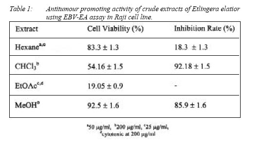
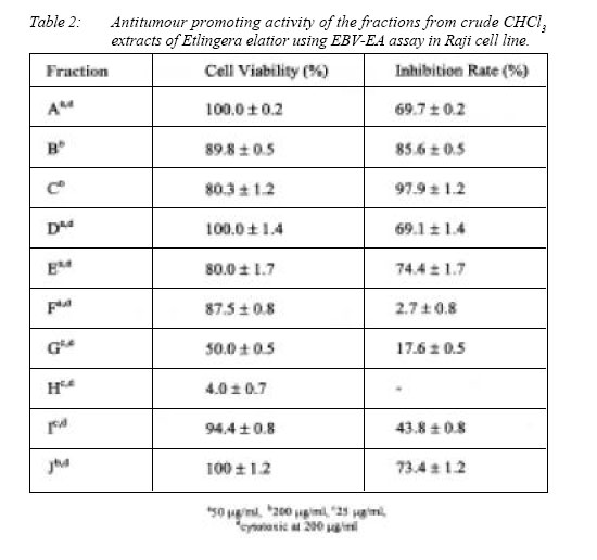
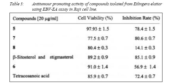
Ten compounds were isolated from E. elatior after extensive chromatography of the crude extracts. Compouds 1-8 (Figure 1), a mixture of stigmasterol and β-sitosterol, and tetracosanoic acid were identified based on spectral data (UV, MS, IR, 1H NMR, 13C NMR, H-H COSY, HMQC and HMBC) and comparison with literature values (7-11, 1315). The antitumour promoting activity of the crude extracts, fractions and compounds is shown in Table 1, 2, and 3, respectively. The cytotoxic activity of the crude extracts is shown in Table 4.
Discussion
The preliminary screening showed both CHCl3 and MeOH extracts of E. elatior possessed high antitumour promoting activity, with 92.18% and 85.9% inhibition rate, respectively. Both hexane and ethyl acetate were cytotoxic against Raji cell at initial concentration (200 mg/ml) (Table 1). Five fractions (fractions A-C, E and J) of CHCl3 extract showed strong antitumour promoting activity. The less polar fractions showed high antitumour promoting activity compared to the more polar fractions (Table 2). Seven compounds (5, 6 and tetracosanoic acid from the hexane extract; 7, 8 and a mixture of stigmasterol and sitosterol from CHCl3 extract) were screened for antitumour promoting activity (Table 3). Among them, 5, 7, a mixture of b-sitosterol and stigmasterol, and tetracosanoic acid showed high antitumour promoting activity, with inhibition rate of 78.4%, 80.6%, 85.1% and 72.4% respectively. Compound 6 only showed moderate activity with inhibition rate of 56.9%. Compound 8 and the mixture of stigmast-4-en-6a-ol-3-one and 8 did not show any significant activity. Our finding suggested that the Δ4(5)-3-keto steroids ( 5-7) displayed high antitumour promoting activity. The activity of the Δ4(5)-3-keto steroids increased due to the b-hydroxy group at C-6 as in the case of 7. The activity decreased when this 6-hydroxy group was in its oxidized form as in the case of 6. In a related study, a Δ8(9)-11-keto steroid, 5α,14α-dimethylergosta-8,24(28)-dien-11-one from Euphorbia chamaesyce also displayed a potent inhibitory effect on EBV-EA (15). This finding suggested that both Δ4(5)-3-keto and Δ8(9)-11-keto steroids could act as potent antitumour promoters. Four extracts of Etlingera elatior rhizome were tested for their cytotoxic activity against CEM-SS and MCF-7 cell lines (Table 4). The in vitro cytotoxic assay was based on modification of Monsman’s method (11). The ethyl acetate extract was found to show a significant cytotoxic to both CEM-SS (IC50 4 mg/ ml) and MCF-7 (IC50 6.25 mg/ml). From the ethyl acetate extract, we successfully isolated three diarylheptanoids, 1-3, which showed strong antioxidant activity (4). However the cytotoxicity of each diarylheptanoid could not be evaluated because of insufficient amount. It was reported that demethoxycurcumin had cytotoxicity effect against ovarian cancer OVCAR-3 cells (7), displayed DPPH free radical scavenging activity and showed significant hepatoprotective effects on tacrine– induced cytotoxicity in human liver–derived Hep G2 cells (17). The other extracts, including the hexane, CHCl3 and MeOH extracts, also showed significant cytotoxicity against both MCF-7 and CEM-SS cell lines. It was also reported that the ethanol aqueous extract of the young flower shoots was cytotoxic against HeLa cell line with IC50 value of 10 mg/ml (2). This implied that besides the young flower shoots, the rhizome is also a potential source for cytotoxic compounds.
Acknowledgements
References
The authors wishes to thank the Ministry of Science, Technology and the Environment Malaysia for the fund provided under the Intensified Research in Priority Areas Research Grant (No. 09-02-040067). HM thanks the University College of Science and Technology Malaysia for granting her study leave as well as N. Nakatani and H. Kikuzaki for their assistance and gratefully acknowledges the Program for Promotion of Basic Research Activities for Innovative Biosciences (BRAIN).
© Copyright 2005 - Malaysian Journal of Medical Science The following images related to this document are available:Photo images[mj05003t3.jpg] [mj05003t4.jpg] [mj05003t2.jpg] [mj05003t1.jpg] [mj05003f1.jpg] |
| |||||||||
