 |
| About Bioline | All Journals | Testimonials | Membership | News |
|
||||||
|
||||||
ORIGINAL ARTICLE SERUM LEPTIN AND CORTICOSTERONE LEVELS AFTER EXPOSURE TO NOISE STRESS IN RATS Chandralekha. G, *Jeganathan R, **Viswanathan, ***Charan J.C. * Department of Anatomy, USM, Kelantan. ** Ragas Dental College, Uthandi, Chennai. Pharmacology, Madras Medical college, Chennai. *** Sri Ramachandra Medical College, Deemed University-Chennai. Code Number: mj05009 Even though extensive studies have been conducted on the effect of noise exposure on hearing apparatus / auditory system, information on the effect of noise on the other body functions is sparse. The present study examined the effect of exposure of albino rats to acute and chronic noise stress on two important interlaced endocrine levels. In acute experiments the animals were exposed to 120 dB noise for a duration of 1, 2, 3 hrs. In chronic experiments the animals were exposed to noise for one hour daily for 30, 60 and 90 days. Plasma corticosterone and leptin levels were measured in these animals. There was significant elevation in the levels of corticosterone and leptin after exposure to noise stress. The elevation in corticosterone level after noise stress is in agreement with earlier reports. So noise acts like a stressor and elevates the secretion of the corticosterone, the stress hormone and leptin, the product of the ob gene there is an elevation in leptin levels after noise stress. Key words: Leptin, corticosterone, noise stress, Wistar rats Introduction
Noise is considered a kind of stress, which produces significant physiological and biochemical changes in animals as well as in humans (1). The damaging effect of noise on hearing has been extensively studied (2, 3). However, very little information is available regarding the effect of noise on other body functions. Similar to other types of stress, noise stress has also been shown to increase levels of stress hormones like corticosterone and norepinephrine (4, 5). Recent studies indicate that corticosterone can stimulate the secretion of adiposite-derived hormone, leptin (6, 7). Leptin is an important hormone concerned with food intake, metabolism and reproduction (8). As noise increases corticosterone secretion it may be proposed the exposure to stressors like noise could induce alterations in serum leptin levels. Such a possibility has not been reported in the literature. In the present study an attempt has been made to monitor serum leptin and corticosterone levels in male albino rats after exposure to acute and chronic noise stress. Materials and Methods Animals: Male Wistar-Kyoto rats weighing 100 – 120 g were housed in polyprophylene cages with access to laboratory chew and water ad libitum. They were maintained in an animal experimental laboratory where night and day temperatures varied from between 24° C to 32° C with a relative humidity of 70 to 80% and a 12-hour light and dark cycle. Animal groups: The animals were randomly grouped into control and experimental groups. Each group had acute and chronic subgroups. The rats of the acute subgroup were exposed to noise of 120db for 1, 2 and 3 hours. The rats of the chronic group were exposed to noise of 120 dB of one hour duration every day for 30, 45 or 60 days. Each subgroup had 6 animals. Control groups were treated like the experimental group, but not exposed to noise. Induction of noise:
The animals were exposed to noise stress of 120 dB, which was generated by a noise generator (Suguna 100 M HP 2). The generator was connected to an amplifier (Inkel, Korea) to amplify the sound at an intensity of 120 dB. A decibel meter (C. R 303 Philips, India) with a measurement range of 35 to 135 dB was used to measure the sound level. Blood collection and analysis:
Immediately after the experiments were completed blood was collected by retro orbital puncture. Serum was stored at -20° C until assayed for leptin and corticosterone. Leptin concentration in serum was determined using leptin colorimetric EIA Kit (Assay Design Inc., U S A). Serum corticosterone was estimated using commercially available RIA kits.
Statistical Analysis:
Results were analyzed using ANOVA followed by Dunnet’s test and a ‘P’ level of <0.05 was considered statistically significant. Results
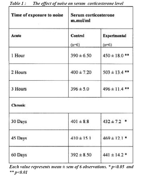
Changes in serum corticosterone levels after exposure to noise
Exposure of rats to acute noise stress for 1, 2 and 3 hour periods resulted in a significant elevation in the level of serum corticosterone compared to the control animals (Table 1). Corticosterone was significantly higher in rats exposed to noise for 2 and 3 hours compared to its level in rats exposed to noise for 1 hour. Serum corticosterone levels were not significantly different between rats exposed to noise for 2 and 3 hours. Similar to acute exposure, a significant increase in serum corticosterone level was observed in animals that were exposed to noise stress for 30, 45 and 60 days compared to the respective control values. A maximum increase was noted after 45 days and there appears to be a downward trend in the elevation of serum corticosterone at 60 days. Changes in serum leptin levels after exposure to noise.
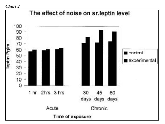
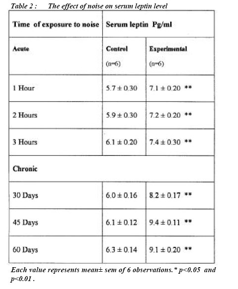
Serum leptin concentrations were significantly higher in animals that were exposed to noise stress for 1, 2 or 3 hrs when compared to the control animals. There was no significant difference in serum leptin levels between animals exposed to different durations of acute stress (Table 2). Significantly elevated serum leptin levels were also evident in animals that were exposed to chronic noise stress for 30, 45 and 60 days. Serum leptin levels were significantly higher in rats exposed to stress for 45 days compared to those exposed to stress for 30 days. However, no significant difference was evident in serum leptin levels of rats exposed to stress for 45 days and those exposed to stress for 60 days. Discussion
The results of the present study indicate significantly higher levels of serum corticosterone and leptin after exposure to both acute and chronic noise stress. Raised serum corticosterone levels following noise stress in rats have been reported before (9, 10, 11). In addition, elevation of glucocorticoid levels following many types of stressors is also well known. The precise mechanism for this remains unclear but it may be related to altered activity of the hypothalamic-pituitary-adrenal axis secondary to the noise stress and may involve alterations in the secretion of corticotropin releasing hormone (CRH), ACTH and proopiomelanocortin (POMC) gene expression. The increase in glucocorticoid secretion during stress appears to be important for the appropriate defense mechanism to be put into place. Significantly higher levels of corticosterone were also evident in rats exposed to chronic stress for 30-60 days indicating poor or absent adaptation of the rats to noise stress. This is in contrast to what has been observed before by others where a somewhat decreased corticosterone response to noise was observed on chronically stressed rats (11, 12). The reason for this difference is unclear. There are several reports indicating that glucocorticoids are capable of stimulating the synthesis and secretion of adipocyte-derived leptin (6, 13, 14), which regulates food intake and energy expenditure. Leptin secretion is under the influence of hormonal and neural control. (6, 12, 15). The results indicate significant elevation in leptin levels both after acute as well as chronic exposure to noise stress. Heimen et al (1997) (16), in an earlier study examined the influence of exogenous administration of leptin on plasma corticosterone and ACTH in animals subjected to restraint stress. They reported that leptin was able to inhibit the release of corticotrophin releasing hormone from the hypothalamus in vitro and also blunted the plasma ACTH and corticosterone elevation due to restraint stress. They also speculated a possibility for reduction of leptin level during acute and chronic stress and thus facilitating the responsiveness of hypothalamic-pituitary-adrenal axis. However they failed to demonstrate any reduction in leptin levels in their study on restraint stress and thus the speculation remains unsubstantiated. In fact the data indicated an elevation of serum leptin levels after restraint stress though the levels were not statistically significant. In another study chronic subcutaneous leptin infusions have been shown to diminish responsiveness of the hypothalamic-pituitaryadrenal axis in female rhesus monkeys (17). It therefore seems that there is a significant interplay between leptin and the hypothalamic-pituitaryadrenal axis. The results of the present study in rats subjected to acute and chronic noise stress clearly indicate simultaneous elevation of corticosterone and leptin levels during both acute and chronic stress. It appears that the inhibitory effect of leptin on corticosterone secretion was somewhat absent during noise stress in this study. The reason for the variation between our observation and that in the mentioned studies is unclear but may be due to species variation or the different nature of stress. Nevertheless our study suggests that one arm of the hypothalamic-pituitary-adrenal-leptin axis appears disabled during noise stress, which permits for increase corticosterone secretion during stress. In conclusion, the present results clearly indicate that sustained exposure to noise stress results in a significant elevation of corticosterone and leptin. These two hormones have wide ranging effects on metabolism, growth and reproduction (8, 18). The elevated levels of leptin and corticosterone even after exposure to continuous noise stress for a period of 90 days, indicates that the expected adaptation is absent. Hence continuous exposure to noise stress may have many adverse effects on some of vital physiological functions (19, 20, 21) in which the alteration in the levels of these two hormones may play a significant contributory role. Acknowledgements
References
Authors gratefully acknowledge the Deans of Stanley Medical College and Madras Medical College for their support and encouragement. The Madras Medical College Animal Ethics Committee approved the study. Special thanks to the animal house of the Madras Medical College for the supply of animals and support.
© Copyright 2005 - Malaysian Journal of Medical Science The following images related to this document are available:Photo images[mj05009f2.jpg] [mj05009t2.jpg] [mj05009t1.jpg] [mj05009f1.jpg] |
| |||||||||
