 |
| About Bioline | All Journals | Testimonials | Membership | News |
|
||||||
|
||||||
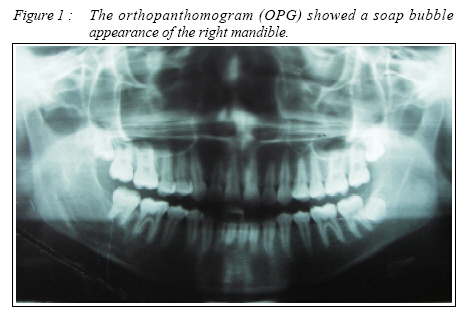
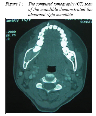
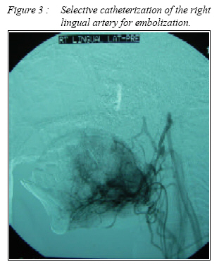
CASE REPORT ARTERIOVENOUS MALFORMATION OF THE MANDIBLE: A RARE BUT LIFE-THREATENING DISEASE Baharudin Abdullah, Abdullah Pohchi* & Abdul Rani Samsudin* Department of Otorhinolaryngology ORL - Head and Neck Surgery School of Medical Sciences, *School of Dental Sciences, Universiti Sains Malaysia, 16150 Kubang Kerian, Kelantan, Malaysia Corresponding Author : Dr. Baharudin Abdullah MBBS(Mal), MMED ORL-HNS (USM) Department of Otorhinolaryngology ORL - Head and Neck Surgery(ORL-HNS), School of Medical Sciences, Universiti Sains Malaysia, Health Campus, 16150 Kubang Kerian, Kelantan, Malaysia Tel: + 609-7664110 Fax: +609-7653370 Email: baharudin@kb.usm.my Submitted-20-02-2004, Accepted-03-12-06 Code Number: mj07011 AVM in the mandible is rare. It may present with recurrent episodes of unexplained gingival haemorrhage, bony swelling, tooth mobility or facial asymmetry. We reported our experience in managing a case of a 15 year old Malay girl who presented with a life threatening bleeding from her mandible. Keywords: Arteriovenous Malformation, Mandible Introduction AVM in the mandible may present with recurrent episodes of unexplained gingival hemorrhage, bony swelling, tooth mobility or facial asymmetry. The most unfortunate presentation however follows dental extraction or bone biopsy when severe, exsanguinating and torrential hemorrhage may occur. Occasionally, a patient may only present with a sentinel molar bleeding which precede the hemorrhage and this fact should not be overlooked (1). Sometimes a bruit can be heard on auscultation over the involved area and a sensation on the region innervated by the mandibular branch of the trigeminal nerve (V) may be altered. Case Report A 15 year old Malay girl from Ipoh, Perak who experienced persistent severe bleeding from the lower right gum was referred to our hospital. Her problem started when she was seven years old whos, she experienced bleeding from the lower right first premolar tooth. The bleeding was minimal but recurred which prompted her parents to bring her to a private dental clinic in Ipoh several times. The bleeding was about 4 to 5 times per year. Unfortunately had the bleeding got worse and never resolved. She developed persistent bleeding from the same region at the age of 15 years and was brought to Ipoh General Hospital. The bleeding occurred spontaneously without any trauma to that region. Each bleeding filled up 1 to 2 cupfuls. Applying pressure will only stop the bleeding temporarily. On examination there was a diffuse swelling on the outer aspect of the right mandible. Intraorally there was a bluish lesion in the right retromolar trigone which extended anteriorly to the premolar region. No bleeding was seen. The throat and the neck were normal. Other systemic examination was unremarkable. No lesion seen in the nasopharynx, oropharynx and hypopharynx on flexible endoscopy. Her orthopanthomogram (OPG) and CT scan showed a soap bubble appearance of the right side of the mandible extending from the right ramus to the body of the mandible up to the 1st molar area figure 1 and figure 2. A diagnosis of AVM of mandible was made which was subsequently confirmed by magnetic resonance imaging (MRI) and magnetic resonance angiography (MRA). MRI brain with gadolinium showed no intracranial involvement. Selective embolization of the right lingual artery was performed prior to the right segmental mandibulectomy (figure 3). A median lower lip split and right submandibular incision was made. The carotid triangle was identified where the external carotid artery and its branches were located. A silk string was placed under the external carotid artery to secure bleeding in case hemorrhage occurs intraoperatively. The periosteal flap of the mandible was raised. A tumor mass was seen involving the lateral surface and cortex of the ramus of the right mandible extending to the second molar tooth region. Feeding vessels were noted along the condyle. A segmental mandibulectomy was performed by removing part of the right mandible between the right second premolar and first molar teeth to the right condylar head. The defect was reconstructed by using a 7cm rib graft taken from the right fifth rib. The graft was placed over the defect and fixed to the mandible with reconstructive titanium plate and screws on both sides. Postoperatively the patient was managed for 1 day in the intensive care unit. She recovered well and was discharged on day 7 postoperatively. DiscussionArteriovenous malformations (AVM) arising within the mandible is exceedingly rare but potentially life threatening. The lesion is more often located in the horizontal portion of the lower jaw than in the condylar process or in the temporomandibular joint (2). An OPG may disclose multilocular radiolucent areas with ‘honeycomb’ or ‘soap bubble’ appearance or unilocular osteolytic images or simple radiolucency without peculiar characteristics; sometimes with cortical expansion, dental dislocation and root resorption of the teeth (2). Less common presentation includes extension of bony spicules extending at right angles into the lesion (sunburst or sunray appearance) which is highly diagnostic of AVM (1). Resection has been advocated both as primary therapy and as salvage treatment after failure of conservative measures (3). The indications for resection suggested are obstruction of visual axis; large lesion with thrombocytopenia; obstruction of luminal structures; uncontrollable ulceration, hemorrhage or infection; atypical growth suggesting alternative diagnosis; cardiopulmonary decompensation from arteriovenous shunting and; small lesions that can be excised without cosmetic or functional risk (3). In this patient resection of the mandible (segmental mandibulectomy) proved to be life saving. References
© Copyright 2007 - Malaysian Journal of Medical Science The following images related to this document are available:Photo images[mj07011f1.jpg] [mj07011f2.jpg] [mj07011f3.jpg] |
| |||||||||
