 |
| About Bioline | All Journals | Testimonials | Membership | News |
|
||||||
|
||||||
ORIGINAL ARTICLE The Routine Histopathological Examination of Tonsillectomy Specimens at Hospital Universiti Sains Malaysia – Retrospective Study and its Implication Irfan Mohamad, Shahid Hassan, Rosdan Salim Department of Otorhinolaryngology-Head and Neck Surgery,
School of Medical Sciences, Universiti Sains Malaysia, Health Campus
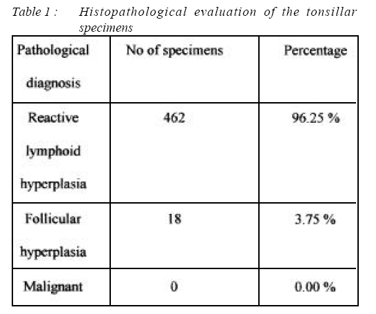
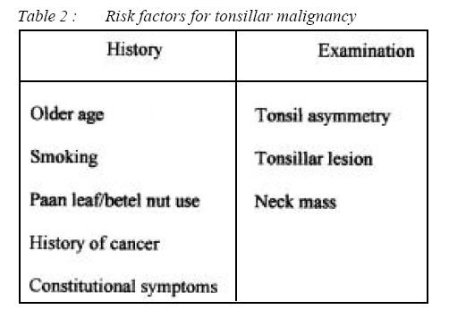
16150 Kubang Kerian, Kelantan, Malaysia Submitted-20-02-2004, Accepted-03-12-06 Code Number: mj07020 Tonsillectomy is performed for several indications, the two commonest in practice are infective ( chronic tonsillitis ) and obstructive symptoms such as sleep apnea. The objective of this study is to determine the necessity of routinely performed histopathological examination of specimens post-tonsillectomy . In this paper, a retrospective evaluation of 480 specimens from 241 patient who has undergone tonsillectomies in Hospital Universiti Sains Malaysia between January 2004 and October 2005 was done. It was found that 462 ( 96.25 % ) were reactive lymphoid hyperplasia and 18 ( 3.75 % ) were follicular hyperplasia. None of them were found malignant. The result of this study indicate that routine histopathological examination of tonsillectomy specimens are unnecessary and results only in added cost and a loss of man hours. Key words : Histopathology , tonsillectomy specimens. Introduction Tonsillectomy is defined as surgical removal of palatine tonsils. It is a long practiced procedure. Tonsillectomy is performed for a wide range of indications which can be divided into therapeutic , diagnostic and as an access for other procedures. Most common indication for tonsillectomy is infective cause. The other indication for tonsillectomy is patients with obstructive symptoms such as snoring and sleep apnoea. Regardless of the indication, tonsillectomy specimens are routinely sent for histopathological evaluation because of the concern that the tonsils might harbor malignancy. As efforts are being intensified to reduce medical costs, especially in developing countries where patients often pay for their own healthcare, the necessity for this routine evaluation is now being questioned (2). This study was undertaken to assess the outcome of histopathological examination in routine tonsillectomy surgeries. Materials and Methods This is an observational ( retrospective ) study. All patients who had undergone tonsillectomy at Hospital Universiti Sains Malaysia between January 2004 and October 2005 were identified from operation theatre registration book. The histopathological evaluation reports were reviewed from the patients’ case notes. Any patients with clinical suspicion of malignancy will be excluded. Results A total of 241 patients age ranging from 2 to 64 years old fulfilled our criteria and were included in this study. Majority of patients ( 68.1 % ) were in the pre-school and school-going age. 46 % of the patients were males. The most common indications for tonsillectomy in our study population were recurrent infection ( 64.3 % ), obstructive symptoms such as snoring and sleep apnea ( 34.9 % ). One of the patient had unilateral tonsillar enlargement that caused foreign body sensation during swallowing. One case of tonsillar cyst operated also included in the series. None of the patients in our study population had clinical suspicion of malignancy. All 480 tonsillar specimens were submitted for gross and microscopic histologic evaluation, as routinely performed in our hospital. The results were given in Table I. DISCUSSION Clinically, malignant tonsils are quite distinguishable from benign counterparts. From history and clinical examination alone, the clinician should be able to exclude benign lesion with the suspected or malignant cases. The risk factors for malignancy are listed in Table II. It is a common practice to send tonsillectomy specimens routinely for histopathological examination to screen for occult malignancy. Proponents suggest that among other reasons, missing an important diagnosis such as occult malignancy or granulomatous disease and possible medicolegal consequences argue in favor of routine histopathological examination. Recently, more authors worldwide came up with evidence that concluded specimens that was found to be positive malignancy was already diagnosed or at least suspected during consultation prior to the surgery. Microscopic examination of tonsil specimens generally correlates well with preoperative clinical impressions, and pathology findings rarely alter patient management (1). Alvi and Vartanian (2) found only one malignancy in a pathologic review of 576 posttonsillectomy specimens. This patient was from the older population study group with the mean age of 21.6 years. Beaty et al, in their retrospective review of adult (age 18 year and older) tonsillectomy tissue, found a higher rate of positive specimens: 25 malignant tumors in 476 samples. However, these 25 samples had been obtained from patients with several definite and identifiable risk factors that had already been detected prior to surgery. 23 of them have two or more risk factors, whereby the asymmetric tonsil being the commonest besides the age factor itself (3). Based on their findings, both authors recommended that evaluation be performed only on specimens of patients who had certain risk factors . Williams et al (4) noted that three out of 4070 post tonsillectomy specimens to be malignant on histopathological examination and all of them were already diagnosed during pre operative consultation. As the increase in age was described to be one of the risk factors, some authors specifically look into the paediatric age group patient who undergone tonsillectomy. A review of 2743 post tonsillectomy specimens from paediatric patients revealed that none of the specimens were positive for malignancy (5). Younis RT et al review of 2099 samples from pediatric patients who has undergone tonsillectomy were negative for malignancy (6). Another study showed that the routine histopathological examination of tonsillectomy specimens in paediatric patient were unnecessary (7). In Malaysia, almost all general hospitals send tonsillectomy specimens for routine histopathological examinations. However, the study shows that routine histopathological examination of all tonsillectomy specimen is not justified. It should be restricted only to cases where there is clinical suspicion of malignancy. Conclusion The result of this study, combined with evidence from our review of the literature, lead us to conclude that in the absence of clinical suspicion of malignancy, routine histologic examination of tonsillectomy specimens is not necessary, especially in paediatric age group patients. References
© Copyright 2007 - Malaysian Journal of Medical Science The following images related to this document are available:Photo images[mj07020t1.jpg] [mj07020t2.jpg] |
| |||||||||
