 |
| About Bioline | All Journals | Testimonials | Membership | News |
|
||||||
|
||||||
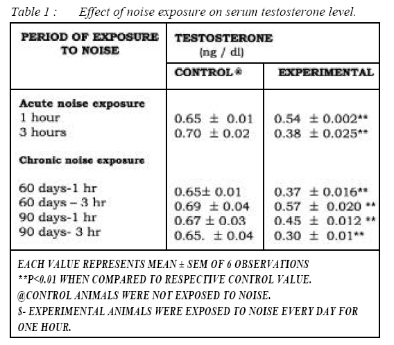
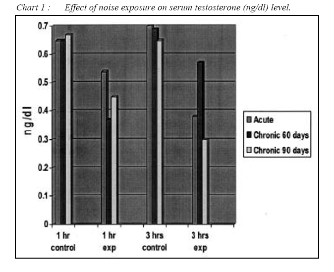
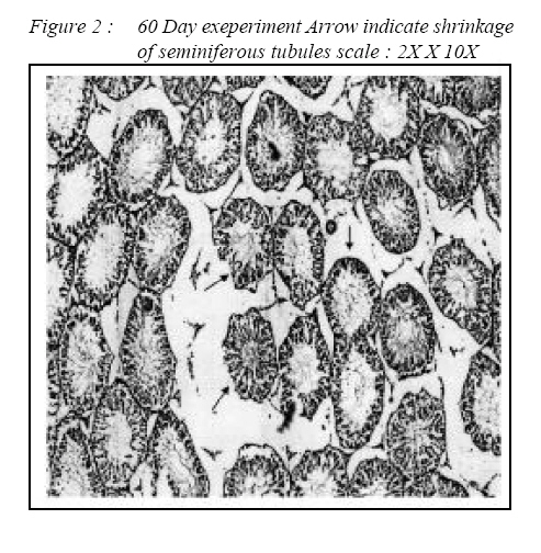
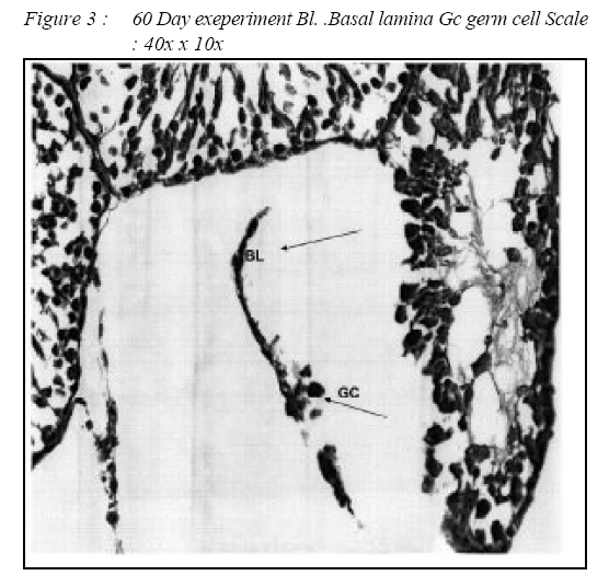
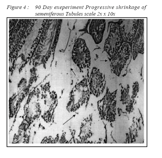
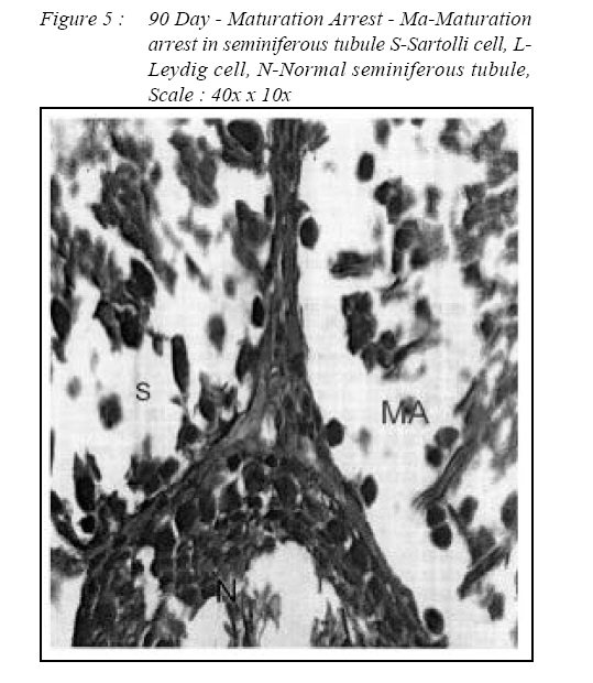
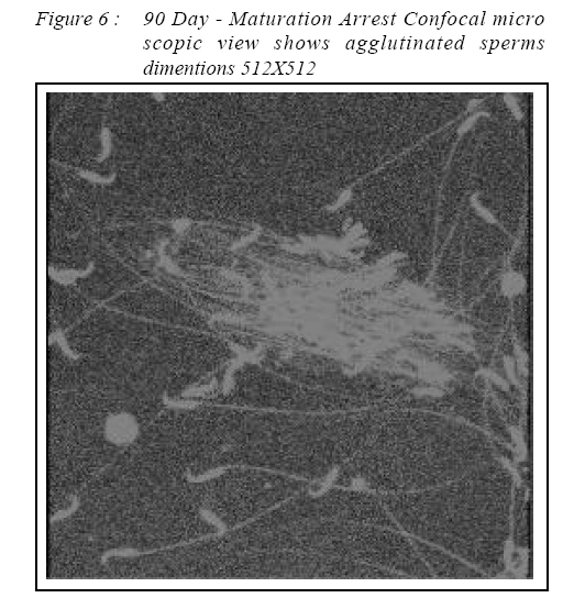
ORIGINAL ARTICLE Noise Exposure Effect on Testicular Histology, Morphology and on Male Steroidogenic Hormone Chandralekha G.Swami , Jeganathan Ramanathan*, Charan Jeganath.C** Department of Anatomy, School of Medical Sciences, Universiti Sains Malaysia, Health Campus 16150 Kubang Kerian, Kelantan, Malaysia. Submitted-20-02-2004, Accepted-03-12-06 Code Number: mj07022 The noise stress, after it passes through the hearing apparatus, not only affects the auditory apparatus but also other body functions. The alterations in the levels of cortical hormone, adrenocorticosterone, nor-epinephrine hormone (which are primarily considered as stress hormones) on follicular stimulating hormone, testosterone, and lutinizing hormone were reported in relation with stress . Male albino rats weighing 200 to 250 grams were exposed to 100 dB of noise for one hour and three hours in acute group and daily one hour exposure for 60 day, and 90 day in chronic group. The serum testosterone levels were measured in these animals. There was significant reduction in serum testosterone levels and this was similar with earlier reports.The tissues were collected for light and confocal microscopic study. 100dB of traffic noise exposure of varying duration had definite permanent effect on testicular histology and morphology and on the male sex hormone . The adaptation mechanism was noticed at the hormonal level only but the structural changes noticed were definite and permanent. The agglutinated dead sperms revealed the possibility of infertily when chronically exposed to noise stress. Key words : noise stress, spermatogenesis¸ sex hormone Introduction & Review of Literature Of all types of environmental pollutants, noiseis the most prevalent and insidious natural pollutant which causes deleterious physiological and structural effects. Noise is also partially responsible for reduced reproductivity (1) Detrimental effect of noise/music on video
players on long term effects of noise exposure are
well documented as being harmful (2).
Abnormalities in reproduction were also reported
and there is a significant decrease in pregnancy
rate and lethal effect in mice embryos exposed to
high frequencies of noise The teratogenic and
embryofetotoxic potential of a broad band of highfrequency
(16-42 kHz), high-intensity (110 dB
sound pressure level), temporally uniform noise was
evaluated in CD-1 mice (3). Noise stress was related with neuroendocrinological response (4). Most of
the studies documented revealed the alterations in
the levels of cortical hormone, adrenocorticosterone
and nor-epinephrine hormone levels which are
primarily considered as stress hormones and also
on testosterone, follicular stimulating hormone and
lutinizing hormone levels (5). The testosterone
response was impaired by water restriction, heat
exposure and immobilization exposure. Acute
noise stress of 80 dB increased the testosterone
level (6). Testosterone production was adversely
affected by nutritional factors (7). Glucocorticoid
mediation of suppressed testosterone biosynthesis
was also seen in male mice exposed to
immobilization stress (8). The pituitary-gonadal
response to stress would be a more sensitive index
of abnormalities induced by protein calorie deficit,
basal concentration of LH,follicular stimulating hormone (FSH) or testosterone (9, 10). Tamoxifen each period of immobilization stress (12). Chronic decreased the serum testosterone level and disrupted immobilization stress provoked an increase in serum the testicular seminiferous epithelium (11).The corticosterone which caused the decline in influence of immobilization stress on testicular germ testosterone concentration (13). Pituitary adrenal cell apoptosis was investigated in rats. A transient activity in rats were affected by 85 dB chronic increase in serum corticosterone and a transient noise stress (6). Stress is believed to influence male decrease in serum testosterone were observed during reproductive activity (14, 15 ). Five minutes of high Method The male Sprague Dawley rats weighing 200-250gm were exposed to 100 dB of recorded traffic noise for one hour and three hours in acute group and daily one hour and three hours for 60 days and and treated like experimental groups in all aspects 90 days exposure in chronic groups. Their normal except exposure to noise. Upon completion of all habitat was maintained. The animals were exposed noise exposure regimens, the animals were to a pure tone noise of sine waves with a frequency anesthetized with an intraperitoneal injection of of 10,000 Hz and an intensity of 100 dB. sound level intraval sodium (60 mg/kg body weight) and decibel meter was used to measure and maintain exsanguinated. Approximately 5 ml of blood was the intensity of noise. The controls were maintained collected from each rat for testosterone hormonal assay, using Elisa method. The DRG Testosterone ELISA KIT IBL HAMBURG, GmbH, GERMANY, was used which was based on the competition principle and the micro plate separation and the kit was stored at 4°C. The separated sample serums were stored at -20º C. The reagents and the specimens were brought to the room temperature before use. On the stipulated day after the collection of the blood for hormonal assay, laparotomy was performed and the testis and epididymides were removed and separated. Tunica vaginalis was carefully removed and the testes were dissected out and cleaned with cold physiological saline to remove blood and the adhering tissues. The samples were then fixed in 10% formaldehyde/in fresh alcoholic Bouin’s fluid for 8 hours, and then processed and embedded in paraffin wax .The testis histology was performed according to the method used by Murthy et al (14) The sections were observed under light microscope for qualitative study. This study was approved by the Universiti Sains Malaysia Health Campus Animal Ethics Committee, Kelantan. Ref:PPSG/07(A)/044,2 Disember 2002, (19). The data of the results was subjected to ANOVA followed by Dunner’s “t” test for statistical significance. A p level of <0.05 is considered as statistically significant. Results There was a significant reduction in serum testosterone level in acute as well as in chronic noise exposed groups (Table 1, Chart 1). The testis showed normal histological pattern in acute groups (Figure 2). Each testis is subdivided by connective tissue septa into lobules. Each contained numerous highly coiled semeniferous tubules. Each seminiferous tubule is lined by the stratified germinal epithelium, consisting of two major types of cells, the proliferating spermatogenic cells and nonproliferating supporting sustentacular cells or sertoli cells. Surrounding the seminiferous tubules were numerous blood vessels, various connective tissue cells, clusters of epitheloid cells and the interstitial cells of Leydig. Changes were seen in the histological pattern in 60-day (Figure 3,4)) and 90-day (Figure 5,6) exposure groups. The seminiferous tubules were shrunken so that there was a space between the septum and the tubules. This type of change was noticed in the central part of the tubules only. The peripheral parts of tubules were compact and normal. Moreover maturation arrest in the germinal layers were noticed in some tubules. The basement membrane was broken and the germ cells sloughed into the interstitium. This was more advanced in 90 day exposure group. The sperms in the epididymis showed agglutination both under the light microscopy and confocal microscopy (Figure 8). Discussion There was a significant decrease in the serum testosterone level after exposure to 100dB of traffic noise. The secretory response of Leydig cells of the mutant rats were slightly decreased since plasma concentration of testosterone did not increase in response to high concentration of plasma LH (20). This is similar to the invitro testosterone production by Leydig cells of singly housed, timid mice were lower in comparison with those aggressive mice housed in groups. Normally, leutinizing hormone acts on the testes and increases the plasma testosterone level. The follicular stimulating hormone controls the secretion of androgen-binding protein by the sertoli cells. This protein increases the concentration of testosterone in the seminiferous tubules for proper spermatogenesis. Testosterone and follicular stimulating hormone (FSH) suppression induced spermiation failure (21) is similar in the present study. It may be due to the feed back action of testosterone on the hypothalamus. In chronic noise exposed rats there was maturation arrest in the germ cells due to reduced testosterone level. The Leydig cell proliferation is likely to be a compensatory mechanism to increase testicular steroidogenesis triggered by testosterone insufficiency (22). During stress, the hypothalamic-pituitary-adrenal axis is activated, the glucocorticoid secretion increases (23) and consequently, circulating testosterone levels are decreased via glucocorticoid receptors in Leydig cells. In noise exposed rats the serum testosterone level was reduced which is similar as in immobilization test where the stress did not alter plasma luteizing hormone (LH) levels, but plasma testosterone (T) levels were reduced by 82% (24) which is similar to that of Bambino and Hsueh (25). Transient increase in serum corticosterone and transient decrease in serum testosterone were observed during each period of noise and immobilization stress. Also enhanced testicular germ cell apoptosis was seen. (23, 26, 27). Low testosterone production (28) adversely affects the quality of ejaculates and subsequent fertility. The decrease in testosterone level is also associated with the marked reduction in epididymal sperm number (22). Moreover the epididymal sperms were agglutinated in noise exposed chronic group and the number of dead sperms were increased (29). So alterations in the normal testicular morphology associated with maturation arrest in the germinal cells was found and is similar to those findings following DPP treatment (20, 22, 30, 31). Conclusion Hence, 100dB of traffic noise exposure of varying duration has definite permanent effect on testicular histology and morphology and on the serum testosterone levels. Alteration in the testosterone levels produces structural changes in testicular tissues during long exposure of 100 dB noise . The adaptation mechanism described by Ogale was noticed in sex hormonal levels in the present study. But there were structural changes in different duration of time in chronic groups seems to be irreversible. Once the structural changes occur, they may not return to normalcy. So, 100 dB of traffic noise has made definite structural changes and maturation arrest in the proliferating germinal cells in testis and increased the dead sperms and agglutinated sperms. This is a sign of infertility. So the agglutinated dead sperms revealed the possibility of infertily in chronic exposure to noise of higher decibels. As such “Noise is a silent killer” (32). Acknowledgements This project was carried by short term grant no: 304/P PSP/6131269. I acknowledge the Research Creativity and Management Office, Chancellory, Universiti Sains Malaysia, 11800, Penang, the R and D department, and the Animal Ethics, School of Medical Science Committee. The Dean, Deputy Dean of Research, Universiti Sains Malaysia, 16150, Kubang Kerian Kelatan Malaysia for their kind help and cooperation. My special thanks to Mr. Harrissal, technologist, Department of Anatomy for his cooperation and effort to finish this project. But not least, I thank my husband and my son for their help, moral support , valuable suggestions and contributions to finish this project successfully. References
© Copyright 2007 - Malaysian Journal of Medical Science The following images related to this document are available:Photo images[mj07022f6.jpg] [mj07022f1.jpg] [mj07022c1.jpg] [mj07022f4.jpg] [mj07022f3.jpg] [mj07022t1.jpg] [mj07022f2.jpg] [mj07022f5.jpg] |
| |||||||||
