 |
| About Bioline | All Journals | Testimonials | Membership | News |
|
||||||
|
||||||
Malaysian Journal of Medical Sciences, Vol. 15, No. 4, October, 2008, pp. 19-27 ORIGINAL ARTICLE Correlations between subdural empyema and paraclinical as well as clinical parameters amongst urban Malay pediatric patients Saiful Azli Mat Nayan,Mohd Shafie Abdullah*, Nyi Nyi Naing** Mohd Saffari Mohd Haspani*** and Ahmad Razali Md Ralib**** Department of Neurosciences, School of Medical Science, Universiti Sains Malaysia, Health Campus
16150 Kubang Kerian, Kelantan, Malaysia & Department of Neurosurgery, Hospital Sungai Buloh,
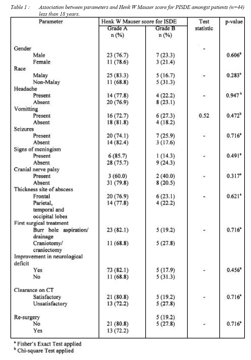
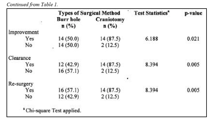
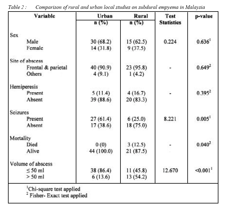
Jalan Hospital, Sungai Buloh, Selangor Malaysia. Submitted-21-04-2008, Accepted-31-08-08 Code Number: mj08035 Pediatric subdural empyema are frequently seen in developing Asean countries secondary to rinosinusogenic origins. A cross sectional analysis on the surgical treatment of intracranial subdural empyema (PISDE) in Hospital Kuala Lumpur (HKL), a major referral center, was done in the period of 2004. A total number of 44 children who fulfilled the inclusion criteria were included into this study. The methods of first surgery, volume of empyema on contrasted CT brain, improvement of neurological status, re-surgery, mortality and morbidity, as well as the demographic data such as age, gender, sex, duration of illness, clinical presentation, probable origin of empyema, cultures and follow-up were studied. Chi-square test was performed to determine the association between surgical methods and the survival of the patients, neurological improvement, clearance of empyema on CT brain, re-surgery and long morbidity among the survivors. If the 20% or more of the cells were having expected frequency less than five, then Fisher’s Exact test was applied. The level of significance was set at 0.05. SPSS version 12.0 was used for data entry and data analysis.There were 44 patients who were less than 18 years. Their mean age was 5.90 ± 6.01 years. There were 30 males (68.2%) and 14 females (31.8%) involved in the study. Malays were majority with 28 (63.6%) followed by Indian 8 (18.2%), Chinese 5 (11.4%) and others 3 (6.8%). The variables which were under interest were gender, race, headache, vomiting, seizures, sign of meningism, cranial nerve palsy, thickness site of abscess, first surgical treatment, improvement in neurological deficit, clearance of CT and whether re-surgery was necessary. All variables were found not to be associated with Henk W Mauser Score for PISDE grading. Comparison between this urban study and a rural setting study by the same corresponding author in the same period on subdural empyema was done. Common parameters were compared and it was found out that seizures was more prevalent in urban study where the patients were more than one year old (p=0.005). Mortality was much higher in urban study than the rural one (p=0.040). The larger proportion of urban group had volume of abscess less than or equal to 50 ml (p=< 0.001). Key words : Subdural empyema, paediatric, urban , rural ,management Introduction Pediatric intracranial subdural empyema (PISDE) is still a fulminant form of an intracranial infection, accounting for 15-25% of pyogenic pediatric intracranial suppuration. If left undiagnosed and untreated, PISDE is rapidly fatal; hence, early recognition is critical. Before effective antimicrobial therapy was introduced, the mortality rate among patients with PISDE, even with surgical drainage approached 100%. Presence of modern antibiotics and sophisticated scanning techniques for localization of empyema in the subdural space had contributed a significant improvement in the outcome of the patients with PISDE. Appropriate management of this condition has lead to good results and has been proven by the remarkable decrease in the mortality rates from 100% in the middle of the last century to 12.2% in the most recent series (1-8). The decline in the mortality rate has been attributed to the increased in diagnostic accuracy with the advent of computed tomography (CT), early surgical intervention and the use of intravenous broad spectrum antibiotics. Difficulties are still encountered in diagnosing PISDE mostly due to nonspecific nature of the early presenting symptoms such as fever and headache and also due to subtle CT changes that are often overlooked. The basic principles of treatment of ISDE are early diagnosis, prompt surgical drainage of pus, with simultaneous eradication of the primary source and high-dose intravenous therapy . The definitive methods of surgical drainage are still conflicting (burr hole or craniotomy) and until now there is no large randomized controlled study done that can show what is the most ideal method. Many recent publications had proposed that a large craniotomy with removal of the pus collection was preferable to burr hole and drainage (24-26). There were also some authors who believed that burr hole was as good as or better than craniotomy in the treatment of PISDE (10-14). Despite the advent of magnetic resonance imaging (MRI), uncertainty continues with respect to the best surgical management in practice. Improper or delay in the definitive treatment cause significant morbidity including re-surgery, permanent neurological deficit, disabling seizures and even death. The purpose of this study was to evaluate our management and outcome of the patients with PISDE whom had been treated surgically with either burr hole or craniotomy as their first surgical treatment. Methodology We performed a cross sectional analysis on the surgical treatment of PISDE in HKL in the period of 2004. A total number of 44 patients who fulfilled the criteria were included into this study. Their case notes and films were studied specifically on the methods of first surgery, volume of empyema on contrasted CT brain, improvement of neurological status, resurgery, mortality and morbidity. Apart from that, demographic data such as age, gender, sex, duration of illness, clinical presentation, probable origin of empyema, cultures and followup were also recorded. Chi-square test was performed to determine the association between surgical methods and the survival of the patients, neurological improvement, clearance of empyema on CT brain, re-surgery and long morbidity among the survivors. If the 20%or more of the cells were having expected frequency less than five, then Fisher’s Exact test was applied. The level of significance was set at 0.05. SPSS version 12.0 was used for data entry and data analysis. Statistical analysis Descriptive statistics was presented by using frequencies and percantages. The outcome was devided into grade A and grade B based on Henk W Mauser score for ISDE. Association between parameters and outcome was determined by using chi-square or Fisher’s Exact test. If less than 20% of cells had expected frequency more than five, chisquare test was applied. If equal or more than 20%, then Fisher’s Exact test was applied. Level of significance was set at 0.005. multiple logistic regression was performed by using backward stepwise method. Likelihood ratio test was used to determine significance of the model. Odds ratio with 95% confidence interval and corresponding p-value from Wald test were applied to determine significance of the variables. SPSS version 12.0 was used in data entry and analysis. Comparison of this study to local rural study published in the same period by the corresponding author was made. Chi-square and Fisher’s Exact test were applied to detect the difference of proportion of parameters between rural and urban setting studies. Results There were 44 patients who were less than 18 years. Their mean age was 5.90 ± 6.01 years. There were 30 males (68.2%) and 14 females (31.8%) involved in the study. Malays were majority with 28 (63.6%) followed by Chinese 5 (11.4%), Indian 8 (18.2%) and others 3 (6.8%). Amongst 44 patients, 18 (40.9%) had headache, 43 (97.7%) had fever, 22 (50.0%) had vomiting, 6 (11.4%) had blurring of vision, 5 (11.4%) had hemiparesis, 27 (61.4%) had seizures. A total of 10 (22.7%) had lethargy, 20 (45.5%) had high grade fever, 23 (52.3%) had moderate fever and only one (2.3%) was afebrile. There was no comorbidity reported among all patients. Duration of illness was reported as 5.3 ± 2.3 days. On physical examination, the level of consciousness at the time of surgery was reported as 22 (50.0%) as grade I , 19(43.2%) as grade II and 3 (6.8%) as grade III. For fundoscopic findings 19 (43.2%) were reported as normal, 12 (27.3%) had papilloedema and 13 (29.5%) were unsure of findings. Ear discharge was reported in only 2 (4.5%) patients. Thirty five (79.5%) had abnormal motor power. On radiological examination,sources of empyema was detected as paranasal sinus 3 (6.8%), post meningitis 15 (34.1%), otogenic 2 (4.5%) and 24 (54.5%) were unknown sources. Computed tomography (CT) scan of the brain showed that thickness site of abscess were frontal 26 (59.1%), parietal 14 (31.8%), temporal 3 (6.8%) and occipital 1 (2.3%). Pre-operative volume of empyema was reported as 38.3 ± 12.3 ml whereas post-operative maximal residual volume was 19.0 ±7.2 ml. For blood investigations, total pre-operative white blood cell count was reported as less than or equal to 10,000 cells per cubic millimeter of blood was 10 (22.7%) and the rest 34 (77.3%) had more. Erythrocyte Sedimentation Rate (ESR) on admission was found as 17 (38.6%) were normal and 27 (61.4%) were elevated . Blood for culture and sensitivity results revealed that only 5 (11.4%) isolated organisms whereas 39 (88.6%) had no growth. Types of surgery done were 28 (63.6%) burr hole aspiration & drainage and craniotomy 16 (36.4%). Pus for culture and sensitivity taken during operation revealed that 14 (31.8%) isolated organisms whereas 30 (68.1%) had no growth. Henk W Mauser score for Intracranial Subdural Empyema (ISDE) was reported before surgery was 34 (77.3%) grade A and 10 (22.7%) were grade B. After surgery, it was found that 28 (63.6%) had improvement of neurological deficit. Repeated CT scan showed satisfaction clearance of empyema in 26 (59.1%) patients. Resurgery(craniotomy) was performed in 18 (40.9%) patients who had burr hole aspiration & drainage done before. With regard to mortality, all patients survived and had good clinical HWM grade. The variables which were under interest were gender, race, headache, vomiting, seizures, signs of meningism, cranial nerve palsy, thickness site of abscess, first surgical treatment, improvement in neurological deficit, clearance of CT and whether re-surgery was done. All variables were found not to be associated with Henk W Mauser Score for ISDE grading (Table 1a & b). Multivariate analysis was performed and none of the variables were remained in the final model for interpretation. Comparison between this urban study and a rural setting study by the same corresponding author in the same period of 2004 on subdural empyema was done. Common parameters were compared and it was found out that seizures was more prevalent in urban study where the patients were more than one year old (p=0.005, Table 2). Mortality was much higher in urban study than the rural one (p=0.040, Table 2). The larger proportion of urban group had volume of abscess less than or equal to 50 ml (p=< 0.001, Table 2). Discussion In our study, we reviewed a total of 44 patients with PISDE which was purely supra-tentorial in location and non-traumatic in origin who had undergone surgery in HKL in the period around 2004. We analyzed the data to determine the association between two surgical methods used with outcome of the patients, improvement of neurological status, clearance of empyema on CT brain, re-surgery and morbidity among survivors at 3 months follow-up. PISDE was the second most common type of intracranial suppuration operated in our neurosurgical unit in HKL after the brain abscess, accounted for about 27.6% of surgery for intracranial infection. Most of the PISDE cases were related to trauma or surgery (42.4%), however, these cases were not included in this study. The purpose of excluding these cases was to avoid difficulty performing the data analysis due to co-founder effects. The prevalence of PISDE among patients with intracranial infection in HKL was higher than a study done by Narendra Nathoo and their colleagues in Durban, South Africa in 2001which was 17.6% (24). The explanation for the high prevalence was that HKL is the only neurosurgical centre in the middle of West Malaysia covering a large area of five million populations. Male was more commonly affected than female 30 (68.2%) with the ratio of 2.1:1. This findings was almost similar with most series of ISDE in the world. The local Malaysian study (9) conducted in Hospital Universiti Sains Malaysia, Kelantan reported the similar gender distribution of male dominance (62.5%) in nature which was conducted in urban area of Kuala Lumpur. However, the reason for this male predominance is still not clear. It had been suggested that larger sinuses, more vigourous nose-blowing habits, and rapid growth of frontal sinuses in male subjects from seven to 20 years of age may account for this striking gender difference. In this study, we found that ISDE had occurred in patients aged from one month to 16 years old. The median age was 2.5 (11.4) (IQR of 33.6). This result is comparable with most of the published series of PISDE (1-6). As stated earlier, the growth of frontal sinus in pubertal boys has been proposed as an explanation for the uneven sex and age distribution (17). Majority of the cases (27 patients, 30.0%) were involving children below the age of 5 years old and meningitis was the most common cause of ISDE in this particular group of patients. We found that in our centre, ISDE in children were detected much earlier than adults. Malaysian study (7) reported in Hospital Universiti Sains Malaysia, all of the children diagnosed as subdural empyema were less than one year significant elder by age group of children than a previous study. There was a significant difference of mean age (p<0.001) between previous Malaysian study (7) conducted in rural area (5.6 ± 3.1 months) and this study which was done in urban setting (5.9 ± 6.0 years). With the presence of ultrasound in rural hospitals, subdural collection was easily be detected early in infants whom presented with signs of intracranial infection with bulging fontanelle. Ultrasound of the infant cranium through an open fontanelle easily picked up any subdural collection which later confirmed the diagnosis of ISDE. Urban PISDE usually presented late because the children were older and developed seizures first before the children were rushed for CT brain which is present in most of the referring hospital.This is the reason why there were more seizures and larger volumes of empyema in the urban area since these older children had a larger volume od empyema to cause seizure and other neurological deficits before specialist medical treatment was seeked. In most Western and African series, paranasal sinusitis was responsible for 55 to 67% of the PISDE (21,25,26). This finding was different from our study, there were only 6.8% of PISDE had the primary cause originating from the paranasal sinuses..We believe that PISDE related to paranasal sinusitis are still high among Malaysia population. In this case, there is a possibility that in patients whom the origin of empyema were unknown, the organisms might still be coming from the sinuses but it were not detected clinically or radiologically during the acute period of illness. Low index of suspicion, inadequate history taking and clinical examination by the attending doctor might be the real cause. A through examination of all patients with ISDE including inspection of paranasal sinuses should always be done during the first presentation and referral to ENT is always mandatory if the cause of infection is not found. As mentioned before, the mechanism of spread from the paranasal sinuses can be direct or indirect as proposed by Courville and Kubik and Adams. The direct spread is due to erosion of the posterior wall of frontal sinus and indirect spread is by retrograde thrombophlebitis involving the valveless venous system in the dura. After the infection reaches the subdural space, it may spread widely over the brain convexity and interhemispheric fissure. The pus might not be apparent during the first surgery and exploratory of the cranial bases are mandatory because of potential recurrence of empyema. A study done by Quraishi et al in 2006 in United States found that the morbidity and mortality of PISDE as a complication of sinusitis remained high in the pediatric group despite adequate access to medical care (5). PISDE appeared to arise in the setting of subacute rather than acute frontal sinusitis. There may be an underdiagnosis and delay in treatment of patients with frontal sinusitis, resulting in subsequent intracranial complications (25). Pus C&S in seven patients with recent history of meningitis grew Haemophilus influenza (4 patients), E coli (1 patient) and Streptococcus pneumoniae (2 patients). Three children who underwent lumbar puncture had Haemophilus influenza (HiB) isolated from their CSF and one child had a positive blood culture of HiB. It seems that, Haemophilus influenza meningitis was still a cause of PISDE in our population despite of availability of immunization against HIB. The etiologies of PISDE among infants and children were different from that in adults in several studies. As reported by many authors (10-13) PISDE in young children was more likely to be secondary to otitis or meningitis. Twelve percents of infants with meningitis have subdural effusion (14) and it has been hypothesized that some cases of ISDE represent the ultimate sequela of an infected effusion (15). The two most common organisms causing PISDE in meningitic patients were Streptococcus pneumoniae and Haemophilus influenza (16). The incidence of meningitis as a cause of ISDE was 5% across studies surveyed although it accounts for a larger percentage in paediatric populations (10, 16, 22). In all our cases with PISDE, we empirically put them on at least two type of broad spectrum antibiotic immediately upon diagnosis. The antibiotics of choice were the third generation Cephalosporine and Metronidazole which usually cover almost all the susceptible organisms causing PISDE in our populations. Most of our patients responded well to these antibiotics treatment as evidence by the drop in the temperature and their clinical improvement. Huang et al had mentioned in their report that prevalence of meningococcal disease among young infants was high, and often presents an invasive clinical manifestation. Clinicians should be aware of meningococcal infection in young infants because the initial presentations may be difficult to distinguish from viral syndrome, and may rapidly progress to clinical deterioration. Patients with subdural empyema acquire encephalomalacia which increases the risk of permanent neurologic deficit (17). As mentioned in the literature, post-infective seizure was one of the main morbidity in children who had PISDE. It was also noted that children who developed seizures had a higher chance of having permanent neurological deficit as compared to those who had no seizures (14, 18). Most pediatricians have recommended to start anti-convulsant in all children with PISDE as a prophylactic measure (1922). In our set up, we routinely start all our patients with PISDE on prophylactic anti-convulsant as soon as they are diagnosed however it was not yet proven to reduce the rate of developing post-infective seizure. On the CT brain, site of maximum thickness of empyema was at the frontal region. The site of empyema may give a clue to the origin of PISDE. In this study, we could not demonstrate any significant association between the site and origin of empyema. However, since PISDE is commonly due to paranasal sinusisis, we believe that those patients with frontal empyema may had asymptomatic sinusitis and further study should be done to prove this statement. Five patients (5.6%) had their empyema located in the interhemispheric region and four of them died after the first surgery. Study have shown that interhemispheric ISDE carried the worst prognosis of all and should be treated with craniotomy to achieve maximum decompression and clearance of empyema (21). The mean estimated volume of empyema on preoperative CT scan was 40.81 ml (SD=13.43). Volume of empyema was one of the factors to decide for surgery. In our set up, patients with empyema volume of > 20 ml all needed to undergone surgery. The type of surgical methods in our unit was based on clinical, radiological findings and neurosurgeon preference. Out of 44 PISDE patients in this study, 28 (63.6%) patients underwent burr hole and drainage as their primary surgery for PISDE which accounted 55.6% of the cases. Another 16 (36.4%) had craniotomy and evacuation of empyema as their first surgical treatment. Previously burr hole and drainage was the surgical method of choice and craniotomy was done for cases of refractory PISDE . In term of improvement of neurological status, we found that majority of the patients (55.2%) who improved after surgery was belonged to the craniotomy group. Statistically, there was significant different between surgical method used and improvement in neurological status p=0.021. Patients who undergone craniotomy and evacuation of empyema had a higher chance to improve neurologically compared to burr hole and drainage after surgery. This result is comparable with another study done by Narendra Nathoo in 2001. He emphasized that outcome in terms of neurological improvement and survival is closely related to wide-exposure surgical procedure (21). The precise reason for this is craniotomy allows removal of the empyema mass and decompresses the brain adequately. Large cranial opening allows re-expansion of the brain and returns of the cerebral blood flow thus improves the neuronal function. Surgical methods was also found to be statistically bearing on the clearance of empyema on CT brain in this study p=0.005. Patients who undergone craniotomy 14 (87.5%) had been noted to have a better clearance of empyema on the CT scan. Among these patients, 85% had the empyema located in the frontal region. Subdural collection which is located in the hemispheric region especially frontal and parietal is usually accessible with either burr hole or craniotomy. The advantage of craniotomy over burr hole procedure is that its allow direct visualization of loculated empyema and complete removal of the membrane which is usually adherent to the dural surface.The bigger the volume of empyema created more mortality in the urban group. The type of empyema seen on the CT scan is also particularly important o determine the mode of surgery. In the acute stage the empyema is fluid and forms a thin, extended layer in the subdural space, whereas in the chronic stage the empyema is encapsulated in pockets.However, in most of cases the loculations of empyema were not always seen on the CT scan making the decision to operate difficult. In this cases, craniotomy should be the preferred method because multiple pockets of empyema are more difficult to remove with burr hole and catheter drainage (21). In terms of re-surgery, more patients in the burr hole group had to undergone another operation in this study. There was significant difference between surgical method used and re-surgery (p=0.005). Patients in the burr hole group were noted to have higher rate (57.1%) of re-surgery compared to the craniotomy group (12.5%). This finding was similar with a study done by Yilmaz et al published in February 2006. In their retrospective study among 28 infants and children with PISDE, they found that children who underwent burr hole as their surgical treatment had a significant higher rate of re-surgery (23). Re-surgery was closely related to the amount of empyema left over, clinical response of the patients to the surgical treatment and accurate placement of the catheter. The more amount of empyema remains, the higher the chance of resurgery. Recurrence of pus collection is probably attributable to ongoing seeding of pathogens from a septic thrombophlebitis into the subdural space (21). It is more common in ISDE related to otolaryngogenic causes and removal of the primary source by otolaryngologist is always mandatory. Long term morbidity among pediatric survivors has no significant statistical association with method of surgery in this study (p = 0.46). Patients with PISDE had equal chance of having permanent disability and post-infective seizures later in life regardless of their primary surgical method. As stated in the literature, other factors like age, level of consciousness, duration of illness, co-morbid illness and location of pus also play a very important role in the long term outcome of these patients which was not seen in our series. Klein et al in 2006 advocated an aggressive surgical treatment of PISDE in children with a large bone flap to allow the surgeon to remove pus and membranes as much as possible, even in the interhemispheric fissure, followed by intravenous appropriate antibiotherapy and eradication of the source of infection. However, even this “aggressive” treatment may sometimes not avoid re-operation. A careful follow-up is mandatory, because of the high risk of recurrence (20). Conclusion Type of surgical method used was determined to be associated with the treatment results of ISDE in terms of improvement of neurological status of the patients, clearance of empyema and re-surgery. However, survival of the patients and long morbidity were not determined by the type of procedure used (either burr hole or craniotomy) to evacuate the empyema. From this study, we concluded that there was no difference between craniotomy and burr hole and drainage. In the practical aspect, craniotomy should be used as the method of choice to treat PISDE because it is more cost effective than burr hole as it reduces the hospital stay, intensive care needs and repeated surgical intervention. Urban children were older when they had their PISDE and had significant seizures and mortality as well a larger volume as compared to a similar study conducted by the correponding author in an rural area in the same period .An older child with fever and seizure must alert a paediatrician to a probable ongoing empyema in an urban setting as volume of more than 50 ml must alert the neurosurgeon to do a craniotomy as compared to a burr hole aspiration for infants in rural areas. The absence of older children in the rural study (9) is interesting to follow up since the repeated exposure of children to infections may have caused immunity or due to it’s biphasic phenomenon (24) even though no proof is present. Audited unpublished hospital records from both the urban and rural center indicate till mid 2007 that older PISDE children tend to dominate in urban versus younger children in rural area A larger group study in the urban/rural area with bacteriological & immunological data would shed some light into the reason for the glaring age difference. Acknowledgements Dr. Saiful Azli Mat Nayan’s publication is part of the partial fullfillment for the Master of Surgery (Neurosurgery) USM Programme. References
© Copyright 2008 - Malaysian Journal of Medical Science The following images related to this document are available:Photo images[mj08035t1b.jpg] [mj08035t2.jpg] [mj08035t1.jpg] |
| |||||||||
