 |
| About Bioline | All Journals | Testimonials | Membership | News |
|
||||||
|
||||||
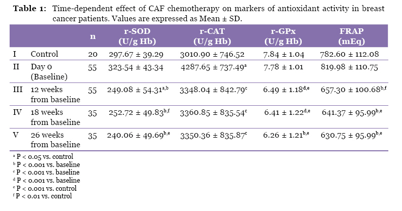
Malaysian Journal of Medical Sciences, Vol. 17, No. 2, 2010, pp. 24-28 Original Article Effect on Antioxidant Levels in Patients of Breast Carcinoma during Neoadjuvant Chemotherapy and Mastectomy Gurpreet SINGH1, SK MAULIK2, Amardeep JAISWAL2, Pratik KUMAR1, Rajinder PARSHAD3 1Medical Physics Unit, Institute of Rotary Cancer Hospital, All India Institute
of Medical sciences, New Delhi-110029, India Correspondence: Dr Gurpreet Singh, PhD (AIIMS), Medical Physics Unit, Institute of Rotary Cancer Hospital, All India Institute of Medical Sciences, New Delhi 110029, India, Tel: +44-79 0931 7947 E-mail: guraiims@gmail.com Submitted: 17 Sep 2008 Code Number: mj10017 Abstract Background: Breast cancer is the most common cancer in Indian women. The
aim of this study was to assess the levels of red blood cell (RBC) superoxide
dismutase (r-SOD), RBC catalase (r-CAT), RBC glutathione peroxidase (r-GPx)
and the ferric reducing ability of plasma (FRAP) in advanced breast cancer
patients post mastectomy before and after chemotherapy. Keywords: antineoplastic combined chemotherapy protocols, antioxidants, breast neoplasms, medical sciences Introduction Breast cancer is reported to be the most commonly occurring cancer, with an annual age-adjusted incidence of 22–28 in 100 000 women per year in Indian urban areas, and 6 in 100 000 women per year in rural areas. More than 75 000 new cases of breast cancer are reported in India each year, and the majority of breast cancers in India (50–70%) present with locally advanced disease (1). Free radical-induced oxidative stress in cancer patients has attracted a great deal of scientific attention in the last two decades. Free radicals are chemical species possessing an unpaired electron and are generally very reactive. They are produced continuously in cells either as by-products of metabolism, or during phagocytosis in the extra-nuclear compartment by the mitochondrial respiratory chain and the mixed function oxidase system. Free radicals can be detected by electron spin resonance spectroscopy, however this is not possible in vivo. Detection of free radical activities in vivo can be determined using antioxidants as markers. All aerobic organisms have mechanisms by which they can minimise free radical toxicity, for example reaction of superoxide radical with the enzyme superoxide dismutase (SOD), breakdown of hydrogen peroxide (H2O2) to water and oxygen (O2) by catalase; and glutathione-mediated detoxification. Thus the markers for antioxidant defence system include r-SOD, r-CAT, and r-GPx. Neoadjuvant chemotherapy is currently a common approach for treatment of cancer patients. Despite the fact that the vast majority of patients show a clinical response to chemotherapy, its benefit is only realised in a small number of patients who achieve optimal tumour burden reduction. At our institute, every breast cancer patient presenting with a locally advanced tumour undergoes mastectomy after three cycles of chemotherapy. Following mastectomy, these patients received three further cycles of chemotherapy. Most patients with breast cancer are treated with a combination of the anticancer chemotherapy drugs cyclophosphamide, doxorubicin, and 5-florouracil (CAF) (2–3). These antineoplastic agents cause a reduction in antioxidant levels because their toxicity increases the peroxidation of the unsaturated fatty acids of membrane phospholipids (3). The aim of the present study was comparison of antioxidant enzymes levels in breast cancer patients at different intervals of treatment with normal subjects. Materials and Methods Female breast cancer patients who were admitted to the Department of Surgery of the All India Institute of Medical Sciences in New Delhi were enrolled in this study. The study included two arms: a control group consisting of healthy age-matched females (n = 20), and patients undergoing treatment with CAF: cyclophosphamide 500 mg/m2 + doxorubicin 50 mg/m2 + 5-fluorouracil 500 mg/m2 treatment + mastectomy (n =55). No treatment was given to the control group. The patient group received CAF treatment at weeks 0, 3, and 6 and underwent surgery on week 9 followed by CAF treatment on weeks 12, 15, and 18. As per protocol, a drug free interval of three weeks was given between two chemotherapy cycle treatments. Patients were between 27 and 65 years of age (mean age 42.8 + 10.4 years). Of the enrolled patients, eleven were pre-menopausal and 14 were post-menopausal. Patients with associated illness that are known to alter free radical levels in cancer patients such as diabetes, hypertension, myocardial ischemia, myocardial infarction, renal disorders, pancreatic disorders, pulmonary disease, and pregnancy; and patients with fibroadenomas or with any previous treatment were excluded from this study. The criteria for inclusion in this study was presentation of a palpable mass in the breast that was observed with mammography and further confirmed by fine needle aspiration cytology (FNAC) at our institute. The study was conducted after obtaining the appropriate clearance from our institutional ethical committee. Blood samples taken from 20 healthy females ranging from 26 and 60 years old (mean age 42.8 + 9.8 years) were used for control measurements. Blood samples were collected in heparinised vials and centrifuged at 5000 rpm in order to separate erythrocytes from plasma. Plasma was stored at -80 ° C, and the erythrocytes were washed three times with normal saline and used for estimation of the endogenous antioxidants r-SOD (4), r-CAT (5), and r-GPx (6). Plasma was used for estimation of the FRAP as per the procedure laid by Benzie and Strain (7). Chemicals All chemicals used were of analytical grade and were obtained from Sigma Chemicals (St. Louis, USA). Double distilled water was used for all biochemical assays. Statistical Analyses Data are expressed as Mean ± SD. One-way analysis of variance (ANOVA) followed by a post-hoc test was used for comparison of experimental values. Values of P < 0.001 was considered statistically significant. Results The present study was conducted to investigate the effect of mastectomy and chemotherapy regimen on antioxidant status in red blood cells in breast cancer patients. The antioxidant status was determined using r-SOD, r-CAT, r-GPx, and FRAP as markers of antioxidant activity. Erythrocyte r-SOD levels At week zero, there was significantly higher r-SOD activity in breast cancer patients (323.54 ± 43.34 U/g Hb) in comparison with the control group (297.67 ± 38.29 U/g Hb). After 12 weeks of treatment the r-SOD level in patients undergoing CAF treatment was observed to decrease to 249.08 ± 54.31 U/g Hb, which is lower than the r-SOD activity observed in healthy controls. In CAF-treated patients, r-SOD activity was lower than in controls at 18 weeks (Table 1). After completion of CAF treatment and mastectomy, r-SOD activity was decreased to 240.06 ± 49.69 U/g Hb. All patients showed a statistically significant (P < 0.01) decrease in r-SOD activity in comparison to the control group. Erythrocyte r-CAT levels At week zero, a statistically significant difference was found in r-CAT levels in CAF-treated patients (4287.65 ± 737.49 U/g Hb) in comparison to the control group (3010.90 ± 746.52 U/g Hb) as shown in Table 1 (P < 0.05). As treatment continued, r-CAT levels remained decreased in CAF-treated patients in comparison to control subjects; r-CAT activity levels were measured at 12 weeks (3348.04 ± 842.79 U/g Hb), 18 weeks (3360.85 ± 835.54 U/g Hb) and at 26 weeks (3350.36 ± 835.87 U/g Hb). Erythrocyte (r-GPx) levels At week zero, no statistically significant difference was found in r-GPx levels in CAF-treated patients (7.78 ± 1.01 U/g Hb) in comparison to the control group (7.84 ± 1.04 U/g Hb). After beginning CAF treatment, r-GPx levels were observed to decrease; activity levels were measured at 12 weeks (6.49 ± 1.18 U/g Hb), 18 weeks (6.41 ± 1.22 U/g Hb), and at 26 weeks (6.26 ± 1.21 U/g Hb). These activity levels are significantly decreased (P < 0.001) in comparison to baseline levels measured at week zero as shown in Table 1. r-GPx activity levels show a statistically significant difference in comparison to the control group at 12 weeks, 18 weeks, and at 26 weeks ( P < 0.001). Plasma FRAP levels No statistically significant difference was found for FRAP levels CAF-treated patients at week zero in comparison to the control group (819.98 ± 110.75 mEq vs. 782.60 ± 112.08 mEq) as shown in Table 1. Here “mEq” stands for milliequivalents of Fe2+. After beginning CAF treatment, FRAP levels began to decrease in the plasma of breast cancer patients in comparison to week zero levels; FRAP levels were measured at 12 weeks (657.30 ± 100.68 mEq , P < 0.001), 18 weeks (641.37 ± 95.99 mEq, P < 0.001) and at 26 weeks (630.75 ± 95.99 mEq, P < 0.001). FRAP levels were significantly lower in CAF-treated patients in comparison to the control group at 12 weeks (P < 0.01), 18 weeks (P < 0.001) and at 26 weeks (P < 0.001). Discussion In the present study, we have investigated levels of various enzymes with antioxidant activities at different intervals—after 12 weeks of chemotherapy (after three cycles of chemotherapy and mastectomy), after 18 weeks of chemotherapy (after five cycles of chemotherapy were given), and after 26 weeks from the beginning of chemotherapy (i.e., two months after mastectomy and completion of the chemotherapy regimen). Baseline measurements (week zero) of antioxidant activities were also obtained before beginning chemotherapy. Changes in the activity levels of r-SOD, r-CAT, r-GPx, and FRAP were observed during chemotherapy treatment and mastectomy in comparison to baseline values measured before treatment. Under conditions of oxidative stress, an increased concentration of reactive oxygen species may cause damage to many biomolecules including antioxidant enzymes (8). It is believed that increased H2O2 production in breast cancer patients may be due to an increase in production of superoxide anion (O2-) and elevated SOD activity. This increased H2O2 production may lead to accumulation of damage through formation of OH• and other highly toxic reactive oxygen species which may form from metabolism of H2O2. H2O2 may be detoxified by transformation into water by the catalytic activity of r-GPx and r-CAT. r-GPx is a selenium-dependent enzyme that catalyses the reaction of glutathione and H2O2. A study has reported enhancement of lipid peroxidation and antioxidant status along with significant elevation in both enzymatic and non-enzymatic antioxidants in breast cancer tissues from patients with breast tissue adenocarcinoma in comparison to adjacent uninvolved tissues (9). The authors of this study suggest that upregulation of antioxidant activities induced by oxidative stress confers a selective growth advantage to tumour cells over adjacent normal counterparts. In our study, r-SOD, r-CAT, and r-GPx activities and FRAP levels were observed to decrease after CAF treatment. These chemotherapeutic drugs are hydrophilic and cannot penetrate into the inner membrane of cells where they would be reduced by NADH located on the inner membrane surface (10–11). Chemotherapeutic drugs, particularly doxorubicin used in CAF treatment are able to enter the outer mitochondrial membrane and enter the cytosol. Intramolecular rearrangements result in formation of a lipophilic deoxyaglycone that can penetrate the inner membrane of the mitochondria. There doxorubicin competes with coenzyme Q10 as an electron acceptor and diverts electrons to molecular oxygen resulting in formation of super oxide radicals (11). Doxorubicin intercalates DNA coils and interferes with normal cellular metabolism through a diverse set of biochemical mechanisms that may explain its toxicity. It causes an increase in peroxidation of unsaturated fatty acids of membrane phospholipids which leads to a decrease in the level of antioxidants and generates a high level of oxidative stress. In addition, doxorubicin is able to divert electrons from the mitochondrial electron transport system in addition to generating reactive oxygen species (ROS) at the cellular sites. Studies have shown that chemotherapy causes thiobarbituric acid reactants to increase significantly, and that retinol and alpha-tocopherol levels are lower at the end of chemotherapy (12). Another study found that the concentration of blood glutathione, plasma glutathione peroxidase activity, and plasma zinc and selenium levels were decreased in patients with cancer but were not further modified by chemotherapy (13). Antioxidant levels are significantly decreased in chemotherapy-treated breast cancer patients compared with control groups (14–15). Another study found that r-CAT activity was significantly decreased after chemotherapy along with higher oxygen free radical production (16). Following chemotherapy, both stimulated and unstimulated human polymorphnuclear leukocytes were observed to generate increased amounts of superoxide anion and hydrogen peroxide, accompanied by increased formation of lipid peroxidation products measured by thiobarbituric acid assay. Results from this study confirm that many anti-cancer drugs augment free radical generation and lipid peroxidation in vivo where the erythrocytes are under continuous oxidative stress (17). FRAP levels in our patients are lower after chemotherapy and mastectomy in comparison with pre-chemotherapy baseline levels, indicating that chemotherapy induces lowering of plasma antioxidant levels that may be due to the failure of antioxidant defence mechanisms to respond to the oxidative damage induced by commonly used anticancer drugs. This failure probably results from both the consumption of antioxidants caused by chemotherapy induced-oxidative stress as well as renal loss of low molecular weigh, water-soluble antioxidants such as uric acid (18). The results from the present study show that a change in the enzyme antioxidant systems in patients after chemotherapy and mastectomy causes an overall decrease in antioxidant levels. Chemotherapeutic agents induce oxidative stress that damages many cellular targets. This major side effect of chemotherapeutic agents may be due to generation of superoxide and hydroxyl radicals during treatment. Oxygen free radicals, particularly OH•, are thought to be produced from genomic material and attack it directly. It will be very useful to study the effect of antioxidant supplementation to alleviate the depletion of antioxidant enzyme levels in CAF-treated patients. Acknowledgements We are thankful for financial support from Indian Council of Medical Research (ICMR), India (Project No. 3/2/2/98/NCD-III/2006). Authors’ contributions Conception and design, obtaining of funding: SKM, PK References
© Copyright 2010 - Malaysian Journal of Medical Science The following images related to this document are available:Photo images[mj10017t1.jpg] |
| |||||||||
