 |
| About Bioline | All Journals | Testimonials | Membership | News |
|
||||||
|
||||||
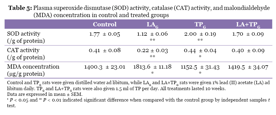
Malaysian Journal of Medical Sciences, Vol. 17, No. 3, 2010, pp. 13-18 Original Article Lycopersicon esculentum (Tomato) Prevents Adverse Effects of Lead on Blood Constituents Emmanuel O Salawu Department of Physiology, Faculty of Basic Medical Sciences, Ladoke Akintola University of Technology, Ogbomoso, Nigeria Correspondence: Emmanuel O Salawu, BTech (Physiology), Department of Physiology, Faculty of Basic Medical Sciences , Ladoke Akintola University of Technology, Ogbomoso, Nigeria, Tel: +234-(0)8056916409 Email: seocatholic@gmail.com Submitted: 24 Oct 2009 Code Number: mj10027 Abstract Background: Lead is known for its adverse effects on various organs and systems.
In this study, the ability of lead to adversely affect blood parameters was
investigated, and Lycopersicon esculentum, or commonly known as tomato (a source
of antioxidants), was administered orally in the form of tomato paste (TP)
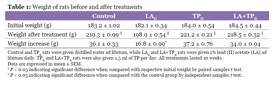
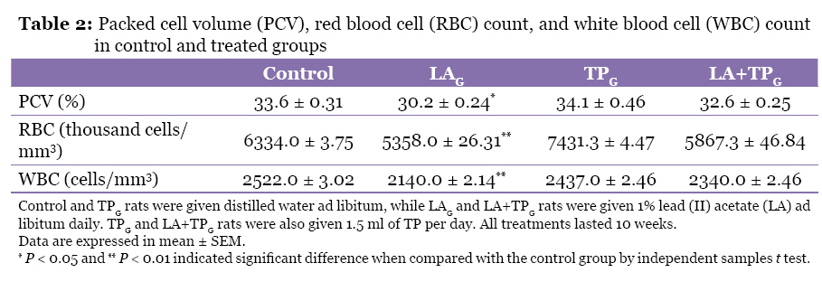
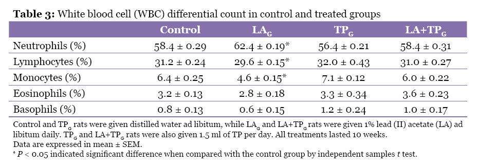
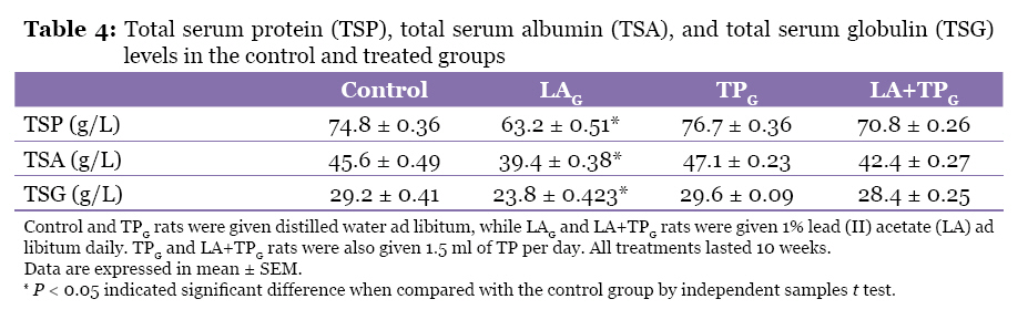
to reduce the adverse effects of lead. Keywords: anaemia, antioxidants, biochemical marker, lead, Lycopersicon esculentum, medical sciences Introduction Lead, a heavy metal, is harmful even in small amounts. Nevertheless, humans are exposed to lead through their environments and diets (1). Lead can be inhaled in dust from lead-based paints (2,3) or from fumes of leaded gasoline. It is found in trace amounts in various foods, notably in fish, which are heavily subjected to industrial pollution. Some old houses may have lead water pipes, which can easily contaminate drinking water (3). The manifestations of lead poisoning in humans are non-specific and may be hard to detect during the initial stages. Symptoms include weight loss, anaemia (4), memory loss (5), nephropathy, and infertility (6). Previous research has established that oxidation accompanies lead toxicity (7). This finding suggests that an ample source of antioxidants could reasonably prevent (or at least suppress) the manifestations of lead toxicity. Lycopersicon esculentum, commonly known as tomato, is a good source of antioxidants (8). It contains nutrients that prevent illness, e.g., by detoxification (8, 9), promoting growth (10) and proper immune system functioning (11), as well as increasing the haematocrit, RBC, and WBC content (4). Cooking helps to liberate the beneficial components of the tomato (lycopene, glutathione, vitamin C, and vitamin A) and also facilitates their absorption in the gastrointestinal tract (12,13). There is a possibility that oral administration of cooked tomatoes could reasonably prevent (or at least suppress) the manifestations of lead toxicity. Therefore, this study focused on the potential of oral administration of cooked tomatoes in preventing or suppressing lead toxicity. Materials and Methods Animals A total of 56 adult male Sprague–Dawley rats (average body weight 183.45 ± 0.903 g) were used for this study. They were inbred at the Animal House of the Faculty of Basic Medical Sciences, Ladoke Akintola University of Technology, Ogbomoso, Nigeria. The rats were transferred to the research section and acclimatised over a period of 2 weeks. The research section was maintained at a temperature of 24–26 °C, relative humidity of 70%–75%, and a light/dark cycle of 12/12 hours with adequate ventilation throughout the period of research. The rats were fed with commercially available rat pellets. The maintenance, care, and treatments of the rats complied with National Institutes of Health guideline for the humane use of laboratory rats and the guideline of Ladoke Akintola University of Technology. Preparation of tomato paste Since no published method is available for concentrating the tomato paste, the following in-house method was adopted. Furthermore, cooking has been shown to liberate the beneficial/active components of tomatoes (lycopene, glutathione, vitamin C, and vitamin A) and facilitate their absorption in the gastrointestinal tract (12,13). Tomato paste (TP) was prepared by grinding tomatoes and heating them in a water bath at 80 °C for 45 to 60 minutes. The obtained TP was removed from the water bath when its relative density was 1.124. It was then allowed to cool to 25 °C, and then stored at 4 °C. The quantity of TP to be administered was thawed to 25 °C 1 hour prior to administration. Treatments The rats were randomly divided into 4 groups: 1) control given distilled water, 2) LAG given 1% lead (II) acetate (LA), 3) TPG given distilled water and 1.5 ml of TP per day, and 4) LA+TPG given 1% lead (II) acetate (LA) and 1.5 ml of TP per day. All groups consisted of 14 rats each. All treatments lasted 10 weeks. Sample collection and processing Each animal was weighed and sacrificed by cervical dislocation 24 hours after the last treatment. Blood samples were collected via cardiac puncture. The blood obtained from each rat was injected into 2 tubes; a plain (no-additive) tube and an EDTA tube. The blood samples in the EDTA tubes were mixed thoroughly (although gently) with the EDTA. All the blood samples were then stored below room temperature (10–14 °C) prior to the measurement of haematological and biochemical parameters. Measurement of haematological and biochemical parameters and data analysis Packed cell volume (PCV) was determined using plain capillary tubes filled with anti-coagulated blood and centrifugation at 3000 rpm for 20 minutes. Red blood cell (RBC) and white blood cell (WBC) counts were determined using an improved Neubauer counting chamber following the procedure documented by Cheesbrough and McArthur (14). Field’s stain A and stain B were used for the differential WBC counts. Plasma and serum were obtained by centrifugation of whole blood at 3000 rpm for 20 minutes. To serve as a guide, total serum protein (TSP), total serum albumin (TSA), and total serum globulin (TSG) levels were first estimated using the Levibond comparator (i.e., the Biuret method) following the method documented by Cheesbrough and McArthur (14), and accurate (reported) values for TSP, TSA, and TSG were then determined using photo-electric colourimeter. The total plasma protein (TPP) was on the other hand determined using the following formula derived by Kagan (15): P = 340(G-1.0099) where P is the total grams of protein per 100 cm3 of plasma and G is the specific gravity of plasma at 25 °C. Plasma superoxide dismutase (SOD) activity was determined using the method described by Fridovich (16). Plasma catalase (CAT) activity was determined using the method described by Sinha (17). Plasma malondialdehyde (MDA) concentrations were determined using the procedure described by Varshney and Kale (18). SOD, CAT, and MDA were chosen because many previous studies have reported their reliability in the measurement of antioxidant activities and level of lipid peroxidation (19–22). The data obtained are presented as mean ± SEM. The control and the treatment groups were compared using the independent samples t test using SPSS version 14 (SPSS Inc, Chicago, IL). The level of significance was set at a P < 0.05. Results Weight gain Comparison of the initial and final weights of rats showed that there was significant weight gain (P < 0.05) in all the groups at the end of 10 weeks of treatment (Table 1). There was no significant difference in weight gain in the TPG, LA+TPG, and control groups. However, the increase of weight in the LAG group was significantly lower than that of the control group (P < 0.05). Haematological parameters Haematological analyses indicated that LAG rats (unlike TPG and LA+TPG rats) have significantly lower PCV, RBC count, and WBC count (P < 0.01 each) (Table 2), as well as lower percentages of lymphocytes and monocytes (P < 0.05 each), but significantly higher percentages of neutrophils (P < 0.05) when compared with the control group (Table 3). The percentages of eosinophils and basophils for each of the groups were, however, not significantly different from those of the control group. Protein content TSP, TSA, and TSG levels for LAG rats were significantly lower (P < 0.05) than those of control group, while the levels in TPG and LA+TPG rats were not significantly different from the control group (Table 4). Antioxidant activities and lipid peroxidation LAG rats had significant decreases in plasma SOD and CAT activities (P < 0.01); whereas TPG animals had significantly higher SOD (P < 0.01) and CAT (P < 0.05) activities when compared with the control group (Table 5). LAG rats had significant increases in plasma MDA concentration (P < 0.05), while TPG had significant decreases in MDA concentration (P < 0.05) compared with the control group. On the other hand, LA+TPG rats had no significant differences in SOD activity, CAT activity, and MDA concentration compared with the control group. Discussion The results showed that chronic exposure to lead significantly reduced weight gain, which support the findings of Wadi and Ahmad (23) and can be linked to less-efficient metabolic processes due to lead toxicity (24). Administration of 1.5 ml TP/day, however, eliminates the adverse effect of lead on weight gain. This result would be chiefly due to presence of protective antioxidants in TP, such as lycopene, vitamin C, and vitamin A (7), even though it has low protein content and low caloric value (18 kcal/100 g). Similar reasons could be attributed to the significant decrease in PCV observed in rats exposed to lead (LAG) and the non-significant difference in PCV observed in rats treated with TP and lead (LA+TPG) when compared with the control group. The finding that lead decreased PCV agrees with the findings of Anetor et al. (25) that some indices of erythropoietic activity, such as haemoglobin (Hb) concentration, packed cell volume (PCV), and mean corpuscular haemoglobin concentration (MCHC), were significantly decreased in workers exposed to lead compared to the control group. However, Franson et al. (26) reported that PCV is not altered in birds after supplementation of 50 ppm of lead in their feed, and Arvind and Chopra (27) also reported no changes in PCV in calves after supplementation of 100 ppm of lead in their diets. This may considerably be due to the low concentration of lead (50 ppm or 0.005%, and 100 ppm or 0.010%, respectively) administered, and/or the different species used in the experiments. There was no significant decrease in RBC counts in animals co-treated with TP and lead. In contrast, the RBC counts in LAG rats were significantly lower than those of control group. The reduced in RBC counts may be caused by lead interference in the energy metabolism of erythrocytes (28), which affects the ATP concentration in erythrocytes, thus, shortening their life span and lowering the blood counts. Morse et al. (29) reported that acute lead toxicity in mice produced transient erythroid hypoplasia and impaired utilisation of RBC 59Fe for haem synthesis. The administered TP would therefore be responsible for preventing these effects of lead on RBC probably by preventing the adverse effects of lead on the energy metabolism of erythrocytes and the utilisation of RBC 59Fe for haem synthesis and/or by elevating RBC glutathione level (a function of vitamin C component in tomato), which protects RBC against damaged caused by hydrogen peroxide (a toxic by-product of many metabolic reactions) by reducing the peroxide to water (30). In a similar manner, WBC counts in rats co-treated with TP and lead were not significantly different from those of control group. Conversely, rats treated with lead had significant decreases in WBC counts. This result suggests that TP combats the adverse effects of lead on WBC, resulting in no significant differences in WBC counts observed in control group and in rats co-treated with TP and lead. This finding was most likely due to the detoxification effect of some antioxidant components of TP. Similar detoxification/protective effects have previously been documented for varieties of antioxidants, such as carotene, retinol, and bioflavonoids (31, 32). Lead caused significant decreases in lymphocyte and monocyte counts but increased neutrophil counts. These results support the finding by Sembulingam and Sembulingam (33) that lead (like some other chemicals and drugs, e.g., mercury, camphor, benzene derivatives, venoms, and some vaccines) causes neutrophilia. Di Lorenzo et al. (34) also reported that mean absolute neutrophil counts were significantly higher in workers exposed to lead compared to workers who were not exposed to lead. TP prevented lead-induced changes to WBC and differential counts. The percentages of eosinophils and basophils in rats exposed to lead alone and those of rats treated with TP after lead exposure were not significantly different from those of control group. The significantly lower TSP, TSA, and TSG levels in rats exposed to lead could result from the fact that lead causes disturbances in metabolism, reduces the efficiency of gastrointestinal tract digestion and absorption processes, and negatively affects protein synthesis (4,25). TP, in contrast, was able to annul the adverse effects of lead on TSP, TSA, and TSG levels of rats co-treated with TP and lead. This effect was perhaps due to some protein (sources of amino acids) components of TP (which might be used as raw materials for, or facilitate, aminogenesis/proteogenesis) and could, better still, be traced to its vitamins and lycopene components and TP ability to modulate gastrointestinal tract digestive and absorptive functions (9). There were significant decreases in plasma SOD and CAT activities in lead-treated rats, while in rats co-treated with TP and lead, no significant differences were observed in comparison with the control group. These findings are in agreement with those of Hsu and Guo (35). Although plasma MDA concentration was significantly increased in lead-treated rats, no significant difference was observed in rats co-treated with TP and lead in comparison with the control group. This confirms that TP, as a source of antioxidants (7, 8), reduced the oxidative stress caused by lead exposure in this animal model. Conclusion Exposure to lead significantly reduced weight gain; PCV, RBC count, and WBC count; percentages of lymphocytes and monocytes; TSP, TSA, and TSG levels; and plasma SOD and CAT activities. In contrast, it caused significant increases in the percentages of neutrophils and eosinophils, and plasma MDA concentration. TP, however, significantly reduced these adverse effects of lead. It would be good if subsequent studies could examine if human consumption of considerable (high) amount of TP could have protective effect against lead toxicity, and if prolonged consumption of TP has any potential adverse effects in humans. References
© Copyright 2010 - Malaysian Journal of Medical Science The following images related to this document are available:Photo images[mj10027t5.jpg] [mj10027t1.jpg] [mj10027t2.jpg] [mj10027t4.jpg] [mj10027t3.jpg] |
| |||||||||
